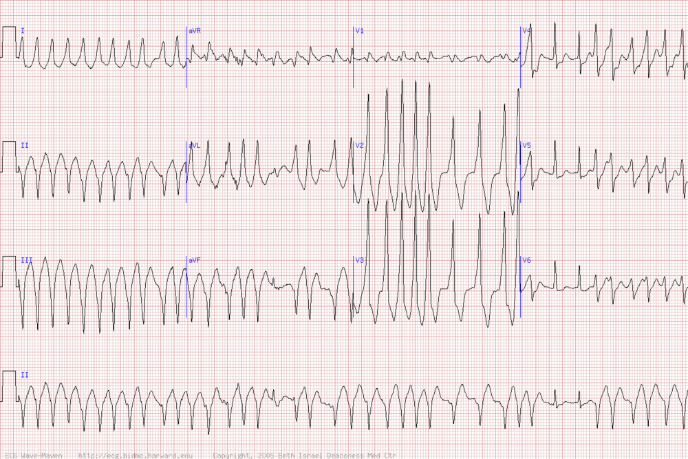
4/2/14 Conference notes

Dr. Girzadas is at CORD, poor substitution by C Kulstad

8-9: CV Study guide by Dr. C Kulstad

9-930: Safety lecture by Dr. Balogun

Presentation of 4 cases with various chief complaints united by delayed urine testing. 89% of patients with GI/GU complaints have documented UCG at ACMC. Takes 2.5-4 hours after registration to get UCG performed here.  Reasons UCG is delayed- patient can’t urinate, sample there but not processed, not considered important, or ED busy (staff overwhelmed, sample lost).

Important to avoid delay in imaging, psychiatric placement, medication choices.

Ideas for improvement 1) expand and enforce triage UCG orders 2) order UCG for patient of childbearing years who might need medications or imaging before you see them 3) talk with nurse/tech 4) give pt urine cup and instructions and/or perform urine hcg 5) straight cath when UCG is critical to management

Pushback- physicians don’t need to take on additional roles unless emergent need. Compromise- give cup and instructions then notify tech/RN that pt is giving sample

930-12 U/S lecture and workshop- Dr. Lambert

Lambert finds 1st trimester pelvic ultrasound most meaningful ED ultrasound  for patient management.

Anatomy reminders- pelvis tilted 45 degrees anteriorly when upright. Just behind symphysis pubis will be the bladder. Uterus tends to be horizontal. There is peritoneal reflection between bladder and uterus- not normal to have fluid here. Another peritoneal reflection between uterus and rectum (aka pouch of Doulgas)- very common to have small amount of fluid here in normal patient.


Ovaries are next to external iliac vessels, just anterior to ureters.  Bowel gas can obscure visualization of uterus, helps if bladder is distended.

Yolk sac should be visible by 5 wks. Often see cardiac flicker just afterwards, at  5-6 wks.

Ways to image pelvis- Transabdominal- place probe just above symphysis pubis. Use bladder as acoustic window. Less invasive and good field of view. Lower frequency probe means lower image quality. Uncomfortable to press on distended bladder.

Obtain transverse and sagittal views. Sagittal- indicator points to umbilicus. You will see bladder (triangular at top of screen) then uterus (pear shaped). At inferior part of screen- see vaginal stripe towards left screen. Should see endometrial and vaginal stripe in one view, that shows you are in midline. Transverse- Indicator to right. Top of screen will show bladder (rectangular) with uterus (oval) posterior. Ovaries may be seen inferior to bladder at edges of uterus (often are not seen).  


Sagittal                                                                            Transverse

 Transvaginal- better because probe is closer to organ of interest, and the high frequency probe gives better images. Not than uncomfortable, especially compared to pressing on full bladder. Get wide field of view but not as much depth. Anatomic relationships can be confusing. Obtain sagittal and coronal views.

Sagittal- indicator up. Think of flipping the sagittal transabdominal view 90 degrees counterclockwise. See long axis of uterus with cervix towards right of screen (opposite of indicator side). May need to tilt probe by moving back of handle down (tip of probe to ceiling). Bladder may be visible at top, left of screen.

Coronal- see slices of uterus from cervix to fundus in short axis- back of handle down to see fundus. May get better images by backing probe out a little bit (make sure you don’t allow air gap which would degrade image quality a lot).

Ovary – 2 x2 x3 cm- should be oval and have peripheral follicles, may have to slide probe lateral to cervix and a bit deeper. They should be inferior/medial to iliac vessels (can use color flow to identify vessels).

Cystic structure on ovary in pregnant patient is corpus luteum- usually a couple of cm but can get up to 6 cm. Starts regressing at 6 wks

Endometrial stripe- hyperechoic inner part of the uterus, has 3 layers. Decidua is the same thing as endometrium, just in a pregnant patient. Double decidual sac is endometrium over embryo, seen at 4 wks.

At 5 wks, see yolk sac which looks triangular inside decidual sac.

60% of u/s done in ED for r/o ectopic in first trimester will show IUP clearly. Greatly decrease time to patient dispo. Improve patient satisfaction as you spend more time with patient. Plus, unstable ruptured ectopic patients can’t go to ultrasound.

Diagnostic criteria for 1st trimester ultrasound

Live IUP- gestational sac at least 5 mm internal diameter within the endometrial echo of uterus with 1) fetal pole and 2) heart beat

IUP- same criteria but without cardiac activity.

Abnormal IUP- same criteria but 1) gestation sac > 10 mm w/o yolk sac or 2) gestational sac > 16 mm and no fetal pole or 3) obvious fetal pole w/o cardiac activity

Extrauterine gestation- gestational sac at least 5 mm internal diameter- outside endometrial echo and one of the following 1) yolk sac or 2) fetal pole.  This is why landmarks are so important! Ectopic pregnancies often look like they’re in the uterus if you do a cursory ultrasound.

No definitive pregnancy- 1) normal uterus or 2) sac that isn’t big enough yet or 3) gestational sac with yolk sac or fetal pole.

15-20% of ultrasounds end up being no definitive IUP. About 30% of those will end of being ectopic- rate increases with free fluid or mass seen. Must have good follow-up arranged.


Hands-on practice

Feb 12, 2014- CKulstad subbing for Dr. Girazadas who is attending AAEM

8-9 Procedural sedation Study guide-  Lovell

Minimal sedation = anxiolysis. Spontaneous breathing, airway unaffected.

Moderate sedation- purposeful response to verbal or light tactile stimuli. Standard examples- midazolam/fentanyl

Deep sedation- what actually happens during procedural sedation most of the time. Responds only to painful or repeated verbal stimuli. Airway reflexes may be lost, respiratory effort may be inadequate.

General anesthesia- all reflexes lost. Must support airway and possibly CV system

Dissociate sedation- trancelike, cataleptic state. Airway reflexes maintained

Sedation is a continuum- be prepared to treat someone on a stage deeper than you intend.

NPO-ACMC: No solids 8 hrs prior, no clear liquids 2 hrs before. Anesthesia national guidelines say 2 hrs npo for clear liquids, 4 hrs for breast milk, 6 hrs for solids. Harwood adds these guidelines originally derived from c-section data on term pregnant women who have very high vomiting risks

This is out-dated. ACEP guidelines say fasting rules not supported by evidence.

ASA classifications- procedural sedation generally only for patients in category 1-3. Add “E” to the category for “emergency” and you’re saying you have to do procedure (pulseless limb s/p dislocation)

1-      Healthy

2-      Mild systemic disease that is well controlled

3-      Severe systemic disease- eg symptomatic wheezing in COPD

4-      Life-threatening illness

5-      Dying patient

6-      Brain death

Tips for safe sedation- use monitoring (ECG for hx of cardiac patients). You’re in charge of everything. If you use Demerol, it can cause CNS excitation (seizures). Fentanyl can cause respiratory depression, esp in elderly. Generally better to give meds slowly and titrate doses.

Dosing

Midazolam (Versed) 1mg (0.025 mg/kg)

Fentanyl (Sublimaze) 25-50 mcg (0.5-1 mcg/kg)

Flumazenil for benzodiazepines only 0.1-0.2 mg

Narcan for opioids only- suggested dosing- dilute 0.4 mg in 10 ml normal saline, then 1 ml/dose

-5 ways to decrease pain of local anesthesia- add bicarb, warm it, inject slowly, use small needle, distract patient, inject through wound margins

-Eutectic mixture of local anesthetic is what EMLA stands for. Other fast acting topicals- LET (lidocaine- epinephrine-tetracaine) for not-intact skin, ELA-Max-liposomal lidocaine for intact skin

-Side effects of ketamine- rare laryngospasm. More likely with suctioning or with lots of secretions. Usually able to bag someone through it. Can use in kids 3 months or older. OK to use in head injury but not in hydrocephalus or known increased ICP. Additional benzos not recommended for kids, is for adults.

-Treatment with opioids in ED is not the same for all racial/ethnic groups (JAMA 2008). Be aware of your biases.

-Can treat benign headache with injection of local anesthetic (bupivacaine) in paraspinous muscles in lower cervical spine. See EMRAP or Youtube for more details (Dr. Mellick).

-Local anesthetic allergy: 2 classes amides (lidocaine, bupivacaine, prilocaine- all have ”i” before “caine”) esters (procaine, benzocaine, tetracaine). If allergic to both, can inject diphenhydramine as local

-Toxicity of lidocaine- CNS (seizure, coma) then CV (dysrhythmias, myocardial depression). Before that get symptoms that sound like anxiety (perioral numbness, not feeling right). Treat with benzos then amiodarone. Short lived toxicity which Andrea has never seen. Intralipid for bupivacaine overdose.

-Nitrous oxide needs to have a well-ventilated room, can go into gas filled cavities so avoid in ptx, sbo, balloon-tipped catheter. Altered patients should not get NO as patients control their dose.

-Discharge requirements- ambulate, responsible person to watch (no driving for 24 hours), normal vital signs. See modified Aldrete score for more details

-Max dose local anesthetics: lidocaine 4 mg/kg plain 7 mg/kg with epi. Bupivacine 3 mg/kg plain, 5 mg/kg with epi


9-930: Geriatrics in ED – Beckemeyer

Geriatric population is increasing- 20% by 2039, and 25% in 2050. And >65 fill out surveys.

Every ED visit  at age >65 is a sentinel event for further decline so transitions in care critical. Social work, home visits, prompt PMD visit, action plan for decompensation.

4/2014 roll out date for new geriatric ED experience. Goal- age >65 is no wait for all complaints. Go to “senior care area” in former GC front area. Have specific assessment protocols (ADL/med questions, fall risk, social support), and specially trained techs.

Will also have specific protocols so care can be started prior to MD eval for common geriatric problems.

Discharge packet will include senior specific support info (eg meals on wheels).

Will use grants for inexpensive changes- eg clocks with large numbers, magnifying glasses, bedside commodes, more comfortable carts, more pillows and blankets available, possible carry Ensure in ED.

Try to speak more slowly and loudly- face patient at eye level. Elderly patients often overwhelmed/scared in ED.

930-10: Safety lecture- Cash

Had unscheduled downtime early Jan- Firstnet, allegra, pacs, phones all down. Code triage called, went on bypass. Concern for safety issues.

Issues- unclear if orders when through prior to downtime. Delay in identifying which patients needed to be seen. Hard to track workup for find results. Hard to identify acuity, when patient for which team. Patient lost to system. No one familiar with paper system.

Scheduled downtime for computer maintenance- extra staff and materials ready. There are few downtime computers throughout ED- very basic list of patients. One for main room adults, one for fasttrack and peds.

Use whiteboards and paper packets. Whiteboards will list patient by color (red, blue, and black for gold team). Make sure papers have patient stickers. Lab results are faxed to ED and placed in physical chart- you have to keep checking.

To discharge patients find a downtime computer and hit depart. Discharge instructions under “patient ed” which pulls up your usual discharge instructions. Can also use uptodate patient instructions.

10-1030: Advanced DKA

Brian Febbo is a second year resident.

DKA defined by serum or urine ketones, glc usually >250, anion gap acidosis

Euglycemic DKA (below 250) exists. It is not equivalent to mild DKA.  Associated with continued insulin use as DKA develops, pregnancy, and starvation.

If a patient has low albumin, the anion gap may be falsely low. Correct by adding 2.4(4.4 – [albumin]).

Mixed acid-base disorders are common- vomiting can falsely normalized pH.

Serum ketones would be very helpful but are not available in ED at ACMC. Urine ketones have other causes, so less specific.

HHS- older, sicker, more dehydrated. Will need much more fluids. Give IVF alone for first couple of hours as they will significantly drop glucose and potassium. These patients need extensive workup.

Look for precipitating factors. Usually infection, lack of insulin, or other critical illness (medications, pancreatitis, MI, PE, other endocrine abnormality).

But non-specific lab abnormalities are very common- especially leukocytosis and lactate- so difficult to diagnosis.

Management- Fluids early and aggressively. Use isotonic fluids, will need to change additives throughout.

Insulin- 0.1 U/kg/hr or 0.14 U/kg/hr gtt. If you want to give a bolus, it is also 0.1 U/kg but utility is questionable.

SubQ  insulin used to good effect in mild or moderate, stable DKA patients. They need an IV or IM bolus dose.

Remember about pseudo hyponatemia- use corrected to calculate AG

Hyperchoremic acidosis common with large volume IVF resuscitation so consider using LR

Must check potassium before starting insulin- can trigger malignant arrhythmia if it was low and you give insulin.

DKA in ESRD- total body water is near normal, most in extracellular space. Treat with insulin gtt alone.

Pediatric DKA- no evidence that aggressive IVF repletion causes cerebral edema but standard practice in US is to replete fluids over 48 hours.

1030-1230 Sedation small groups


Conference Notes 11-5-2013

Last guest conference notes, Dan’s back in country soon. This week back to Christine Kulstad.


8:00-9:00- Oral Boards- Felder vs Wise

Case 1- Thyrotoxic paralysis. Critical actions- treat hypokalemia and thyrotoxicosis. This is rare disorder with painless muscle weakness- this is a subcategory of periodic paralysis. Usually in SE Asians, more often men, usually young adults. Respiratory and bulbar muscles are not affected, and arrhythmias are uncommon. Weakness is proximal > distal, legs >arms. All symptoms resolve after treatment.  Treat with potassium, but high dose can cause rebound so give lower initial than you might think. Propranolol will move potassium into serum where it is lacking (total body potassium is fine, just shifted into muscle cells during attacks), dosing is 1 mg q10 to max of 3 mg/kg. Treat hyperthyroidism as usual. Have patients avoid high carbohydrate meals, fasting and heavy exercise as they can precipitate episodes. Chronic potassium supplementation is ineffective.

Case 2- Toxic shock syndrome from skin abscess. Treat sepsis as usual, drain abscess. TSS is caused by either Staph aureus or group A strep. Was associated with tampon use in past, now more with wound-packing or skin infections. Staph will rarely be found in blood cx (GAS does yield + blood cultures). Symptoms arise from exotoxins that staph produce- commonly see fever, hypotension, skin manifestations. May also have chills, malaise, v/d, sore throat, headache. DDx of ill young patient with rash- TSS, RMSF, meningococcemia. Treat with clindamycin and vancomycin. Remember to treat septic oral boards patients with EGDT as you would in the ED.

Case 3- FB aspiration in a child with prolonged cough. Found with air trapping on decubitus x-rays. May also see atelectasis, mediastinal shift, pneumonia.  If FB is left long enough, air in alveoli will be absorbed causing atelectasis. Infection often develops- first pneumonia and then pulmonary abscess and bronchiectasis are possible. Take-home point: do more evaluation (at least xrays) in child with prolonged cough.


9:00-10:00- EMS Study Guide- Motzny (and How to Survive the Zombie Apocalypse)

Triage: When triaging during a disaster, green is walking wounded. Remember triage is dynamic and will require reassessment.

START and SAVE are 2 triage systems. SAVE (secondary assessment of victim endpoint)- determines who will benefit significantly from austere field interventions. Patients who will die if not treated in field (vs those whose fate will not change if treated because too critical or not that bad). START (simple triage and rapid treatment) uses a quick assessment of respirations, perfusion, and mental status. Assess respitations first, if repositioning airway dosen’t fix it they are black/dead. If it does or breathing more than 30- red (immediate rx). If not, check radial pulse. If absent, red. If normal, check MS. If patient can respond they are yellow. Otherwise red.

Disasters: Internal disaster- catastrophic problem in the hospital (power failure and generator failure). External or just a disaster is defined by the capacity of the surrounding hospitals to deal with patients. E.g. 40 victims in a rural area is a disaster while 100 in a large urban area is not.

JCAHO requires a hospital’s disaster plan to be activated twice a year for accreditation.

Communications is consistently identified as a problem during disaster drills. Causes can be failure of equipment and unclear terminology, chains of commands. The first step when you are notified of a disaster is to verify the information (before activating the disaster plan).

Mass gatherings are defined as 1000 people in one site for a common purpose. The most common complaint of patients at a mass gathering is dermal injury (cuts/scrapes).

Medical control from the EMS physician is indirect and direct. Direct is real-time contact with paramedics, whether over the radio or at the scene. SMO (standing medical orders) give indirect guidance for many common problems. Physicians cannot assume control of a scene if unknown to EMS UNLESS you have proof of licensure.

HAZMAT command centers should be set up uphill and upwind. Many toxins are heavier than air (so go uphill), if lighter than air they will soon rise above your head. When dealing with patients with significant chemical exposure, eyes take precedence in decontamination. No procedure (including airway) should be done without decontamination unless you are wearing the appropriate protective gear (HAZMAT suit) and then get decontaminated yourself.

Weapons of mass destruction include chemical, biologic, nuclear, radiologic, and explosive agents. Biological agents classified as Class A agent of concern by CPC are smallpox, anthrax, plague, tularemia, viral hemorrhagic fevers, botulism- all the agents that you think of as really bad. Others are class B if they could be used but not effectively or not that toxic (e.g. salmonella).

Radiation- alpha, beta, and gamma. Alpha particles will not penetrate skin or clothes, only dangerous if ingested. Beta goes a couple of centimeters (lead protects). Gamma goes all the way through (like xrays) and will give whole body irradiation. The good news is you don’t have to decontaminate gamma since its still going. The earliest indicator of radiation injury is lymphopenia. 

Hypoxemia and hypoperfusion are not caused by helicopter EMS transport generally because they don’t fly very high, can factor in fixed wing aircraft transport (planes). Decreased air pressure in planes can 1) increase fluid rates in medications given in glass bottles as gas expands 2) increase pressure of pneumothorax increasing the size 3) tracheal cuff on ETT may pop (use saline instead of air). If you are approaching a helicopter, avoid the rear.

NIMS (National Incident Medical System) standardized terminology and procedures. It states material managment is logistics section responsibility. For more info, www.fema.gov/emergency/nims

10:00-10:30- Discharge Instructions- Kiernicki-Sklar

Don’t make the mistake of thinking discharged patients are not sick or someone’s problem. CRICO 2011 benchmarking report summarized 200k EM malpractice cases, they found #2 cause of malpractice cases related to missed/delayed diagnosis (majority of cases) was development of discharge plan.

Think of d/c instructions as sign-out to the patient. Other functions as a patient care summary, contract with patient (if patient doesn’t follow instructions and suffers harm, care contract is breached).

Patients don’t remember verbal instructions well, make sure you write down important information too.

Mnemonic to remember how to provide good d/c instructions. WTF DR DC?

W- What we did or didn’t find. Include incidental findings (lung nodules, elevated BP).

T- Treatment/tests that are still needed (have BP rechecked, have a stress test in 72 hrs, have sutures removed). Include risks of non-compliance.

F- Follow-up required. Timed f/u when undifferentiated abd pain (24-28 hrs), significant wound checks (1-2 days), chest pain (72 hrs). Stress that patient needs to call to make appointment (or if specific time arranges). Make effort to contact f/u physician in high-risk cases.

D- Drug warnings- legally it is your responsibility to provide warning and check for allergies/interactions.

R- Restrictions. Things that may worsen a current or undiagnosed condition (undifferentiated CP and exertion, seizures and swimming/bathing/driving). Not only could patient sue you, but 3rd party could (driver who seized and hurt someone else).

D- Diagnosis. Don’t feel need to make a diagnosis when it is not clear (abdominal pain vs GERD). Two reasons, easier to sue for misdiagnosis in case 2 and causes anchoring bias in physician who next sees patient.

C- Comeback (what to return for). Highlight specific concerns based on that patient’s problems, don’t assume that they will read all the pages of the pre-formatted d/c instructions. Include contingency plan if f/u cannot be arranged (patient can always return to ED as last resort and should be encouraged to do so).

?- Final checks. Re-check vital signs, reassess pain. Use simple words (6th grade level), and avoid medical terms and abbreviations.

Brief discussion of AMA- think of as informed non-consent. Must discuss and document 1) risk/benefit/alternatives 2) Pt has decision making capacity 3) Patient has understanding of #1 4) patient can ask questions and get answers. Give them good discharge instructions and any treatment they will accept.

10:30- Musculoskeletal Exam- Hands-On Skills Station

You had to be there

Conference Notes 10-29-13

Today's guest for conference notes is Elise Lovell. Don't worry, only one more Tuesday until Dan's back.

Bonus, a tox lecture by alumni and tox fellow Janna Villano.

00 am  Orbital Trauma/Oculoplastics-Dr. Hassan Shah; he’s happy to be involved with any Oculoplastics questions!

Ruptured Globe:  How to tell?  Peaked/irregular pupil, full hyphema, 360 degree subconjunctival hemorrhage, low IOP.

Most important reason to fix orbital fractures?  Prevent diplopia.

What does optho need to know about intraocular foreign body?  High pressure à systemic steroids.  Ruptured or not ruptured.  Visual acuity.

Blunt eye injury: “Blown pupil” actually not helpful to tell if optic nerve injured...can have temporary pupil dilation simply due to blunt injury.  APD however IS a sign of optic nerve dysfunction.  If APD, high pressure, loss of vision, need to perform lateral canthotomy/cantholysis.  Dr. Shah believes cantholysis important; simple canthotomy usually not enough, and most common mistake is to think you’ve relieved the pressure but you haven’t done enough.  He can fix any eyelid laceration that you cause!
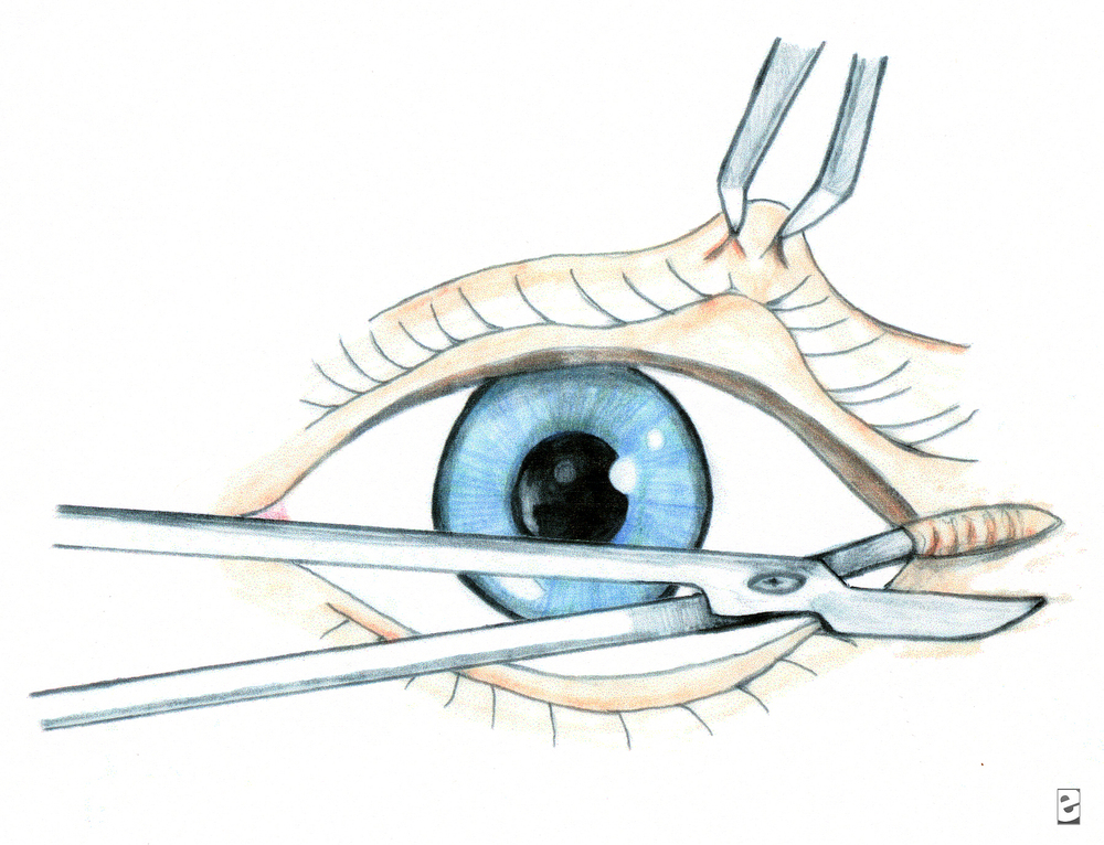
 

This is lateral canthotomy.  You also need to cut up and down (3 total cuts) for complete cantholysis.

Be sure to cover the eye in the setting of eye trauma.  Use ointment, try to bring lid back down over eye.  Best covering for globe is the conjunctiva.

Blow out fractures.  Adults almost never needs to be repaired emergently.  Even if “entrapment”, usually due to soft tissue swelling.  Usually wait for one week before repair to allow swelling to resolve.  Big fractures much less likely to get true entrapment than small fractures.  Big fractures = lots of swelling, but also room for movement.  For kids, they get “greenstick” type fractures of the orbit; softer bones, the fracture is often very small, with mild swelling, but with restricted ROM up and down due to trapped muscle àthis is case for same day surgical repair.  If you don’t have diplopia on Day 1, you will not develop it!

Facial fractures and antibiotics:  He doesn’t usually give antibiotics unless contralateral sinus disease, but there is controversy, not one correct answer.  He has never had an infection.

9:00 am  Janna Villano: - Tox-Uses of Sodium bicarb, and some controversial Antidotes

Most important 4 things in Tox:  Vital signs, Mental Status, Skin and Neuro exam


Many Sodium Channel blockers out there:  TCAs, cocaine, some antiepileptics, dysrhythmics...lots of others.

3 Uses for Sodium Bicarb:  Cardio toxicity, Prevent acidosis, Urinary alkalinization

Bicarb:  Treat cardiotox:  helps with prolonged QRS, wide complex tachycardia, hypotension.  Give 1 to 2 meq/kg bolus over 1-2 minutes.  Boluses better than infusions.   Give boluses and sit there watching for QRS to narrow.

Bicarb:  Prevention of academia:  maintain pH >/= 7.4.  Prevents drug distribution, may be useful peri-intubation of tox patients.

Bicarb: Urinary alkalinization:  enhances elimination.  Maintain pH 7-8.5, ion trapping, must maintain normal K levels for this to work-aggressive K replacement!


Anticholinergics:  antihistamines, TCAs, vertigo meds, antipsychotics, Jimson weed...etc.

Physostigmine:  To treat anticholinergic toxicity.  Will allow history when patient’s mental status clears.  Should see nearly complete resolution of symptoms in order to attribute presentation to anticholinergic  toxicity.  Allows diagnosis, limits workup.  Can cause seizures, GI distress/rest of cholinergic symptoms.  Reports of physostigmine associated with cardiac arrest , but these in patients with TCA OD, also cases of seizure in patients with prior seizure disorder.  If giving physo, watch for bradycardia/heart block,  seizure, cholinergic side effects.  Give 0.5 mg over 2-5 minutes, may repeat up to 3-4  doses.  If necessary can “reverse” physo with atropine.  ***Janna doesn’t give in patients with known seizure disorder, patient who took pro-convulsant, or patients with sodium blocker OD (eg TCA)***


Flumazenil:  Don’t use drug screen to help you decide whether or not to give it-drug screen misses many benzos.  Some studies have shown avoidance of intubation.  Why not to use it in a documented benzo OD?  Inconsistent clinical response, precipitation of benzo withdrawal (rare, and can give benzos), may precipitate seizures (seen in pts who took proconvulsant, or in patients using benzos for sz disorder), maybe risk of complications outweighs harm from OD.

Pick the right patient-no seizure disorder, pro-convulsant use, not if chronic benzo users, not for undifferentiated coma or prophylactically.  Often used in kids, pure benzo OD, procedural sedation.   If patient has seizure after flumazenil, then just flood patient with benzos (competive inhibitor).


10:00 am Dr. Khatiyar-  Chemical Asphyxiants

Fires:  Simple Asphyxiants, Chemical Asphyxiants, Chemical Irritants

Carbon Monoxide:  from partial combustion:  broken furnace, blocked car or boat exhaust, grilling in a garage, heating homes with gas ovens, car idling in garage...etc.  Also from Methylene Chloride, in degreasers/lacqueer thinner, bathrub refinishesàmetabolized to CO.

CO toxicity:  high affinity for hemoglobin forming CO-Hgb, but there are other toxic effects as well.  Also binds to myoglobin (heart tox, rhabdo), direct organ toxicity due to increased NO production and lipid peroxidation.  CO attachment to cytochrome oxidase (inhibits aerobic metabolism)

Think about it in headaches, “flu” in the fall when heaters turned on, kids with “colic”., gastro, viral syndrome, ..great imitator.

Diagnosis depends on a great history.  Ask about recurrent sx, others at home with same sx, time of year, in setting of Headache, flu sx, “hangover” symptoms, chest pain, neuro sx.

Diagnosis:  Breath sampling, CO level:  VENOUS just as good as ARTERIAL blood gas.  Don’t use O2 sat!  Check renal function, pregnancy test as changes management dramatically.  CPK, cardiac monitoring.  CO levels also can be unreliable, depending on treatment with O2, time since ingestion.  Levels:  < 10% no biggie, > 50% likely fatal, everything in between open to discussion.

Delayed neuro sequelae:  days to weeks later, incidence variable (1% - 47%), needs cognitive testing  (think mini mental status) and re-testing on followup.

Red flags:  pregnant women, kids, cardiac patients.

Half life 4-6 hours on room air, 60-90 minutes on 100% O2, 20 minutes with HBO.

Treatment:  HBO controversial:  but if level > 25%, any neuro issues/syncope, coma, Cardiac sx, abnormal cerebellar exam, symptomatic pregnancy or pregnancy with fetal distress à  call HBO chamber and discuss with specialist.  Put patient on 100% O2 as soon as you suspect CO.  Local chambers:  Advocate Lutheran General or Loyola, LCOM, Illinois Masonic, others.

Cyanide:  neuro sx, acidosis, seizures.    House fires, burning plastics, Nipride (sodium nitroprusside), certain foods-cassava, apple seeds, fruit pits, Laetrile.

Toxicity:  Neurotoxin, Enhances NDMA receptor activity, inhibits aerobic energy metabolism (blocks cytochrome oxidase).


Diagnosis:  Symptoms between 3 – 24 hours if oral, few minutes with gas exposure, sicker with premorbid conditions, kids, and dose dependent.

Presentation:  LOC/AMS, metabolic acidosis, GI, SOB, CV instability.  Be worried with rapidly developing coma, lactic acidosis, CV instability, symptomatic industrial worker/chemist.

Treatment:  decontamination (clothing, skin decon), 100% O2.  Old antidote:  Lilly cyanide antidote kit (amyl nitrate, sodium nitrite to cause methemoglobinemia which will bind cyanide, sodium thiosulfate to form thiocyanate which will be excreted in urine).  This kit usually available in ED, and this toxicity very time sensitive.  Cons:  causes hypotension, methemoglobinemia, multiple steps.

New antidote:  Hydroxocobalamin binds with cyanide, conversion to cyanocobalamin (vitamin B12):  safe, no methemoglobinemia, no hypotension, can penetrate to tissues.  Does cause flushing./urine discoloration.  Will mess up color change sensitive testing for hours.  Not available in ED but we have it in pharmacy.


11:00 am : Dr. W.A. Bret Negro - Cold Weather exposure/Hypothermia: 

Core Temp < 35 degrees

Causes:  exposure, EtOH, sepsis, burns, massive resuscitation, metabolic problems (low sugar, low thyroid/adrenal), CNS causes

Mild > 34 degrees:  shivering, “responsive” stage, trying to generate/maintain heat

Mod 30-34 degrees: depressed CV function, stupor, afib with slow ventricular response is classic

Severe < 30 degrees:  low BP, no shivering, coma

Cardiac tox:  sinus tach àsinus bradycardia à slow afib à vfib/asystole.  Heart very sensitive, can cause dysrhythmias with central line placement and tip in heart/rough handling of patient.  Also less responsive to ACLS meds/electricity.  Shock once if T < 30 C, then just remarm until T > 30 before shocking again or giving drugs.  May want to extend intervals between drug dosing.  ECG Osborn wave.
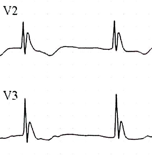


Respiratory depression, messed up ABG values-to keep simple just treat as normothermic when making vent decisions.

Metabolic cold diuresis, rhabdo, ARF, hypokalemia from ion shifts (don’t over correct), pancreatitis, CNS alteration.

Treatment:  remove from environment, dry patient.  Take your time finding a pulse, may be extremely bradycardic.  Don’t start CPR unless absolutely necessary due to cardiac irritability.  Warm fluids (can warm IVF in microwave), warm O2, forced air blanket, Arctic Sun vest/Alsius Icycath central line devices (same devices used to cool patients, but set for warming).  Warm gastric lavage, DPL, Foley fluids, 2 chest tubes with warm fluids, Bypass or thoracotomy as last resorts.

Rewarming shock:  external heat àperipheral dilation, relative hypovolemia and hypotension. 

No firm guidelines on treatment.

Non-freezing cold weather injuries:  Trench Foot in military, direct injury to soft tissues, blistering/anesthesia, treat with heat/dry feet.  Prevent with dry socks/well fitting boots.  Chilblains/pernio:  kids, Raynaud’s, uncomfortable inflammatory lesions of skin by longterm intermittent exposure to damp/nonfreezing ambient temp.  Hands/ears/toes.  Panniculitis:  mild necrosis of subcut fat.  Cold urticaria-sensitivity.

Freezing injuries:  Frostbite, usually < 4 degrees F, frozen tissue, most damage from endothelial damage during thawing.  Clinical diagnosis, warm them up and AVOID RE-FREEZING.  No rubbing-will cause more damage.  Also do not re-warm out of hospital.  Aloe, NSAIDS, tetanus, no antibiotics.  TPA after rewarming to reduce digit amputation.  Heparin/HBO don’t work.  Amputations delayed until final demarcation.


11:30 am:  Dr. Dennis Ryan – MSK Study Guide

Too much fun to take notes

00 am  Orbital Trauma/Oculoplastics-Dr. Hassan Shah; he’s happy to be involved with any Oculoplastics questions!

Ruptured Globe:  How to tell?  Peaked/irregular pupil, full hyphema, 360 degree subconjunctival hemorrhage, low IOP.

Most important reason to fix orbital fractures?  Prevent diplopia.

What does optho need to know about intraocular foreign body?  High pressure à systemic steroids.  Ruptured or not ruptured.  Visual acuity.

Blunt eye injury: “Blown pupil” actually not helpful to tell if optic nerve injured...can have temporary pupil dilation simply due to blunt injury.  APD however IS a sign of optic nerve dysfunction.  If APD, high pressure, loss of vision, need to perform lateral canthotomy/cantholysis.  Dr. Shah believes cantholysis important; simple canthotomy usually not enough, and most common mistake is to think you’ve relieved the pressure but you haven’t done enough.  He can fix any eyelid laceration that you cause!

:756148-799528-584.jpg

This is lateral canthotomy.  You also need to cut up and down (3 total cuts) for complete cantholysis.

Be sure to cover the eye in the setting of eye trauma.  Use ointment, try to bring lid back down over eye.  Best covering for globe is the conjunctiva.

Blow out fractures.  Adults almost never needs to be repaired emergently.  Even if “entrapment”, usually due to soft tissue swelling.  Usually wait for one week before repair to allow swelling to resolve.  Big fractures much less likely to get true entrapment than small fractures.  Big fractures = lots of swelling, but also room for movement.  For kids, they get “greenstick” type fractures of the orbit; softer bones, the fracture is often very small, with mild swelling, but with restricted ROM up and down due to trapped muscle àthis is case for same day surgical repair.  If you don’t have diplopia on Day 1, you will not develop it!

Facial fractures and antibiotics:  He doesn’t usually give antibiotics unless contralateral sinus disease, but there is controversy, not one correct answer.  He has never had an infection.

9:00 am  Janna Villano: - Tox-Uses of Sodium bicarb, and some controversial Antidotes

Most important 4 things in Tox:  Vital signs, Mental Status, Skin and Neuro exam


Many Sodium Channel blockers out there:  TCAs, cocaine, some antiepileptics, dysrhythmics...lots of others.

3 Uses for Sodium Bicarb:  Cardio toxicity, Prevent acidosis, Urinary alkalinization

Bicarb:  Treat cardiotox:  helps with prolonged QRS, wide complex tachycardia, hypotension.  Give 1 to 2 meq/kg bolus over 1-2 minutes.  Boluses better than infusions.   Give boluses and sit there watching for QRS to narrow.

Bicarb:  Prevention of academia:  maintain pH >/= 7.4.  Prevents drug distribution, may be useful peri-intubation of tox patients.

Bicarb: Urinary alkalinization:  enhances elimination.  Maintain pH 7-8.5, ion trapping, must maintain normal K levels for this to work-aggressive K replacement!


Anticholinergics:  antihistamines, TCAs, vertigo meds, antipsychotics, Jimson weed...etc.

Physostigmine:  To treat anticholinergic toxicity.  Will allow history when patient’s mental status clears.  Should see nearly complete resolution of symptoms in order to attribute presentation to anticholinergic  toxicity.  Allows diagnosis, limits workup.  Can cause seizures, GI distress/rest of cholinergic symptoms.  Reports of physostigmine associated with cardiac arrest , but these in patients with TCA OD, also cases of seizure in patients with prior seizure disorder.  If giving physo, watch for bradycardia/heart block,  seizure, cholinergic side effects.  Give 0.5 mg over 2-5 minutes, may repeat up to 3-4  doses.  If necessary can “reverse” physo with atropine.  ***Janna doesn’t give in patients with known seizure disorder, patient who took pro-convulsant, or patients with sodium blocker OD (eg TCA)***


Flumazenil:  Don’t use drug screen to help you decide whether or not to give it-drug screen misses many benzos.  Some studies have shown avoidance of intubation.  Why not to use it in a documented benzo OD?  Inconsistent clinical response, precipitation of benzo withdrawal (rare, and can give benzos), may precipitate seizures (seen in pts who took proconvulsant, or in patients using benzos for sz disorder), maybe risk of complications outweighs harm from OD.

Pick the right patient-no seizure disorder, pro-convulsant use, not if chronic benzo users, not for undifferentiated coma or prophylactically.  Often used in kids, pure benzo OD, procedural sedation.   If patient has seizure after flumazenil, then just flood patient with benzos (competive inhibitor).


10:00 am Dr. Khatiyar-  Chemical Asphyxiants

Fires:  Simple Asphyxiants, Chemical Asphyxiants, Chemical Irritants

Carbon Monoxide:  from partial combustion:  broken furnace, blocked car or boat exhaust, grilling in a garage, heating homes with gas ovens, car idling in garage...etc.  Also from Methylene Chloride, in degreasers/lacqueer thinner, bathrub refinishesàmetabolized to CO.

CO toxicity:  high affinity for hemoglobin forming CO-Hgb, but there are other toxic effects as well.  Also binds to myoglobin (heart tox, rhabdo), direct organ toxicity due to increased NO production and lipid peroxidation.  CO attachment to cytochrome oxidase (inhibits aerobic metabolism)

Think about it in headaches, “flu” in the fall when heaters turned on, kids with “colic”., gastro, viral syndrome, ..great imitator.

Diagnosis depends on a great history.  Ask about recurrent sx, others at home with same sx, time of year, in setting of Headache, flu sx, “hangover” symptoms, chest pain, neuro sx.

Diagnosis:  Breath sampling, CO level:  VENOUS just as good as ARTERIAL blood gas.  Don’t use O2 sat!  Check renal function, pregnancy test as changes management dramatically.  CPK, cardiac monitoring.  CO levels also can be unreliable, depending on treatment with O2, time since ingestion.  Levels:  < 10% no biggie, > 50% likely fatal, everything in between open to discussion.

Delayed neuro sequelae:  days to weeks later, incidence variable (1% - 47%), needs cognitive testing  (think mini mental status) and re-testing on followup.

Red flags:  pregnant women, kids, cardiac patients.

Half life 4-6 hours on room air, 60-90 minutes on 100% O2, 20 minutes with HBO.

Treatment:  HBO controversial:  but if level > 25%, any neuro issues/syncope, coma, Cardiac sx, abnormal cerebellar exam, symptomatic pregnancy or pregnancy with fetal distress à  call HBO chamber and discuss with specialist.  Put patient on 100% O2 as soon as you suspect CO.  Local chambers:  Advocate Lutheran General or Loyola, LCOM, Illinois Masonic, others.

Cyanide:  neuro sx, acidosis, seizures.    House fires, burning plastics, Nipride (sodium nitroprusside), certain foods-cassava, apple seeds, fruit pits, Laetrile.

Toxicity:  Neurotoxin, Enhances NDMA receptor activity, inhibits aerobic energy metabolism (blocks cytochrome oxidase).


Diagnosis:  Symptoms between 3 – 24 hours if oral, few minutes with gas exposure, sicker with premorbid conditions, kids, and dose dependent.

Presentation:  LOC/AMS, metabolic acidosis, GI, SOB, CV instability.  Be worried with rapidly developing coma, lactic acidosis, CV instability, symptomatic industrial worker/chemist.

Treatment:  decontamination (clothing, skin decon), 100% O2.  Old antidote:  Lilly cyanide antidote kit (amyl nitrate, sodium nitrite to cause methemoglobinemia which will bind cyanide, sodium thiosulfate to form thiocyanate which will be excreted in urine).  This kit usually available in ED, and this toxicity very time sensitive.  Cons:  causes hypotension, methemoglobinemia, multiple steps.

New antidote:  Hydroxocobalamin binds with cyanide, conversion to cyanocobalamin (vitamin B12):  safe, no methemoglobinemia, no hypotension, can penetrate to tissues.  Does cause flushing./urine discoloration.  Will mess up color change sensitive testing for hours.  Not available in ED but we have it in pharmacy.


11:00 am : Dr. W.A. Bret Negro - Cold Weather exposure/Hypothermia: 

Core Temp < 35 degrees

Causes:  exposure, EtOH, sepsis, burns, massive resuscitation, metabolic problems (low sugar, low thyroid/adrenal), CNS causes

Mild > 34 degrees:  shivering, “responsive” stage, trying to generate/maintain heat

Mod 30-34 degrees: depressed CV function, stupor, afib with slow ventricular response is classic

Severe < 30 degrees:  low BP, no shivering, coma

Cardiac tox:  sinus tach àsinus bradycardia à slow afib à vfib/asystole.  Heart very sensitive, can cause dysrhythmias with central line placement and tip in heart/rough handling of patient.  Also less responsive to ACLS meds/electricity.  Shock once if T < 30 C, then just remarm until T > 30 before shocking again or giving drugs.  May want to extend intervals between drug dosing.  ECG Osborn wave.


:J-wave-1.jpg

Respiratory depression, messed up ABG values-to keep simple just treat as normothermic when making vent decisions.

Metabolic cold diuresis, rhabdo, ARF, hypokalemia from ion shifts (don’t over correct), pancreatitis, CNS alteration.

Treatment:  remove from environment, dry patient.  Take your time finding a pulse, may be extremely bradycardic.  Don’t start CPR unless absolutely necessary due to cardiac irritability.  Warm fluids (can warm IVF in microwave), warm O2, forced air blanket, Arctic Sun vest/Alsius Icycath central line devices (same devices used to cool patients, but set for warming).  Warm gastric lavage, DPL, Foley fluids, 2 chest tubes with warm fluids, Bypass or thoracotomy as last resorts.

Rewarming shock:  external heat àperipheral dilation, relative hypovolemia and hypotension. 

No firm guidelines on treatment.

Non-freezing cold weather injuries:  Trench Foot in military, direct injury to soft tissues, blistering/anesthesia, treat with heat/dry feet.  Prevent with dry socks/well fitting boots.  Chilblains/pernio:  kids, Raynaud’s, uncomfortable inflammatory lesions of skin by longterm intermittent exposure to damp/nonfreezing ambient temp.  Hands/ears/toes.  Panniculitis:  mild necrosis of subcut fat.  Cold urticaria-sensitivity.

Freezing injuries:  Frostbite, usually < 4 degrees F, frozen tissue, most damage from endothelial damage during thawing.  Clinical diagnosis, warm them up and AVOID RE-FREEZING.  No rubbing-will cause more damage.  Also do not re-warm out of hospital.  Aloe, NSAIDS, tetanus, no antibiotics.  TPA after rewarming to reduce digit amputation.  Heparin/HBO don’t work.  Amputations delayed until final demarcation.


11:30 am:  Dr. Dennis Ryan – MSK Study Guide

Too much fun to take notes

Conference Notes 10-22-13

It seemed a shame to let the conference notes go while Dan took a well deserved break down under. So this week Christine Kulstad attempts to fill his shoes.

8am: King videoscope, King airway (laryngeal airway), and Ambu aScope training


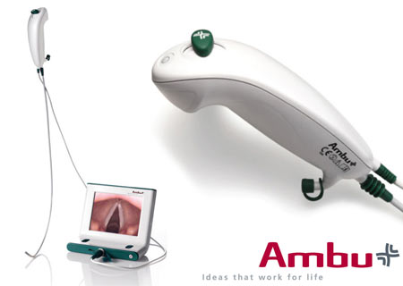
Ambu aScope- video cable (instead of fiberoptic) with articulating tip and cord to attach to video monitor (all disposable- everything except the monitor is disposable). When starting, keep video cable taut which helps function of articulating tip. Currently there is an oxygen port, next model will also have a suction port. Handle has on/off switch and lever that controls tip. Turn on monitor first- has 3 hrs battery life but should generally be charged. Pre-load ETT on tube stop at top of video cable, can go through nose or mouth. Remember when performing nasotracheal intubation, use a smaller tube (7-0). Consider pre-treatment with nebulized lidocaine and lubrication with lidocaine jelly, oxymetazoline (Afrin) spray or phenylephrine drops if time permits. 

9:00-  Tranexamic acid (TXA) and trauma- Rob Mokszycki (PharmD)

Trauma pts are coagulopathic due to multiple causes. During resuscitation, have to transfuse fluids (crystalloids), blood products (massive transfusion protocol potentially), and pro-coagulant agents (TXA and Factor VII).

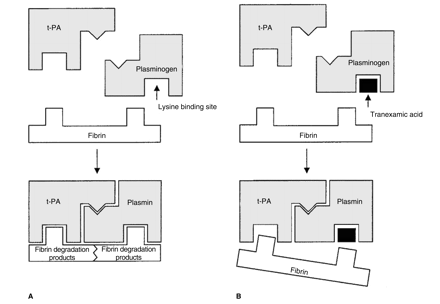
TXA works by inhibiting plasmin and preserves platelet function.

First promise of TXA was in CABG- less blood transfusion, platelets functioned better, less blood loss. Same in spinal surgery.

CRASH-2 [Effects of tranexamic acid on death, vascular occlusive events, and blood transfusion in trauma patients with significant haemorrhage (CRASH-2): a randomised, placebo-controlled trial. CRASH-2 trial collaborators. Lancet. 2010 Jul 3;376(9734):23-32. doi: 10.1016/S0140-6736(10)60835-5. Epub 2010 Jun 14.]

- international (except US) study of trauma pts and TXA. Outcome measure was all cause mortality. 2nd meausres- thrombotic complicatiosn, surgical intervation, transfusion. Inclusion- suspicion of bleeding within 8 hrs of injury, age >16. Exclusion- contraindication -active clotting, or allergy. 20,000 pts, groups well matched and low fall out. Injury severity was not measured. All cause mortality was decreased in TXA group. No difference in vascular occlusive events. Dosing not optimized- guesstimate from infusions given during surgery.

Secondary anaylsis of bleeding patients- benefit was greatest when given <1 hr, good if given 1-3 hrs, and negative if given >3 hrs. This was not a pre-specified subgroup.

NNT- 66 for all cause mortality. If this is true, could save 4000 lives in US.

MATTERs  [Arch Surg. 2012 Feb;147(2):113-9. doi: 10.1001/archsurg.2011.287. Epub 2011 Oct 17. Military Application of Tranexamic Acid in Trauma Emergency Resuscitation (MATTERs) Study. Morrison JJ, Dubose JJ, Rasmussen TE, Midwinter MJ.]

- military retrospective trial- primary outcome mortality. 800 patients. TXA group was more severely injured than control, and overall group had more GSW and blast wounds. 48h and in-hosptial mortality were better in TXA group, more pronounced in massive transfusion subgroup. This study did find a higher rate of PE and DVT in the TXA group.  NNT of 15, 7 in massive transfusion group. Time to TXA treatment and TXA dosage was not often recorded.

Summary- use TXA when <3 hrs and life threatening hemorrhage. Don’t use it if known clot. Possible harm in SAH cases (based on other studies).

Given in two 1 g doses, one as 10 min infusion and one as 8 hr infusion.

9:30- Student Loans- Jim Maletich

Medical school debt is increasing rapidly- median private med school debt is $180,000 and only $155,000 for public schools. Our residents owe from $84,000 to $450,000! As accrued interest is based on principal amount borrowed plus last years interest (capitalization), loan amounts continually increase. During residency, can be in negative amoritization- paying less than new interest accruing.

Deferment- grace period where you are not required to make payments. Usually capitalization does not occur during deferment.

Standard payment is your loan divided into 120 payments (generally about $2000/yr). Forbearance- don’t have to make payments but loans are all capitalized). EG- over 3 yrs capitalization adds $3000 to a $200,000 loan vs other fees and interest of $42,000.

Federal Repayment Plan for subsidized loans (IBR- income base repayment)- payments cap at 15% of discretionary income (your income – 150% of poverty limit[$11,490]) which ranges from $0 and $500/month. Government pays your interest so your interest does not capitalize. 3 year limit.

Consolidation- federal option- save 0.25 to 0.5% interest. Private option loses subsidized perks.

Public Service Loan Forgiveness- Must work for tax-exempt 501(c)(3)- federal, state, local, or tribal government for 120 monthly payments (residency counts). Then they repay the rest of your loans. Question if funding will exist by the time you qualify.

10- Admin Study Guide Part 2- Chintan Mistry

Boarding increases LWBS rates, increases length of stay, and diversion rates.

Parental consent is required except for emergency treatment and EMTALA screening, reproductive health, emancipated minors (court order, married, pregnant, minor who is a parent, Armed Forces).

The medical screening exam required by law (EMTALA) checks for an emergency medical condition, does extend to patients in waiting room (no time frame given). Duty ends if patient voluntarily leaves without being seen (LWBS). If an emergency medical condition (EMC) exists, treatment must be provided. EMC means potential serious injury to patient or organ. Transfer the patient when risk of transfer is outweighed by benefits. EMTALA applies to all facilities that accept Medicare payment. EMTALA violation means fines not covered by malpractice insurance, and potential exclusion from Medicare payments.

In all 50 states, child and elder abuse must be reported- can be done by physician, nurse, or social worker. Don’t assume PMD will do it. You are protected legally when good faith reports made (someone may file suit, but they won’t go anywhere). GSWs and stab wounds are also reportable in all states. 

For a malpractice suit to be successful, it must be proven that a breach of duty occurred (patient was in ED usually enough), actual injury did happen, standard of care was violated, and actions led to injury.

On call physicians can refuse transfer of patient with an emergency medical condition when the receiving hospital does not have the capacity to accommodate the patient. Capacity means facilities, expertise, and space.

Leaving against medical advice (AMA)- the treating physician determines if they are competent to make that decision. It’s nice to have a refusal form on file, but it isn’t necessary and doesn’t help much legally. Alternative care should be provided to patients leaving AMA, and discharge instructions should be given. In a teaching setting, the attending should be involved- also don’t let your nurse do it without your involvement. It IS good to have your nurse witness your discussion and document it independently. 

1030- Dialysis- Abraham Thomas

Nephrology update- can do dialysis for ED patients without requiring admission (can be done in ER, SUU, and HD unit). Call nephrologist early for potential discharges who are missing HD so that it can be rescheduled.

Good practice to check access site, ask about dry weights, dialysis schedule, dialysis center.

For HD patients who receive normal amounts of IV contrast for CT, they do not usually need immediate HD. It will increase their fluid balance by about 0.5 L. No good evidence it worsens anything, but caution may be beneficial for patients with temporary HD or improving renal function.

Gadolinium is contraindicated, but evidence of fibrosis was in patients who received multiple doses. If need, patients will need daily HD for 3 days afterwards.

BNP is not an accurate marker of volume status in HD patients- study shows some correlation with wide amounts of variation when studied in HD patients. However, a low BNP is probably accurate. Better to use clinical judgment to determine volume overload (dyspnea, RR, CXR, hypoxia).

Troponin can be elevated, but don’t ignore an elevated troponin. Change in troponin is more meaningful. Baseline elevation is not that concerning.

Please take down all bandages to look at access sites- especially intravascular catheters. Catheters are tunnels into IJ even though it looks subclavian. For AV grafts and fistulas- feel for thrill and listen for bruit. If it is not there, contact nephrology.

For bleeding vascular access (fistula or graft)- occluded artery for a minute or two. Then place mattress suture through skin ONLY  at bleeding site. You can also use gelfoam or hemcon or similar. Bleeding is a marker of a problem with the fistula/graft so let nephrologist know even when bleeding is controlled.

Aneurysms can form at fistula/graft site. If you see firmness, skin turning white, or ulceration nearby be very concerned. This aneurysm is at high risk for rupture and vascular & nephrology should be contacted.

Acute kidney injury- creatinine increased by 0.5. ATN, obstruction, and pre-renal common causes. Always ensure pt is voiding (consider Foley), image kidneys with U/S or CT, check UA. Avoid NSAIDs, ACE/ARBs, contrast. When you have a patient with hyperkalemia, order repeat lytes in 4-6 hours.

Indications for HD- fluid overload; hyperkalemia, esp. with EKG changes; refractory acidosis; overdose- aspirin, lithium, toxic alcohols; uremia with encephalopathy/pericarditis

Chronic kidney disease- GFR <60 for at least 3 months. Usually from DM and HTN. These patients likely to present with volume overload, anemia, infection, hyperkalemia. Try to avoid pRBC transfusion in someone waiting for transplant as it will make a match harder. Don’t give these patients NSAIDs, gentamycin, or IV contrast if possible. If contrast necessary, give normal saline 1 ml/kg/hr for 6 hours pre/post. No benefit from bicarbonate or Mucomyst. Give instructions for creatinine check in 2-3 days.

Expect to see more kidney transplants at ACMC, we do them here. Call Dr. Chet Desai or Dr. Darshika Chabbra (417100), who are transplant nephrologists, surgeon is Dr. Mittal. Any change in baseline creatinine is critical. Any obstruction is potentially catastrophic. Evaluation after surgery- hematoma or urine leak. Afterwards worry about rejection and infection, drug toxicity. Make sure you check UA, BMP and levels of immunosuppresants which should be back in 2 hours. U/S likely helpful- specify that patient has a transplant.

Many causes of hyperkalemia- final common pathway through aldosterone which opens Na/K pump in tubule. Urgency of treatment depends on cause and clinical condition of patient- ie hydrating a patient who can urinate will lower K by itself.

Calcium-onset is immediate and duration is 30-60 minutes. Give 1 g of CaCl or Cagluconate over 2-3 minutes. Stabilize cardiac membranes.

Insulin/D50- insulin will push K into muscle cells, will drop 0.5-1.2 mEq. Onset in 20-30 minutes.

Bicarbonate- does not do very much. You can use it if pt is very acidotic (7.2 or lower) and you are putting them on a bicarb gtt.

Albuterol- safe and very effective. Must use 10-20 mg over 10 minutes. Effect seen in 90 minutes.

Kayexalate- exchange resin that may bind potassium but there have been cases of bowel necrosis. Do not use in pts with ileus, recent surgery, opioid use, bowel obstruction. Avoid multiple doses or large doses (30 mg OK). Onset in 90 minutes, lasts 4-6 hours.

If patients make urine, treat with IVF (saline) and loop diuretic (furosemide).

Peritoneal dialysis- most common ED presentation is peritonitis- happens about 1x/year/patient. May have all or only one of abdominal pain, n/v, cloudy dialysate. Work up as usual plus add 1 L of 1.5% dextrose dialysate, let it dwell for at least 20 minutes but ideally 60 minutes. Send for cell count and cultures. Peritonitis – 100 cells with 50% PMNs. Give vancomycin 1 g IP if possible, IV otherwise PLUS ceftazidime 1 g IV/IP  or 500 mg ciprofloxacin bid. Can be discharged if not toxic and can be seen the next 1-2 days.

Hypercalcemia- treat levels 12-14 if symptomatic, all over 14.  First, 200-300 ml/hr of normal saline. Do not had furosemide unless volume overload. Add calcitonin (4-6 IU/kg sq over 6-12 hrs x 48 hrs), bisphosphonate (ie zoledronic acid 4 mg iv over 15 min- slow onset but lasts for weeks). 

1130- Amphetamines- Andrea Carlson

Pharmaceutical amphetamines-for ADHD, obesity (short term use), depression, enuresis, parkinsons (selegeline), alcoholism, narcolepsy.

Majority of drug abuse is misuse of drugs prescribed to patients, but illegal sales are rising. Check IDPH website before prescription refills of Adderall, etc.

Mild-moderate effects- agitation, hallucinations, HTN, tachycardia. Unlikely to have serious side effects from small, accidental ingestions.

Methamphetamines: 2nd only to cannabis in use. Significant geographical variation, seems to be increasing in Midwest.  Highest rate of admission in all drug abusers, mostly psych. Crank is dirty, smelly meth. Yaba is meth + caffeine, big in Thailand.

Meth works by releasing norepinephrine and dopamine. Binds to serotonin receptors and effects brain glutamate. Renally excreted, large Vd. Causes neuroexcitation, tachycardia, HTN, mydriasis, hyperthermia, diaphoresis. Like cocaine but less dysrhythmias and seizure, lasts longer, more psychosis.

Meth- get horrible teeth. “Meth bugs”- from tweaking and picking at skin. Gross.  


Meth- increases child neglect and violence, HIV transmission. Montana meth project (now in 7 other states including Illinois) has been very effective at reducing use.

Ecstasy (aka Adam, XTC, Molly)- often not in the pill (can be caffeine, designer drug, decongestants). Lower dose- trouble speaking, agitation, repetitive behaviors. Higher doses- more repetitive/compulsive behavior (bruxism), chorea, hyponatremia. Less cardiac effects. Chronic use- necrotizing vasculitis, psych problems.

Bath salts- cathinones- are newer congeners.

Drug screen- positive for a couple of days. Lots of false +- decongestants, buproprion, selegiline. MDMA and designer drugs false negative. Treat clinically.

Overall treatment- cool, calm room. Watch HR and core temp. Check BMP, CPK, EKG. CT head if seizure or significant CNS depression. Generous chemical sedation- up to 100 mg Valium (10 mg q 10 min). Can add anti-psychotics (newer generation). If sedated but still hypertensive, give phentolamine or nicardipine. 

Conference Notes 4/16/13

Kettaneh/Felder    Oral Boards

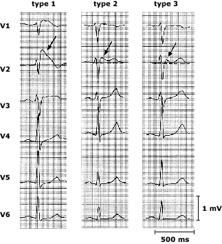
Case 1.  Syncope due to Brugada syndrome.  Pt also fractured humerus due to fall.  Critical actions: Identify brugada and consult cardiology. Splint and manage fx.  Brugada syndrome arrhythmias usually occur at rest.

Three types of Brugada waveform. Three distinct types of ST segment elevation have been described. In type 1, the ST segment gradually descends to an inverted T wave. In type 2, the T wave is positive or biphasic, and the terminal portion of the ST segment is elevated ≥1 mm. In type 3, the T wave is positive, and the terminal portion of the ST segment is elevated <1 mm. Arrows denote the J-waves.

Case 2.   Seizing patient due to hyponatremia from post-partum pituitary necrosis.  Pt had intra-partum hemorrhage with hypotension and transfusion. This resulted in pituitary infarct/necrosis.   Pt has been having difficulty lactating and has been weak.  Critical actions: Hypertonic saline, antibiotics for uti, hydrocortisone.  Consult appropriately.   Sheehan syndrome consists of pituitary necrosis due to ischemia in the peripartum period.  

Case 3.   Seizing NH patient due to hypoglycemia.  Pt accidentally received  a dose of insulin intended for another patient. Critical actions:  Identify hypoglycemia, IV glucose, feed, check serial blood sugars. 

Elise comment: If there is a specific fx that causes a known neuro-vascular injury then specifically check for this injury.  Example: midshaft humerus fx is associated with wrist drop.  Specifically tell the examiner you want to examine for wrist extensor strength.

Harwood comment:  You can get an insulin level on a patient to see if they received too much insulin.  There can be varying severity of Sheehan’s syndrome.  Mild cases can be difficult to diagnose.   There is a website brugada.org that can give you some guidance on how to manage these patients.   Incidental brugada cases identified in the ED should be referred to an EP specialist to see if an arrythmia can be induced.   IF so, they will need an AICD.

Girzadas comment: Be alert for hyponatremia and mild hyperkalemia in a weak or hypotensive patient. This is usually adrenal insufficiency. Get a serum cortisol level and give hydrocortisone in stress doses. 


Chandra    M&M

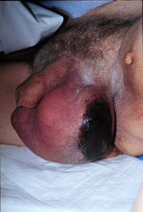
Pt with fournier’s gangrene.   Rapidly progressing necrotizing fasciitis of perineal/perianal/genital region.  Usually in men around age 50-60.   Diabetes and obesity increases the risk.  Indwelling catheters, etohism, immunosuppression also increase risk.

Can be caused by local infections like abscess, fissure, diverticulitis, etc.   10% of cases are idiopathic.

Look for scrotal pain, swelling, fever. Patient’s may have severe genital pain.   Check for subcutaneous crepitation.

94% of cases involve the srotum,  47% involve the penis, and 35% of cases involve the perineum.

Fournier’s severity index: Basically SIRS plus lab abnormalities correlates with increased mortality.   Elise comment: This index looks like unvalidated BS.

U/S can show subQ air but is less sensitive than CT.  CT is most sensitive for subQ emphysema and can show source of infection and extent of spread.   Harwood comment: Get fine cuts through scrotum and perineum.  Less concerned about the rest of abdomen.

Treat with surgical debridement.   Give triple antibiotics.  Hyperbaric o2 is controversial and is used after surgical debridement.   24 hour delay in surgery increases mortality by 11%.  Mean hospital stay is 30 days.

Pitfalls: Have high suspicion for this rapidly progressive diagnosis.  Sign outs increase risk.   Push back against consultant bias that downplays disease process.

Girzadas comment: During sign out make sure there is some back and forth discussion between teams about these high risk patients.   Harwood comment: Be careful to not just say “nothing to do”.  Rather you need to say, you have to re-examine the patient’s scrotum.   Question whether the antibiotics have been written for repeated doses if they are boarding for a prolonged time in the ED.  Elise comment: Identify your sickest patient and highlight the need for further attention.    DenOuden  comment:  Bedside sign out may be helpful in these high risk cases.   Harwood comment:  These guys need serial exams because it can be “awe inspiring to see how fast this can progress”.  Christine comment: Ct is very important in these diagnoses.   Harwood comment: MRI is the most sensitive test for necrotizing fasciitis.   

Paarul comment: Prior to sign out review your orders for critically ill patients.

Harwood comment:  This case was due to a system error.   Our EMR takes so many clicks to place orders, it increases your risk of error.  Elise comment: It is ok to push back professionally against consultants if you feel strongly the patient needs a certain intervention.   Kelly Williamson comment: We train for worst case scenarios and are constantly alert for them.  Consultants frequently down play a disease process.  You have to be aware of this difference in perspective.  Willison comment: Inturruptions also increase our risk for error.  Joan Coghlan comment: signouts are made more difficult also by the arduousness of the EMR system.


Kettaneh    Tranexamic Acid in the ED

TXA is a synthetic analog of lysine.  It competitively inhibits the activation of plasminogen to plasmin.

Common minor Adverse effects: abdominal pain, arthralgia, headache, fatigue.

CRASH-2  study:  40 countries with 20,000 trauma patients.  Showed lower all cause mortality.

MATTERs  study: showed lower mortality in TXA group.

  ICH study: showed trends to less ICH growth and lower mortality.

Re-analysis of CRASH-2  showed the earlier you give TXA the better the effect.  Want to give in less than 3 hours.

Give to Trauma patients with hypotension or tachycardia  or expected  need  for transfusion.   Until further research, don’t give to isolated head trauma patients.

Dose is 1g IVPB over 10min followed by 1g IVPB over 8 hours.

PharmD comment:  This is a benign drug.  It is not a pro-coagulant.  It only stabilizes the clotting system.   Elise comment: This drug provides a modest but real benefit in bleeding trauma patients   There was consensus  that we should be using this in multisystem trauma patients with bleeding, hypotension, tachycardia, or need for transfusion. 


Kettaneh          Case F/U

63yo male with presyncope.  Sob and diaphoretic after lifting a pallet.    His wife made him go to the ER.

Which is a clear marker for bad pathology.  Harwood comment: Another marker for bad pathology is when long time smokers spontaneously stop smoking.  When a patient tells him they recently stopped smoking on their own, he gets worried.    Vitals showed tachycardia.  EKG showed sinus tachycardia with subtle inferior st changes.

Pt became hypotensive in the ED.  Bedside echo shows moderate pericardial effusion with good contractility.  No chest or back pain.   Dimer is elevated.   Second EKG looks better.

To diagnose cardiac tamponade , tachycardia is almost always present.  JVD is commonly present.  Electrical alternans is an EKG finding specific  but insensitive for tamponade.


Electrical alternans.

U/S will show diastolic collapse of RV when tamponade is present.

CTA of this patient showed an aortic dissection and no PE.

Type A in Standford classification involves ascending aorta.   Type B means only descending aorta.

This patient had a painless Type A dissection.  6% of aortic dissection patients have no pain. These patients with painless dissections are more likely to have syncope, stroke symptoms or CHF symptoms.

Patient went to OR and had repair of ascending dissection and aortic valve replacement.

Nick felt the dimer may have saved the patient’s life.   Kari Tekwani recently diagnosed a relatively painless dissection on another patient and she also felt the dimer helped pick up the dissection.

Harwood comment:  If you aren’t sure if the diagnosis is pe or dissection go with the CT PE rather than CTA study.   You probably won’t miss a significant dissection on a CT PE study.   If you do a CTA aorta study, you will miss many pe’s.   

Watts     Vent Management Software Demonstration

Cash    5 Slide F/U

91 yo female with altered mental status.   Dexi was 56 in the field.   Pt was hypothermic and hypertensive.  Cachectic and dehydrated.

Labs c/w urosepsis.   Pt became more hypothermic and became hypotensive.   Diagnosis was urosepsis.

Started on Cetriaxone and Levophed.     IM brought up possibility of myxedema coma.   EM clinicians considered diagnosis but felt the expected doughy appearance of a myxedema patient  was not present.   Pt’s TSH was later found to be 250.  Diagnosis was made of myxedema coma. 

  Hallmarks of myxedema coma are: mental status change, hypothermia, bradycardia, hyponatremia, hypoglycemia, hypotension, and precipitating illness.

Treat with levothyroxine, hydrocortisone to cover possible adrenal insufficiency. Manage hypoglycemia, manage hypotension, provide passive re-warming, and monitor for arrhythmias.

Beckemeyer    5 slide F/U

79 yo female with sudden onset headache and neck pain with emesis.   Pt has afib and is on Coumadin.  

ED clinicians ordered CTA of head and neck.  Pt was found to have a right MCA aneurysm with diffuse SAH.   Pt was given FEIBA to reverse Coumadin  and then went to neurointerventional lab and had aneurysm coiled.  Pt was dc’d home with no focal deficits.  Badabing Badaboom!


3/26/13

Conference Notes 3-26-2013

Please consider donating to our EM Foundation to benefit resident education/development.  Thanks for your consideration/generosity.
/em-foundation/

Herrmann    Trauma in the Pregnant Patient

Bedside U/S will give you a quick assessment of the fetus as far as fetal movement, heart rate and age.   Bedside U/S does not give you adequate info regarding abruption.   You need to do fetal monitoring to determine the frequency of contractions to guage the probability of abruption.  Normal heart rate and no contractions in 4 hours of observation has 100% negative predictive value for abruption.

Pertioneal signs are not as evident in the pregnant patient.   Blunt trauma has a higher mortality than penetrating trauma.  0.5 of fetal deaths result from “mild” trauma.

No single imaging study provides a radiation dose high enough to cause harm in a fetus.  So image as needed but no more than needed.

Kleihaur Betke test identifies fetal-maternal hemorrhage that exceeds the normal dose of Rhogam (300mcg).  If KB test is positive you need to give extra Rhogam.

Left lateral decubitus position when pregnant patient is supine off-loads the inferior vena cava and can treat hypotension.

Peri-mortem C-section: Best results when initiated within 4 minutes of maternal arrest.  Goal is to get kid out by 5 minutes of mom arresting.   C-section off-loads the IVC and may improve maternal survival,  CPR continues during C-section.   Make big midline incision (xyphoid to pubis).  Hysterotomy through upper uterine segment.   Cut through placenta if necessary.   Have two teams if possible to divide resuscitative efforts between mom and baby.

Harwood comments:  KB test is of less value in 2013 because most RH neg mom’s have gotten a rhogam shot as part of their standard prenatal OB care.   Each CT study has to be carefully considered in the pregnant patient.  Do  the studies that need to be done but no more than that.   If doing perimortem C-section make big xyphoid to pubis incision going around umbilicus. 

Girzadas comment: If you are looking for a bright line cutoff of which mom’s  need fetal monitoring after trauma my suggestion:  If a butterfly alights on a patient’s gravid abdomen and the fetus is >20weeks ega, they should have monitoring for 4 hours.    Dr. Omi also felt a very low threshold was indicated. 

Girzadas                    Study Guide   Arrythmias/Pharmacology

I gave the lecture so I couldn’t do detailed notes, but trust me the lecture was Awesome! :)

Antiarrythmic classification

  • 1. Sodium channel blockers
    • 1a   procainamide, disopyramide, quinidine, tricyclics
    • 1b  lidocaine, mexilitine, phenytoin
    • 1c   encanide, flecanide, propafenone
  • 2. Beta-blockers
  • 3. Potassium efflux blockers-  Amiodarone,  sotolol,  ibutilide
  • 4. Ca channel blockers
  • Unclassified-   Digoxin, Adenosine, Magnesium

There was some discussion on management of arrhythmias/QRS widening/hypotension due to TCA overdose

Harwood comment:  TCA caused QRS widening and hypotension can be treated by raising the ph alone. The Na in Sodium Bicarb is not necessarily the key factor.  Harwood then discussed the following abstract:

Abstract
Study objectives: We carried out this study to determine the effects of pH alteration on QRS width with administration of tromethamine, a non-sodium-containing buffering agent, in experimental amitriptyline overdose. Design: Prospective, nonblinded trial. Participants: Adult mongrel dogs. Interventions: Pentobarbital-anesthetized dogs were overdosed with amitriptyline 5 mg/kg followed by infusion at 1.0 mg/kg/minute until the QRS width doubled, then decreased to .5 mg/kg/minute until the end of the experiment. At two defined points of toxicity, the dose of tromethamine required to raise the pH to 7.50±.4 was given. pH and QRS width at a speed of 100 mm/second were measured
over a 30-minute period after each tromethamine dose. Data were analyzed with non-linear-regression analysis. Results: At toxicity 1 the mean pH was 7.32, with a QRS width of 11.6 mm. Two minutes after the tromethamine dose the pH rose to 7.51, with narrowing of the QRS width to 8.4 mm. At toxicity 2 the pH was 7.40, with QRS width of 10.6 mm. Two minutes after tromethamine, the pH rose to 7.49 and the QRS width decreased to 9.7 mm. Regression analysis showed a correlation
between pH and QRS width; as pH increased, QRS width decreased (P=.0001). Conclusion: Cardiac toxicity of amitriptyline overdose, as manifested by QRS widening, is reversible by pH changes alone.

Andrea comment: TCA’s are highly protein bound.  In overdose situations, changing the ph of the blood will increase protein binding and decrease toxicity.  The NA is sodium bicarb can act synergistically on the fast sodium channels. 

Mckean/Kerwin    Oral Boards

Case 1: Werniecke’s Encephalopathy.    Classic triad of Encephalopathy/ataxia/oculomotor dysfunction.is present only 1/3 of the time.  Critical Actions: Rule out toxic alcohols, consider disagnosis, give hi dose thiamine 500mg IV Tid. 

Case 2: Afib with WPW. Critical Actions: IV procainamide 17mg/kg no faster than 50mg/min.  Stop if pt gets hypotensive  or QRS widens by 50%.   Avoid calcium channel blockers or beta blockers or anything that would block the AV node.  That would increase conduction down bypass tract.  If patient is unstable with chest pain, hypotension, altered mental status or severe chf then cardiovert.


Hemodynamically stable monomorphic VT or pre-excited atrial fibrillation (ACLS, 2010): Loading dose: Infuse 20-50 mg/minute or 100 mg every 5 minutes until arrhythmia controlled, hypotension occurs, QRS complex widens by 50% of its original width, or total of 17 mg/kg is given. Follow with a continuous infusion of 1-4 mg/minute. Note: Not recommended for use in ongoing ventricular fibrillation (VF) or pulseless ventricular tachycardia (VT) due to prolonged administration time and uncertain efficacy.

Girzadas comment: consider using only procainamide on the boards for narrow/wide afib or wide complex tachycardia.

Harwood comment: give procainamide 1gram over 30 minutes for your initial managment.  This rate will work safely for the majority of patients. If they get better you are done. If they get worse you can shock them or give more procainamde if they have a higher BMI.

Case 3: Toxic Shock syndrome.  Fever/rash (sunburn)/desquamation/hypotension/3 or more organ systems.     Critical actions: management of shock/abx for staph/remove foreign bodies/surgery for abscesses or localized infections.      Kettaneh comment: pt had criteria also for TTP however  faculty felt platelets weren’t low enough for TTP.   Elise comment: Put the synapse in your brain that shock with a sunburn type rash is toxic shock syndrome.  Girzadas comment: think about TSS or Anaphylaxis in the hypotensive pt with vomiting/diarrhea who doesn’t improve with a liter of fluids.


Toxic shock rash from Up to Date


Toxic Shock Rash from Up to Date


Maletich/Gupta   Oral Boards

Case 1: Benign Intracranial Hypertension.  Critical actions: Do physical exam to find afferent papillary defect and papilledema, pain medication, head ct, LP to check opening pressure.    Treatment with acetazolamide is the medical management.   Nosek comment: It is unusual to see unilateral eye findings.  Rohit agreed.  He said findings are usually bilateral but there may be asymmetry of severity of findings from right to left eyes.

Case2:   Pyloric stenosis.     Critical actions: IV hydration, diagnose pyloric stenosis with U/S, consult surgery, admit patient.   Classic metabolic picture is hypochloremic metabolic alkalosis.  Hypokalemia takes a couple of weeks to develop. 

Case3:  CHF/Pneumonia, ARF with hyperkalemia.  Critical actions: Treat hyperkalemia, treat CHF and pneumonia,  arrange for dialysis.

Purnell      Midgut Volvulus

MGV  can present with vomiting, hyperglycemia, altered mental status.  2 yo with Initial clinical picture that looked like DKA with a blood sugar of 600 and lethargic mental status.  Pt had no ketones in urine.   Abdominal exam when pt was resuscitated demonstrated abdominal tenderness/guarding.  Xrays showed distended bowel loops.  U/S shows free intra-abdominal fluid. Pt ended up with a small bowel resection.  He is awaiting small bowel transplant. 

Nosek   Peripartum Cardiomyopathy (PPCM)

Post-partum patient with SOB/CP/Cough. DDX was mostly between PE and PPCM.  Bedside echo showed poor contractility and BNP was quite high.  Pt started on carvedilol and low dose lasix, lovenox.  PPCM occurs 1 month before or up to 5 months after delivery. 90% occur in the first 2 monts post-partum.   Older patients and multparous patients are at greater risk for this problem.  Treat with b-blockers/lasix/digoxin.  Can’t use ACE-I during pregnancy but ACE-I’s can be useful post-partum.  If while pt is ill EF>30%, it is likely pt will regain good EF.   Girzadas comment: Lung windows can be a hepful adjunct to cardiac echo.  The finding of “headlights in the fog” consistent with extra lung fluid is very reliable.  Chastain comment: U/S findings preceed CXR findings.  Lovell comment: It is true the CXR findings were not impressive.

Conference Notes 3/19

Conference Notes  3-19-2013

Please consider donating to our EM Foundation to benefit resident education/development.  Thanks for your consideration/generosity.
/em-foundation/

Badillo    Ultrasound

Sorry, I missed this lecture.

Lovell   Study Guide  Sedation/Analgesia/Local anesthetic

5 ways to decrease the pain of local infiltration of anesthetic: small guage needle (Harwood uses insulin syringe initially followed by larger meeting), bicarb buffering, warm the anesthetic  (Anneken method: put it on the computer CPU at the beginning of your shift),inject anesthetic  into the wound not through intact skin, slow injection.

Etomidate causes myoclonus up to 30% of the time when used for sedation/induction.

Toxicity of local anesthetics is CNS toxicity (paresthesias followed by seizures) early followed by cardiovascular toxicity (arrhythmias).  Bupivicaine is a great local anesthetic but has the highest risk for toxicity.  Treat seizures with benzos and treat arrhythmias with amidarone.  There is also some case reports on lipid infusions for local anesthetic toxicity. It has been very effective in some case reports.  Risk for local anesthetic toxicity is greater in kids so be careful of dosing and risk also higher if you accidentally inject into an artery/arteriole. 

Sedation levels: Minimal (anxiolysis), moderate (airway reflexes intact), deep (airway reflexes may not remain intact), general anesthesia (airway and cardiovascular issues).

  • Analgesia – Relief of pain without intentionally producing a sedated state. Altered mental status may occur as a secondary effect of medications administered for analgesia.
  • Minimal sedation – The patient responds normally to verbal commands. Cognitive function and coordination may be impaired, but ventilatory and cardiovascular functions are unaffected.
  • Moderate sedation and analgesia – The patient responds purposefully to verbal commands alone or when accompanied by light touch. Protective airway reflexes and adequate ventilation are maintained without intervention. Cardiovascular function remains stable.
  • Deep sedation and analgesia – The patient cannot be easily aroused, but responds purposefully to noxious stimulation. Assistance may be needed to ensure the airway is protected and adequate ventilation maintained. Cardiovascular function is usually stable.
  • General anesthesia – The patient cannot be aroused and often requires assistance to protect the airway and maintain ventilation. Cardiovascular function may be impaired.
  • Dissociative sedation – Dissociative sedation is a trance-like cataleptic state in which the patient experiences profound analgesia and amnesia, but retains airway protective reflexes, spontaneous respirations and cardiovascular stability.  (up to date)

 EMLA is an acronym for eutectic mixture of local anesthetics.

Simple rule: If you want to be cautious, Patient to be sedated should be NPO for 3 hours for any liquid or solid.  If situation is emergent/urgent, you can shorten that time period.  It is controversial whether NPO status does anything to protect patient from aspiration.

Safe dosing for : Lidocaine  4mg/kg plain/7mg/kg with epi      Bupivicaine  3mg/kg plain/5 mg/kg with epi

Can’t use nitrous oxide (NO) in patients with high oxygen requirement,  SBO, pneumothorax, altered mental status.  Nitrous oxide will expand gasses in closed spaces.  Pt’s need normal mental status to cooperate with NO sedation.   Balloon tipped catheters in the body may also expand with the use of NO.

Oral opioids in increasing order of strength: Codeine, morphine, hydrocodone, oxycodone, hydromorphine.    Harwood comment: 100micrograms of fentanyl=1.5mg of dilaudid.   25microgram doses of fentanyl are too small.   Start with 1microgram/kg of fentanyl as a rule of thumb.

Complex regional pain syndrome: Pt  s/p traumatic injury who is getting worse a couple of weeks later.  Painful area may be edematous, allodynia present, diaphoretic, possibly mildly erythematous.  Treat by removing cast, low dose steroids, better pain control, consult orthopedist/trauma.

Benzocaine (hurricane spray, teething gel) and prilocaine can cause methemoglobinemia.

There is research showing that you can treat benign headache with local injections 2 cm bilat laterally to C7, 2 cm deep. Use  1.5 ml of bupivicaine with each injection.  (65% effectiveness for complete resolution of pain)

Abstract:
OBJECTIVE: The primary objective of this retrospective chart review is to describe 1 year's experience of an academic emergency department (ED) in treating a wide spectrum of headache classifications with intramuscular injections of 0.5% bupivacaine bilateral to the spinous process of the lower cervical vertebrae. BACKGROUND: Headache is a common reason that patients present to an ED. While there are a number of effective therapeutic interventions available for the management of headache pain, there clearly remains a need for other treatment options. The intramuscular injection of 1.5 mL of 0.5% bupivacaine bilateral to the sixth or seventh cervical vertebrae has been used to treat headache pain in our facility since July 2002. The clinical setting for the study was an academic ED with an annual volume of over 75,000 patients. METHODS: We performed a retrospective review of over 2805 ED patients with the discharge diagnosis of headache and over 771 patients who were coded as having had an anesthetic injection between June 30, 2003 and July 1, 2004. All adult patients who had undergone paraspinous intramuscular injection with bupivacaine for the treatment of their headache were gleaned from these 2 larger databases and were included in this retrospective chart review. A systematic review of the medical records was accomplished for these patients. RESULTS: Lower cervical paraspinous intramuscular injections with bupivacaine were performed in 417 patients. Complete headache relief occurred in 271 (65.1%) and partial headache relief in 85 patients (20.4%). No significant relief was reported in 57 patients (13.7%) and headache worsening was described in 4 patients (1%). Overall a therapeutic response was reported in 356 of 417 patients (85.4%). Headache relief was typically rapid with many patients reporting complete headache relief in 5 to 10 minutes. Associated signs and symptoms such as nausea, vomiting, photophobia, phonophobia, and allodynia were also commonly relieved. CONCLUSION: Our observations suggest that the intramuscular injection of small amounts of 0.5% bupivacaine bilateral to the sixth or seventh cervical spinous process appears to be an effective therapeutic intervention for the treatment of headache pain in the outpatient setting.

Citation:
Treatment of headaches in the ED with lower cervical intramuscular bupivacaine injections: a 1-year retrospective review of 417 patients.
Mellick LB - Headache - 01-OCT-2006; 46(9): 1441-9
MEDLINE® is the source for the citation and abstract of this record

Diamonds are location of injections.  Circle is the spinous process.


Carlson    Oral Board Test Review

Case 1: Shaken Baby syndrome with bilat subdurals.    Crtitical actions:  CT head, get CXR/identify rib fractures, say abuse, take custody of child, consult neurosurgery for subdurals.  A lot of residents missed skin exam showing trauma.

Case 2: Complex orthopedic injury with posterior elbow dislocation and lis-franc fracture dlx.  Elise comment: nice video youtube by Gromis/Fakhori on how to reduce elbow dislocation. http://www.youtube.com/watch?v=mlAOGgocRnk

Case 3: Acute aortic dissection. Critical actions are: appropriate imaging studies, make diagnosis, control BP and tachycardia (labetalol, nitroprusside with esmolol), and consult vascular surgery.  Giving ASA in this case for chest pain is a dangerous action.  Dissections involving the ascending aorta are surgical.

Case 4: Kawasaki DZ.   Critical actions are: Say Kawasaki, obtain CBC, start ASA, discuss need for IVIG, admit patient.    Cardiac involvement with coronary artery aneurysms starts around 1 week of illness.  Criteria for Kawasaki’s: Fever for 5 days, conjunctivitis, mucous membrane changes, cervical lymphadenopathy, rash, involvement of fingers/toes with swelling/rash.

Case 5: Testicular Torsion.  Common pitfalls were not doing GU exam, and not giving prompt pain medication.  Trosion can occur any time during life.  2 peaks: first year and adolescence.  Recent EM Rap said don’t do U/S for torsion, go right to surgery.   For the boards and probably real life, do an U/S prior to sending patient to OR.   Harwood comment: EM Rap is not a peer-reviewed publication.  They are giving a lot of opinion.   As a practicing EM physician in most places in the country, urologists want an U/S prior to taking a patient to the OR.

Case 6: Myxedema coma.  Critical actions: intubate, identify history of under-treated hypothyroidism,  give thyroxine, warm patient, treat with antibiotics for infection.

Case 7: CO poisoning.  Critical actions: 100% O2 and transfer to hyperbaric O2.  CO half life: RA=4 hours, 100%O2=40 minutes,   HBO=20 minutes.

Case 8: Ruptured ectopic.   Critical actions: IV fluids/prbc’s,   consult OB to take patient to OR,  give rhogam for A- blood type.   Methotrexate is not indicated to treat ectopics who have free fluid or hypotension or pain plus multiple other complications.   Girzadas comment: EP’s should have nothing to do with giving or deciding to give methotrexate.  Ectopic management with methotrexate is fraught with great medico-legal risk. 

Chastain   Ocular Ultrasound

Use hi frequency linear probe.   Scan the closed eye through lid.  Have pt do EOM while you scan.

Papilledema: you can measure optic nerve sheath diameter.   Diameter > 5.7 in adults is abnormal. Measure 3 mm from the retina.   Harwood comment: can you use this to identify idiopathic intracranial hypertension?   Michelle, not sure if there is a report in the literature on this.

Retinal detachment:  Retina is always attached at the optic nerve.  If it isn’t then it is more likely to be a vitreous detachment.  If macula is detached (mac-off) this is more likely to require surgery.  Macula is just medial to the optic nerve.

Vitreous detachment:  Looks like clothes in a dryer.   

Vitreous hemorrhage: layered hyperechoic material in globe.

Negro   5 Slide Follow Up

Elderly man on Coumadin who fell at home.  Pt did have a small stroke which likely caused fall.  AAA identified on U/S.  Ct showed no leak.   He had endovascular repair of aneurysm in hospital.

Kmetuk  5 Slide Follow Up

Elderly female with vertigo and vomiting.  Abnormal heel to shin exam and nystagmus.  CT head negative.  Neuro dx’d pt with  vertebral-basilar stroke.   CT is 26% sensitive for diagnosing cerebellar stroke.  HINTS testing:  head impulse test suggests stroke if pt keeps eyes on your nose, nystagmus, test of skew (check eye alignment after covering one eye).  


Elise's conference pearls from 1-17 (also sent via email)

From Study Guide:
1.  Neutropenic fever and rectal exams:  7th Edition Tintinalli does say digital rectal exam is relatively contraindicated in neutropenic patients, and should be withheld until after antibiotics are started.  It also says to pay attention to the oral exam, perianal exam and entry sites of IV catheters; areas of infection not commonly evaluated in non-neutropenic patients.
2.  Coagulopathy and paracentesis:  7th Edition Tintinalli also says to reverse coagulopathy and thrombocytopenia before doing paracentesis, so correct answer for the test, but probably not the correct answer in real life:
Hepatology. 2004 Aug;40(2):484-8.

Performance standards for therapeutic abdominal paracentesis.

Source

Advanced Liver Diseases Study Group, Division of Gastroenterology and Hepatology and Internal Medicine, Mayo Clinic, Rochester, MN 55905, USA.

Abstract

Large-volume paracentesis, the preferred treatment for patients with symptomatic tense ascites due to cirrhosis, has traditionally been performed by physicians as an inpatient procedure. Our objectives were to determine (1) whether large-volume paracentesis could be performed safely and effectively by gastrointestinal endoscopy assistants and as an outpatient procedure, (2) whether the risk of bleeding was associated with either thrombocytopenia or prolongation of the prothrombin time, and (3) the resources used for large-volume paracentesis. Gastrointestinal endoscopy assistants performed 1,100 large-volume paracenteses in 628 patients, 513 of whom had cirrhosis of the liver. The preprocedure mean international normalized ratio for prothrombin time was 1.7 +/- 0.46 (range, 0.9-8.7; interquartile range, 1.4-2.2), and the mean platelet count was 50.4 x 10(3)/microL, (range, 19 x 10(3)/microL - 341 x 10(3)/microL; interquartile range, 42-56 x 10(3)/microL). Performance of 3 to 7 supervised paracenteses was required before competence was achieved. There were no significant procedure-related complications, even in patients with marked thrombocytopenia or prolongation in the prothrombin time. The mean duration of large-volume paracentesis was 97 +/- 24 minutes, and the mean volume of ascitic fluid removed was 8.7 +/- 2.8 L. In conclusion, large-volume paracentesis can be performed safely as an outpatient procedure by trained gastrointestinal endoscopy assistants. Ten supervised paracenteses would be optimal for training the operators carrying out the procedure. The practice guideline of the American Association for the Study of Liver Diseases which states that routine correction of prolonged prothrombin time or thrombocytopenia is not required is appropriate when experienced personnel carry out paracentesis.

____________________________________________________________
3.  From GI Curbside Consult:  IV erythromcyin now well accepted pre-endoscopy, and probably better than NG in cleaning out upper GI tract to help with visualization:
Aliment Pharmacol Ther. 2011 Jul;34(2):166-71. doi: 10.1111/j.1365-2036.2011.04708.x. Epub 2011 May 25.

Meta-analysis: erythromycin before endoscopy for acute upper gastrointestinal bleeding.

Source

Department of Gastroenterology, Changhai Hospital, Second Military Medical University, Shanghai, China.

Abstract

BACKGROUND:

Studies evaluating the effect of erythromycin on patients with acute upper gastrointestinal bleeding (UGIB) had been reported, but the results were inconclusive.

AIMS:

To compare erythromycin with control in patients with acute UGIB by performing a meta-analysis.

METHODS:

Electronic databases including PubMed, EMBASE and the Cochrane Library, Science Citation Index, were searched to find relevant randomised controlled trials (RCTs). Two reviewers independently identified relevant trials evaluating the effect of erythromycin on patients with acute UGIB. Outcome measures were the incidence of empty stomach, need for second endoscopy, blood transfusion, length of hospital stay, endoscopic procedure time and mortality.

RESULTS:

Four RCTs including 335 patients were identified. Meta-analysis demonstrated the incidence of empty stomach was significantly increased in patients receiving erythromycin (active group 69%, control group 37%, P<0.00001). The need for second endoscopy, amount of blood transfusion and the length of hospital stay were also significantly reduced (all P<0.05). A trend for shorter endoscopic procedure time and decreased mortality rate was observed.

CONCLUSIONS:

Prophylactic erythromycin is useful for patients with upper gastrointestinal bleeding to decrease the amount of blood in the stomach and reduce the need for second endoscopy, amount of blood transfusion. It may shorten the length of hospital stay, but its effects on mortality need further larger trials to be confirmed.


4.  From Joint Peds/EM conference:  This is just a reiteration of an excellent point made during the discussion:  for healthy, self-limited new onset seizure in peds patient, NO emergency neuro-imaging needed unless: focal neuro deficit, prolonged altered state, fever, or focal seizure.  If the kid needs an emergent neuro-imaging study due to one of these reasons, MRI far preferable.  All kids will get EEG, try to arrange within 24 hours for improved predictive value, and EEG results will guide need for outpatient MRI.

7/5/11

Pneumocephalus is rare complication of epidural anesthesia. Usually characterized by acute onset of headache after procedure. +/- neuro deficits. Giving O2 speeds absorption of air. Can also be seen after trauma, cancer, otogenic infection. Prognosis based on cause, but generally good. Remember peripartum headache can be bad.