
Lorenz/Shroff STEMI Conference
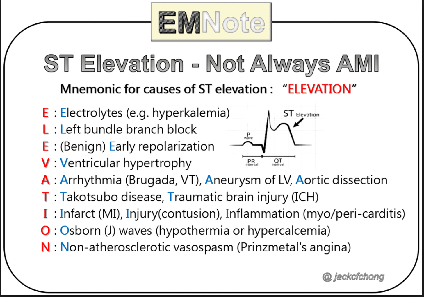
There are numerous causes of ST elevation other than STEMI.
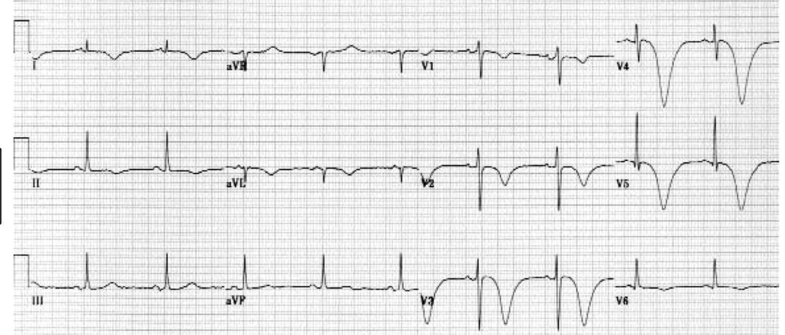
Pictured are Cerebral T waves which can be seen in subarachnoid hemorrhage. Subarachnoid hemorrhage can cause changes on the EKG such as deep t wave inversions or ST elevation.
ECG Findings of Cerebral T Waves
Inverted, wide T waves are most notable in precordial leads (can be seen in any lead).
QT interval prolongation.
Pearls
These are associated with acute cerebral disease, most notably an ischemic cerebrovascular event or subarachnoid hemorrhage.
They may be accompanied by ST segment changes, U waves, and/or any rhythm abnormality.
Differential diagnosis includes extensive myocardial ischemia.
Strongly suspect an intracranial etiology in a patient with altered mental status and these electrocardiographic findings. (Atlas of EM)

Catecholamine surge seems to be the pathophysiologic pathway of Takotsubo Cardiomyopathy. The LV apex is most sensitive to catecholamines. Takotsubo’s causes apical ballooning in response to a catecholamine surge.
Features of takotsubo cardiomyopathy.
Chest pain and shortness of breath after severe stress (emotional or physical)
Electrocardiogram abnormalities that mimic those of a heart attack
No evidence of coronary artery obstruction
Movement abnormalities in the left ventricle
Ballooning of the left ventricle
Recovery within a month
What is it?
Takotsubo cardiomyopathy is a weakening of the left ventricle, the heart's main pumping chamber, usually as the result of severe emotional or physical stress, such as a sudden illness, the loss of a loved one, a serious accident, or a natural disaster such as an earthquake. (For additional examples, see "Stressors associated with takotsubo cardiomyopathy.") That's why the condition is also called stress-induced cardiomyopathy, or broken-heart syndrome. The main symptoms are chest pain and shortness of breath. (Online Harvard Website)
Dr. Lovell comment: Pay attention to the details of managing the post-resuscitated cardiac arrest patient. Protect the airway. Avoid hyper/hypo-oxia and hyper/hypo-ventilation. Provide Targeted Temperature Management. Correct hypotension. Provide ASA and Heparin. Identify STEMI and discuss with cardiologist consideration of Cath lab.

ACC Risk Stratification Algorithm for Comatose Post-Cardiac Arrest Patients. Patients with multiple unfavorable characteristics are less likely to benefit from emergent cardiac cath.

Beware the Hyperacute T wave in the setting of chest pain. Hyperacute T waves may not always have ST elevation. The real key is the T wave’s size in relation to the QRS complex. This EKG was from a patient who went on to develop ST elevation and V-fib arrest. If you are suspicious of hyperacute T waves, get frequent repeat EKG’s to identify ST elevation or other evolution.
Dodd Research Lecture
Reasons to do research: Curiosity, Change the World, Change clinical care, Develop a device or drug, and Career advancement.
Dr. Dodd reviewed all the research opportunities available in our department.
Burns RUSH Exam
Mnemonic to remember all the aspects of the RUSH exam. HI-MAP ED: Heart, IVC, Morrisons, Aorta, Pneumothorax, Ectopic, DVT (common femoral veins)
You will used the phased array probe (cardiac probe) and the linear probe to get your views.

This is the order of images in the RUSH exam following the HI MAP mnemonic. You can add in suprapubic views looking for signs of ectopic pregnancy(pelvic fluid) and images of bilat femoral veins to screen for DVT.
Lambert Transesophageal Echo in Cardiac Resuscitation
TEE has been shown to alter the resuscitation management of the cardiac arrest patient. TEE can improve the performance of CPR by identifying where chest compressions are impacting the heart. TEE will frequently give the resuscitationist information to improve the location of chest compressions on the chest. Without TEE info CPR may be only compressing the SVC or RV and not the LV.
TEE can also identify pericardial tamponade, valvular dysfunction, V-fib, and cardiac rupture.
Team Ultrasound Ultrasound Lab

Write here…

Write here…
