Treatment of Massive Pulmonary Embolism
CASE:
21-year-old male was admitted to saint elsewhere with severe shortness of breath, epigastric discomfort and vomiting. He had recently been admitted to outside hospital with pancreatitis for 10 days and was discharged home with PMD f/u with improving idiopathic pancreatitis. When the patient arrived in the ED his vital signs were:
HR 140, Sat 92% on RA, BP 140/50
CC: epigastirc pain and vomiting
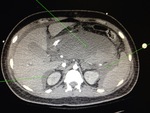
In the ED the patient was triaged to intermediate care had a chemistry panel sent and a CT A/P ordered. His lipase came back at > 3000. The medicine team was consulted and the patient was to be admitted to the floor pending a CT A/P. Image shown below is the most superior image from the abdominal CT which happened to catch the main pulmonary arteries
The patient requires 4L NC to maintain his sats > 92% but is awake and mentating well and his BP was normal. His EKG is shown:
The ED felt the patient was hemodynamically stable to be admitted to the floor and medicine admitted with IR consults for possible thrombectomy and IA tPA due to large RV seen on CTA. About 5 am the next morning while in the angio-suite the patient codes during thrombectomy. The patient is PEA without an airway in IR.
If you are called to this code, what would you do?
What was done:
A Pulmonary artery catheter was in place because of the attempted thrombectomy and the code team pushed 50mg of IA tPA (after about 10 minutes of me looking in the cath lab for the vial of tPA that was not in 2 mg aliquots). The patient has ROSC about 30 seconds later. Unfortunately his first ABG intubated on the vent was as follows:
6.79/90/85 on AC 30/500/100%/5
What would be your next move, and why do you think the pH and CO2 are so high?
We decided to add inhaled nitric oxide (see below) with only mild improvements in the CO2 to 75, pH still 7.0 on 3 vasopressors. What would you do?
CV surgery was called for immediate ECMO, and the patient was cannulated in the angio suite.
2 days later the patient was decannulated he did suffer from hemorrhagic conversion of his pancreatitis, and large RP hematomas and intra-abdominal hemorrhagic ascites, likely due to heparin and tPA administration.
However the patient was weaned off the ventilator, his pancreatitis improved and he was discharged home with no residual deficits on hospital day 21.
SO what can you do in the ED to set the patient with massive PE up for success?
Disclaimer: I should mention this portion pertains to the specific subset of patients who have right ventricular failure and not simply pulmonary hypertension from a PE and are reasonably well compensated. These people have the findings I mention below.
Some tricks of the trade are as follows:
Although hypotension is almost always treated initially in the ED with IVF's if you suspect massive PE the cause of hypotension is RV failure/dilation and the treatment is RARELY IVF.
1. You will not have hypotension from acute massive PE without RV dilation
This is because the thin highly compliant RV whose afterload is the wimpy pulmonary arterial tree is very pressure sensitive. Rapid increases in pulmonary pressure result in massive increases in RV volume (it does not hypertrophy acutely). Since the right and left ventricles SHARE the septum the right ventricle balloons outward and impairs diastolic filling of the LV when pressure overloaded, hence the D shaped LV seen on parasternal short axis. This impairs the LV filling, reducing the mean arterial blood pressure and cardiac output.
Signs of RV failure are:
1. Distended neck veins/elevated CVP/ Dilated plethroic IVC without resp variation
2. Elevated transaminases (usually more chronic)
3. Hepatomegaly, lower ext edema
4. Kussmal's sign (sudden increase in CVP in spont breathing patients with inspiration)
Echo:
Dilated RV with wide-open tricuspid regurgitation (this is because of the stretch of the annulus of the valve NOT valvular dysfunction per se). Adding additional volume will only overload the RV even more and worsen TR. LV is usually hyperdynamic with small chamber size.
-To complicate matters the RV (which is usually a low pressure ventricle) gets coronary perfusion during both systole and diastole (since it is a low pressure ventricle). However as the RV pressure increases the coronary filling is markedly reduced during diastole, this creates RV ischemia. Ischemia results in stiffness of the ventricle making the RV more dysfunctional and so continues the downward spiral of right heart failure.
2. The treatment of significant RV failure:
-Reduce RV Preload
To reduce preload avoid giving volume in the ED and consider diuresis
Commonly diuretics are given if the CVP is significantly elevated because the normal geometry of the ventricles has been distorted due to an enlarged RV bowing the spetum towards the LV, diuresis will result in improved LV performance even with a decrease in intrasvascular volume.
-Reduce RV afterload:
Inhaled Nitric Oxidie or Inhaled Prostaglandins like Epoprostenol. Usually iNO is given starting at 20ppm, despite the fact that 90% of patients will have maximal response at 5ppm or less.
The problem with giving systemic pulmonary vasodilators in a hypotensive patient should be intuitive, the nice thing about giving inhaled pulmonary vasodilators is they only act where there is ventilation and perfusion. Nitric oxide is rapidly metabolized by local RBC's and thus has no systemic effects like hypotension.
iNO is not standard of care per se, but is a trick up your sleeve for that severely dyspneic patient with massive PE who you want to avoid intubation in. AM I saying you can give iNO non-invasively, absolutely. This is the nice thing about iNO is sometimes it will save you an intubation or at worst in can prevent the precipitous RV failure that will ensue after intubation.
-Maintain MAPS > 65
I know this is an obvious point, but it has to be stressed that even a minute of hypotension can throw a tenuous RV into the spiral of death mentioned. Sedatives will often result in rapid hemodynamic compromise if you are not careful.
Epinephrinebetween 0-50 nanos/kg/min or (0- 0.05mcg/kg/min) at lower doses epinephrine acts as an inotrope, and above that tends to act more as an alpha agonist. At low doses epi will likely not increase the pulmonary vascular resistance but at higher doses (> 50 nanos) it may.
Dobutamine2-5 mcg/kg/min can augment RV function without increasing the PVR seen with epi and levophed, however it can cause systemic hypotension from beta 2 agonism and often will be given with another agent.
Vasopressin can augment the mean arterial pressure, and likely has NO effects on the PVR.
My unevidenced based practice is to start Dobutamine and Vasopressin (2 mcg/kg/min and 0.04 U/min) to keep MAPS > 70 and follow serial bedside echos.
3. If someone with a PE is dyspneic should I intubate sooner rather than later, they are eventually going to tire out right?
The problem with intubation in a patient with a PE is positive pressure ventilation increases the afterload via PEEP to the right ventricle. This may tip the RV over into florid RV failure.
There are also the hemodynamic perturbations with giving a sedative during induction, which can cause hypotension, again resulting in rapid hemodynamic compromise.
Initial paralysis will prevent spontaneous ventilation and may make it difficult to match a patient's own intrinsic minute ventilation (young pt breathing RR 40, and large TV of 1L with MiV of 40), try setting the vent to get anything reasonable in these patients afterwards is not ideal. If you have to use something use a shorter acting agent like succinylcholine.
Nothing like someone telling you not to do something every sense in your body is telling you to do. However, if you are forced to intubate them, and you know they have a massive PE, consider thrombolysis up front and:
1. Start iNO, if this process isn't available where you work but iNO is available in house find a way to expedite the process in the ED now before you are really desperate. If you work in a hospital without iNO/CT surgery/IR consider establishing transfer to a facility with higher level of care. Worst thing you can do is admit a patient to an ICU where they won't be seen by an intensivist until the morning and there is minimal backup if the patient decompensates. Without a backup if the patient codes the game has been lost.
2. Have your vasopressors ready to go on a PUMP and push dose pressors in hand epi is the simplest in these situations. I use epinephrine in 10mcg aliquots as a push until I have a MAP > 65 if they drop during intubation. Then I place and titrate a drip as needed.
3. Sometimes flipping into A-fib will result in rapid hemodynamic deterioration and may need cardioversion as the loss of the atrial kick may have significant hemodynamic effects.
4. Call your CV surgeon and IR attending and get them on board early. "I have a young patient with massive PE who is crumping in the ED, is this patient a candidate for surgical thrombectomy and if they crash, would you consider them an ECMO candidate?"
Never be afraid to ask for help and if they are young and salvageable don’t accept NO for an answer.


