Eastvold Pearl #28 - Lithium Toxicity Masquerading as Wellen's Syndrome

Take-home point: Rule out lithium toxicity in all patients on lithium presenting with cardiac toxicity and/or EKG changes.

[1] The case: 48 yo F with history of bipolar disease and depression presents to ED for evaluation of substernal chest pressure radiating to neck with bilateral arm "numbness."  Mild shortness of breath and ROS only positive for mild gradual onset headache.  Symptoms x 7 hours.  Chest pain non-exertional, non-positional, and non-pleuritic.  No fever, focal neurologic deficits, mental status changes, or abdominal pain.  Chest pain 7/10 upon arrival, and improved with 3/10 with nitrate therapy but continued to have ongoing pain/symptoms.  Serial EKG revealed dynamic change, and concern for proximal LAD lesion and Wellen's syndrome was discussed with cardiology.  Cardiology performed a bedside echo which revealed anterior wall motion abnormality, and to a lesser degree inferior-lateral dyskinesia.  Patient was taken to cath lab for elective cath, with knowledge of an elevated troponin of 0.61 (normal < 0.03),  Cardiac cath revealed completely normal coronaries, and no LV aneurysm  Lithium level came back elevated at 2.3, with no history of acute ingestion.  Quick literature review (articles attached) revealed that lithium can mimic ischemic EKGs, especially in the anterior leads.  Further, chronic lithium toxicity can also cause myocarditis.  See EKGs below illustrating deep anterior T wave inversions and prolonged QTc, both of which resolved after lithium level decreased.

[2] Quick skinny on lithium toxicity and EKGs, attached is are 2 review articles as well.

  • Lithium causes toxicity through several mechanisms including competition with many electrolytes (sodium, potassium, calcium, and magnesium) and ion transporters, which can disrupt many cellular functions.  The cellular membrane of concern here is myocytes (and Purkinje fibers....and most people just closed this email!), and these disruptions manifest as EKG changes.   
  • Common EKG changes include [a] QTs prolongation, [b] nonspecific ST segment and T wave abnormalities, [c] T wave inversions (reference #1), [d] transient ST elevations mimicking acute myocardial infarction (reference #2), [e] Brugada changes with possibility of sudden cardiac death (reference #3), [f] SA nodal dysfunction and heart block, and [g] severe bradycardia.
  • ECG changes are more likely to occur with chronic lithium overdoses than acute overdoses
  • Treatment is beyond the scope of this review but some important things to consider are
    • Make sure you get lithium levels on all patients presenting with cardiac symptoms
    • Know the difference between acute vs chronic toxicity, read attached article
    • Know your criteria for hemodialysis
    • Avoid thiazides and ACE-I, as these will increase lithium levels
    • Avoid NSAIDS, can also worsen toxicity.  Often given for myocarditis, but do not given if you think this is lithium-induced myocarditis
    • Check TSH, can be contributing to lithium toxicity
    • Consult toxicology and nephrology early, especially if cardiology is going to manage primarily

Feedback always welcome (joshua.eastvold@gmail.com),

Also, if anyone is interested in publishing this let me know??

Josh 

References:

[1] Cases J. 2008 Sep 17;1(1):156. doi: 10.1186/1757-1626-1-156.

[2] J Med Toxicol. 2008 Sep;4(3):170-2.

[3] Circulation. 2010 Aug 10;122(6):e418-9

[4] Toxicol Rev. 2006;25(4):221-30.

[5] J Am Soc Nephrol. 1999 Mar;10(3):666-74

EKG #1 upon arrival to ED

wellens Li1.png

EKG #2 - concerning for anterior ischemic.  QTc 494.  

wellens Li2.png

EKG #3 - worsening QTc prolongation, still getting PO lithium, don't ask

wellens Li3.png

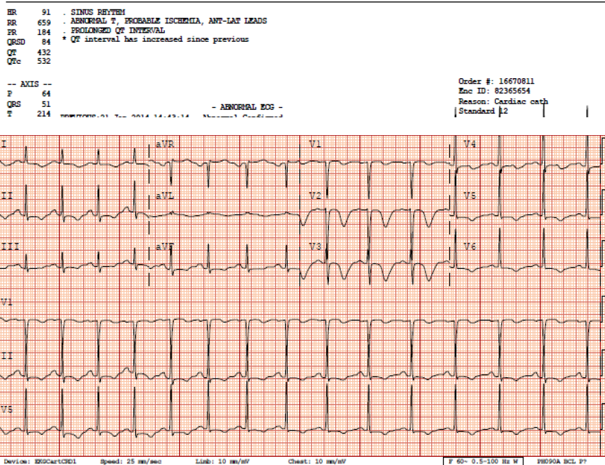
EKG #4 - Courtesy call to cardiology, relayed my thoughts regarding concern for chronic lithium toxicity

-- now after lithium level returned to 1.3, complete resolution of anterior T wave changes and normalization of QTc

wellens Li4.png

Eastvold Pearl #27 - Superficial thrombophlebitis in the lower extremity

Hey guys,

Been a while, and have 2 more coming.  

Superficial thrombophlebitis (ST), aka superficial vein thrombosis

--

Thrombosis of superficial veins has long been considered benign, and deemed a separate entity from venous thromboemolism (VTE)

-- However, multiple studies illustrate a significant association with VTE (DVT and PE).

-- When patient with ST (diagnosed clinically, no ultrasound) are thoroughly evaluated, the degree and the extent of clot are underestimated 75% of the time.  Further, such patients are found to have co-existent DVT or PE 25% of the time and/or rapidly progress to DVT 10% of the time.  

---- Teaching point: get ultrasounds on all clinical superficial thrombophlebitis

-- The risk factors for ST and VTE are the same, and many argue that ST should be treated the same as VTE.  

-- In general,

anticoagulate (as you would for DVT) the patient if they have known clotting risk factors, greater than 5 cm of clot, or clot < 5 cm within the sapheno-femoral or sapheno-popliteal junction.

-- Another way to think about it is that a superficial vein thrombosis is a manifestation of a systemic clotting cascade gone awry.  To even further simplify things, seems pretty pathologic to have any blood vessel clot; ahh hello, you are clotting off blood vessels.

Below is my treatment algorithm on ST in the lower extremity.

Feedback always welcome.

Josh

References

http://www.ncbi.nlm.nih.gov/pmc/articles/PMC3278049/#

    CHEST 2012 guidelines

Eur J Vasc Endovasc Surg 2005; 29:10-17. 

J Vasc Surg 2003; 37:834-8. 

Thromb Haemost 2001 ;86:452-63.

SVT.jpg

* Full anticoagulation with [a] lovenox 1 mg/kg BID (or 1.5 mg/kg QD) for at least 5 days and until coumadin is therapeutic for at least 24 hours. [b] Xarelto or rivaroxaban15 mg BID for the first 3 weeks, then 20mg daily for at least 3 months.  If lifelong anticoagulation planned, would recommend lovenox/coumadin.  2nd reoccurrence consider lifelong anticoagulation but refer to CHEST guidelines for specific permutations. A study by Schulman found a second DVT indicated a need for lifelong anticoagulation and it didn’t matter whether the DVT was provoked or unprovoked.  However, provoking factors vary in their risk of recurrence; surgery patients have the lowest risk of recurrence.  Patients with unprovoked or idiopathic thrombosis or multiple recurrences with lower risk provoking factors will get long‐term anticoagulation. 

** History of DVT or PE, active malignancy (recommend solely lovenox, d/w oncologist), hypercoagulable state.  Other risk factors that warrant consideration of systemic anticoagulation include male gender without varicose veins, history of superficial thrombophlebitis, recent surgery, involvement of saphenous vein, and severe symptoms.

*** Lots of variability in dosing. 

  -- Lovenox: [a] 40 mg daily SC x 4 weeks per CHEST guidelines, [b] 1 mg/kg BID SC x 10 days then 1 mg/kg QD x 20 days per Belgian Society on Thrombosis and Haemostasis. 

  -- Fondaparinux 2.5 mg SC QD x 30-45 days

  -- Xarelto – current trial comparing 10 mg PO QD vsfondaparinux.  Xarelto approved for DVT.  Would consider using if 1st time on anticoagulation given PO form and no need for INRs, but would discuss with PCP before committing to lifelong use of Xarelto.

****

Deep veins = superficial femoral vein, common femoral vein, deep femoral vein, popliteal vein, anterior tibial vein, posterior tibial vein, peroneal vein.

Superficial veins = Greater saphenous vein, small saphenous vein

Eastvold Pearl #26: SCIWORA

This is a long one.  But such an important topic.

The skinny on a case that I had.  80 yo M fell in front lawn while bending over to pick up sprinkler, fell forward striking head on ground (sounded like maybe from a foot or so) and then could not get up. Found by neighbor several hours later, and EMS called with transfer to ED.  Patient ambulatory on scene. Denies any pain or symptoms other than stating he felt weak.  My work-up in ED, negative CT head, EKG, troponin, CK, CXR, UA, and routine labs.  I did note that patient had a very difficult time rolling over in bed due to weakness.  Ambulated (known in the medical world as "leg stuff") without assistance or fall risk.  Recommended admission but the thought of nursing home placement kept this stoic Iowan out of the hospital...until he was brought back into the ED 12 hours later noting difficulty buttoning shirt and eating (known in the medical world as "arm stuff").  CT C-spine attained which was negative, admitted to hospital with MRI several days later revealing central cord syndrome.  Kind of sounds like a classic boards question in hindsight.  So, this was my 2nd SCIWORA case in 4 years.  Both patients injured their C-spines after falling from standing height.  So, what have I learned (apparently not enough yet)...see below.

Spinal cord injuries can be very subtle.  Why?  I think for 2 main reasons; crappy neuro exams and crappy knowledge of presenting symptoms. 

Pearl #1: Patients with spinal cord compression often do NOT have pain

.   My guy had zero neck pain or tenderness on exam.  We all tend to decide on the need to image the C-spine based on pain.  No pain or tenderness, squeeze my hands and wiggle your toes..C-collar off...usually you are okay with this approach.  However, you have to be really sure you are not missing subtle neurologic findings.

  • Neurologic complaints without pain in the extremities are concerning for spinal cord compression. Neurologic complaints with neck pain and/or arm pain are concerning for cervical radiculopathy (i.e., compression on exiting dorsal root nerve)
  • Obviously, neck pain is also concerning for potential fracture with osseous injuries leading to the most devastating injuries.  So the take home message is not that neck pain is benign, but the absence of neck pain does not exclude serious injury.

Pearl #2 - Neuro exam findings are often symmetrical

.  The cursory ED neuro exam misses these because we are so trained on detecting asymmetries, i.e., "brain stuff" or strokes.

  • Motor complaints: Bilateral symmetrical weakness (or spasticity) concerning for spinal cord compression.   Unilateral weakness suggests radiculopathy.  To detect these deficits, you must test all joints that flex or extend, which will effectively test both proximal and distal motor function.

Pearl #3 - Sensory deficits are vague and NOT dermatomal

  • Sensory complaints: vague bilateral non-dermatomal or multiple dermatomes involved is concerning for spinal cord compression.  Sharp demarcation or dermatomal loss is concerning for radiculopathy.

Pearl #4 - Test reflexes

  • Bilateral reflex abnormalities, positive Hoffman's sign, and positive Babinski's sign are concerning for spinal cord compression
  • Slight retraction of lower extremities when testing Babinski does not equal volitional movement.  I made this mistake in the intoxicated patient.  Very well documented false positive so to speak.

Pearl #5 - Autonomic complaints are always concerning for cord compression

  • Impotence or priaprism.  Remember the saying, if they are raising theirs you should raise your eyebrows...maybe that was for Bells palsy
  • Bladder or bowel dysfunction
  • Horners syndrome

Pearl #6 - The "Eastvold Washcloth Test" = Pain and temperature run together via the spinothalamic tract (anterior cord)

  • Testing only pinprick can be [a] difficult to pick up on subtle deficits given as we all know this test is so subjective, "feels a little duller", [b] people malinger
  • Easier and probably much more sensitive (based on my own anecdotal evidence) method is to test temperature with a cold wet washcloth.  Everyone wins, you will pick up on subtle deficits and the patient gets a sponge bath, which is hopefully their last as opposed to a sponge bath from the nurse at neuro rehab.
  • If abnormality detected, then I test pinprick

Pearl #7 - Be Weary of Anyone with a Hyperextension Mechanism of Injury

  • Both patients fell from standing height.  1st guy hit forehead on table with large laceration over forehead.  2nd guy had very small abrasion on chin, that's it.  Both are hyperextension injuries, and the most common subtype of SCIWORA.
  • "Commonly observed in the spondylotic spine in association with low-energy mechanisms, such as fall from standing height, although it can be observed in younger patients in association with higher-energy mechanisms and acute disc herniations.  Clinically these patients often present with minor abrasions or lacerations on the scalp/forehead and a variable degree of neuologic impairment..."  http://www.ncbi.nlm.nih.gov/pmc/articles/PMC2989526/

Pearl #8 - Fractured (aka broken, Gromis) Osteophytes on CT scan May Not Be Insignificant = Cervical spondylosis is a potential red flag

  • Does not equal unstable C-spine injury, true
  • Insignificant, false.  
  • You will read that isolated osteophyte fractures are insignificant.  They are not unstable, but they may represent soft radiographic evidence of serious underlying ligamentous disruption
  • How?  Due to spinal cord compression between a hypertrophied spondylotic disc-osteophyte complex and bulging ligamentum flavum
  • Occurs with hyperextension (as well as hyperflexion) injuries.  There was likely considerable translational movement within the 3 spinal columns and ligamentous disruption at the time of injury, but with muscle spasm and return to normal alignment of the neck this disruption can be very subtle.  See Fig 7 below illustrating this important point.  I have seen this exact case.  Notice how well-aligned the spinal column is.
  • > 80% of adult SCIWORA patients have underlying cervical spondylosis.  I feel pretty good about a negative MDCT scan with recons and no underlying spondylosis...and no neuro complaints.  If spondylosis is present, all patients with subjective or objective neuro findings need an MRI before clearing C-spine (and read pearl #7).  Further, I do a really thorough neuro exam (after my 2nd missed case) on patients with spondylosis without neuro complaints before clearing these patients.
  • Lastly, EMRAP has officially failed on this topic (dammit Mel) spouting comments that if the CT C-spine is negative the chance of surgical intervention is zero.  Almost true, as the majority of these SCIWORA cases are managed conservatively.  I do agree that the overwhelming majority of patients with negative MDCT with recons and no subjective or objective neurologic findings do not need an MRI.  Patients with persistent severe neck pain is a judgment call, and if unsure you should involve your consultants.  Just be really careful about diagnosing someone with hysterical paralysis.
Inline image 1

Pearl #9 - Patients with acute cervical spine injuries need interrogation of entire spine

  • I need to do a better job of this
  • If there is any C-spine injury, the patient needs at least CT of TLS spine and likely MRI

Pearl #10 - If you are doing plain films or flex-ex films, please just stop it

Feedback always welcome.

Next topic is how to manage compression fractures, this one has always bugged me with the lack of recommendations.

Josh

Eastvold Pearl #25: Anterior MI w/o reciprocal cahnge

As we reflect on the year 2012, it is important to know what does not "reflect" (corny stretch) in the world of EKGs.  Fifty percent of anterior STEMIs lack reciprocal changes.  This is crazy but a fact.  I will try to summarize why this occurs, but you may want to read the attached article.  

We have all learned that reciprocal changes are important in diagnosing STEMIs, and their presence increases the likelihood that the EKG pattern is secondary to coronary occlusion.  Further this concept of reciprocal changes is the main focus of my "Swans Reflecting Elephants" talk, whereby localized ST depression is invariably secondary to subtle STEMIs.  

But does the absence of reciprocal change rule out MI?  The answer is no.  As stated above, 50% of anterior STEMIs lack reciprocal changes.  

That said,  inferior STEMIs will almost always have STD (albeit may be subtle) in I/AVL or at least new TWI in AVL.  Lateral STEMIs also will have reciprocal change.   

The pathophysiology of inferior ST segment changes in the setting of anterior STEMI is actually pretty cool.  I have read a lot on this, and I think you will like this article I am attaching.  To summarize, in the setting of anterior STEMIs there are 3 patterns of inferior ST segment changes, [a] ST depression, [b] ST elevation, and [c] No ST change.  Group "c" accounts for 50%, quoted many times in the literature.  

Look at Figures 1, 2, and 3 in the paper, as this is the best explanation on this issue that I have found.

-- Anterior STEMI with inferior ST depression occurs because the occlusion is proximal LAD (proximal to D1 or 1st diagonal), with infarction extending to high lateral wall.  Thus the inferior ST depression actually represents true reciprocal change from lateral ischemia.  Anterolateral STEMI.

-- Anterior STEMI with inferior ST elevation occurs the the occlusion is distal to D1 AND wrap-around LAD (a variant) that supplies the inferior wall, thus causing inferior STEMI.

-- Anterior STEMI with no inferior ST changes occur in 2 settings, which is exemplified in example #3 in the paper.  [1] Occlusion distal to D1 with no wrap-around LAD, so no infarction of lateral (distal) or inferior (no wrap-around) regions.  There is NO reciprocal change for true anterior ischemia.   [2] This setting is the cool one.  If you have a proximal LAD lesion to D1 AND a wrap-around LAD, the ST changes counteract each other.

Josh

Eastvold Pearl #24: False Positive Lactate

Hey guys,

   Worked a shift at UIHC with a resident who got a lactate on a hemodynamically stable asthmatic, and it came back at 4.6.  Repeat arterial lactate similar.  If you practice drone medicine like this internal medicine resident was doing, you'll be heading down the wrong path ..."pretty much means we need to put a central line in, and I guess start antibiotics."

What the resident didn't know was that asthmatic patients on continuous albuterol nebs will have a type B lactic acidemia.  Although the mechanism is not completely understood, β2-agonists are known to stimulate cyclic adenosine monophosphate (cAMP)-mediated glycogenolysis and lipolysis.  Hyperglycemia and free fatty acid inhibition of pyruvate dehydrogenase lead to anaerobic metabolism of pyruvate to lactate, thus causing elevated lactate levels.  Work of breathing has also been hypothesized to contribute as well.

There are many other causes of type B lactic acidemias, just google them.

Eastvold Pearl #23: Swollen Scrotum

Hey guys,

  I have been absent for a while, and hope to send more of these out.  This one was provoked by the conference notes sent (great idea btw).  I ran across a patient with a non-infectious complication from peritoneal dialysis...the massively swollen scrotum.  

65 yo M with h/o ESRD on CAPD presented to ED with [1] massively swollen scrotum (his concern), and honestly [2] facial droop (wife's concern) for 5 days, which turned out to be a Bells palsy or peripheral CN VII palsy.  You gotta love a guy like this.

We have all seen massive scrotal edema in the setting of anasarca, with the work-up really having nothing to do with GU but merely a work-up for anasarca.  

What was different with this guy was that he had no lower extremity or abdominal wall edema, it was all isolated to his scrotum and penis.  No hernia on exam or bedside US.

Quick google search at work, popping out the attached article, then pretending I knew this beforehand as I talked to the nephrologist on-call really changed the work-up and treatment for this patient. 

It turns out that patient with continuous ambulatory peritoneal dialysis (CAPD) have about a 5% risk of dialysate leakage, representing a non-infectious complication of CAPD that we should know.  Why is this important, these patients will need to stop PD and switch to hemodialysis as part of the treatment.  Thus, they will need to be admitted as most do not have HD capabilities.  Why to have to stop PD?  See below and read attached article if time.

PD dialysate leaks are classified as early (1st 30 days of catheter placement) or late (>30 days).  The above complication is a late complication, which is what I'll cover.  Late leaks most often "related to mechanical or surgical tear in the peritoneal membrane, presenting as internal leakage (e.g., pleural cavity, abdominal wall, external genitalia).  Enhanced stress on the supporting abdominal wall structure may lead to" leakage and/or hernia.  The leakage can be considered a mechanical intra-abdominal pressure-related complication due to micro-tears (which may seal up with cessation of PD) or larger tears or hernias (which require surgery).

Diagnosis: Known in the nephrology field as "skinny legs, big scrotum."  May need US to rule out hernia, or CT with PD catheter-instilled contrast to evaluate for leakage based on extravasation of contrast outside peritoneal cavity.

Treatment: [1] Stop CAPD for 1-3 weeks, again this will require most to be admitted. [2] Switch to temporary hemodialysis, which is thought to allow these micro-tears to seal up. [3] Surgery if recurrence after re-initiating CAPD.  Other indications for surgery are concurrent hernia, and some advocate that genital edema is an indication for surgery as these patients fail the PD rest period.

Josh
Reference:  Dialysate Leaks in Peritoneal Dialysis. Semin Dial. 2001 Jan-Feb;14(1):50-4. Leblanc M, Ouimet D, Pichette V. Nephrology Division, Maisonneuve-Rosemont Hospital, University of Montreal, Quebec, Canada.
Dialysate leakage represents a major noninfectious complication of peritoneal dialysis (PD). An exit-site leak refers to the appearance of any moisture around the PD catheter identified as dialysate; however, the spectrum of dialysate leaks also includes any dialysate loss from the peritoneal cavity other than via the lumen of the catheter. The incidence of dialysate leakage is somewhat more than 5% in continuous ambulatory peritoneal dialysis (CAPD) patients, but this percentage probably underestimates the number of early leaks. The incidence of hydrothorax or pleural leak as a complication of PD remains unclear. Factors identified as potentially related to dialysate leakage are those related to the technique of PD catheter insertion, the way PD is initiated, and weakness of the abdominal wall. The pediatric literature tends to favor Tenckhoff catheters over other catheters as being superior with respect to dialysate leakage, but no consensus on catheter choice exists for adults in this regard. An association has been found between early leaks (< or =30 days) and immediate CAPD initiation and perhaps median catheter insertion. Risk factors contributing to abdominal weakness appear to predispose mostly to late leaks; one or more of them can generally be identified in the majority of patients. Early leakage most often manifests as a pericatheter leak. Late leaks may present more subtly with subcutaneous swelling and edema, weight gain, peripheral or genital edema, and apparent ultrafiltration failure. Dyspnea is the first clinical clue to the diagnosis of a pleural leak. Late leaks tend to develop during the first year of CAPD. The most widely used approach to determine the exact site of the leakage is with computed tomography after infusion of 2 L of dialysis fluid containing radiocontrast material. Treatments for dialysate leaks include surgical repair, temporary transfer to hemodialysis, lower dialysate volumes, and PD with a cycler. Recent recommendation propose a standard approach to the treatment of early and late dialysate leaks: 1-2 weeks of rest from CAPD, and surgery if recurrence. Surgical repair has been strongly suggested for leakage causing genital swelling. Delaying CAPD for 14 days after catheter insertion may prevent early leakage. Initiating CAPD with low dialysate volume has also been recommended as a good practice measure. Although peritonitis and exit-site infections are the most frequent causes of technical failure in peritoneal dialysis (PD), dialysate leaks represent one of the major noninfectious complications of PD. In some instances, dialysate leakage may lead to discontinuation of the technique (1). Despite its importance, the incidence, risk factors, management, and outcome of dialysate leakage are poorly characterized in the literature. We will review the limited available information on this topic in the next few sections. PMID: 11208040 [PubMed - indexed for MEDLINE]

Eastvold Pearl: Uveitis

Uveitis

Consensual photophobia is always a red flag!

Case: Pt presented to PMD earlier in the day, dx: conjunctivitis, sent home on topical abx, came to ED later that day for persistent pain.  L eye red, pt in considerable pain, photosensitive even when light shown in other eye, noticeable perilimbal or ciliary flush, anisocoria c affected eye more miotic, 1+ flare in anterior chamber on slit lamp, no fluorescein uptake, visual acuity wnl b/l, IOP < 20 b/l, no FB detected or mechanism, only some of the engorged vessels moved when brushed c Q-tip.

Dx: Uveitis

  1. Consensual photophobia
    1. Definition: severe pain in affected eye when light is shined in unaffected eye
    2. Cellular Flare
      1. Inflammatory cells in anterior chamber (on slit lamp)
      2. May cause blurry vision
      3. Miosis
        1. From ciliary mm spasm, will have anisicora and pain with accomodation (have them follow your finger close then far)
        2. How to evaluate anisicora (http://www.pacificu.edu/optometry/ce/courses/19433/pupilanompg2.cfm)
          1. Basically, note pupil size in the dark and the light, if this difference is constant, it’s physiologic
          2. Scleral Vascular Engorgement
            1. Conjunctival and episclera vessels move when touched with a Q-tip; the deep-seated scleral vessels do NOT
            2. Conjunctival and episcleral vessels blanch c topical phenylephrine gtts, scleral vessels do not
            3. Conjunctivitis: superficial vessel inflammation, Scleritis: deep vessel inflammation, Uveitis: both affected
            4. Ciliary Flush
              1. 2/2 dilation of radial vessels
              2. Often perilimbal flushing, whereas conjunctivitis has more peripheral conjunctival injection (uveitis on left below, conjunctivitis on right)

Book Stuff

Uveitis

  • Inflammation of the middle portion of the eye.
    • Anterior Uvea consists of iris and ciliary body = iritis (iris only) & iridocyclitis (both)
    • Posterior Uvea consists of the choroid - choroiditis, does not cause red eye but included for completeness purposes

Causes

  • Trauma (blunt or penetrating), corneal or scleral injury
  • Systemic microbial infection
    • Syphilis, brucelosis, herpes simplex, Lyme dz, TB
    • Immune-mediated
      • HLA-B27-associated diseases, sarcoidosis, JRA
      • Idiopathic in 50% of cases

Presentation

  • Red eye, unilateral in most cases
  • "Real" photophobia
  • Deep boring pain

Treatment

  1. Ophthalmologist referral within 24 hours
    • Topical steroids + intermediate-acting cycloplegics (i.e. cyclopentolate) for the ciliary spasm
    • Antibiotics not needed
  2. Expectant secondary glaucoma (often due to debris blocking normal drainage). Treat accordingly if IOP > 20.
  3. w/u can be performed by the ophthalmologist in an outpatient setting in 24 hrs, and may include a CBC, ESR, ANA, RPR, VDRL, PPD skin testing, lyme titer, etc.