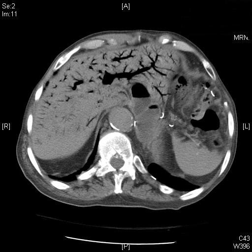
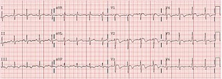
STATUS ASTHMATICUS

23y/o F presents form EMS with "SEVERE ASTHMA". Patient is hypoxic satting 78% on 15L NRB in the rig. Patient is obtunded on presentation diaphoretic and cyanotic. Her sats are in the low 80s on an ambubag at this point.

What would you do?


This patient weighs approximatel 260lbs, and is clearly in distress. She is barely moving air and is extremely tight. Here's what we did: 4g magnesium, ambubag with PEEP valve and 2LNC for increasing PEEP. 150mg of IV ketamine and 2mg of IV versed. Ventilator to the bedside and get her sats up. With the NC and ambu bag sats come up to 100%, color returns patient is clearly anxious fighting and agitated, rather than obtunded now. She will NOT tolerate bipap.


Decision is made for intubation but she is obese. RAMP positioning (thanks dr. g), head is elevated 30 degrees. Nasal cannula is left on DURING the intubation as patient is RAPIDLY desaturating when the BVM is taken off face (she has no cardiopulmonary reserve, due to her intrinsic lung disease and reduced FRC). Patient gets an awake look(this was right after ketamine and versed were given) to see what airway looks like and after cords are visualized patient begins to gag, decision to push paralytics is made. Patient intubated and initial vent settings are!?


AC 8/450/100%/0

We are now hearing air move patient is no longer auto-peeping, she is not hypoxic and her end-tidal is about 70 on waveform capnography (dont forget to change settings to see the graph suually only goes as high as 50).

But about 6 minutes after paralytics are pushed patient is getting agitated here rr increase to 18 and her sats come down. her PCO2 begins to drop and her peak pressures SKY ROCKET to the 80s, clearly she is under sedated.


An insp hold for plateau pressure is completed and a sickening SIGH and wheeeze is heard from the ET tube as the patient expires all the auto peep. Boluses of fentanyl and versed are given while we attempt to paralyze patient with nimbex in order to gain complete control of resp status. BUTTT we cant give nimbex in the er because nurses are NOT trained apparently in this. So instead a bolus of rocuronium was given to buy time with high doses of propofol were given for sedation (BP was very elevated from sympathetic response).


Initial ABG after propofol and a push of rocuronium is: 7.0/102/406/25, clearly a resp acidosis.

We decide bicarb drip is the way to go and allow her to increase rate to 10 with repeat gas of

7.1/96/87/29 on AC 8/450/50%/5


In the ICU managemnet is continued with train of fours for sedation on nimbex, and patient is given heliox 20/80. Patient doing well much improved day one in ICU.


Classic - Tylenol Overdose

Sooooo...it's been a bit and I thought I'd throw up a case I had recently.  Nothing special, but with a lot of tox stuff coming up I thought it would be nice to throw up a classic tox case with a good result.

Ok.  36 yo male presents with abdominal pain.  On history, guy notes he took close to 200 pills of 500mg of Tylenol.  The first 100 he took 36 hours prior to arrival, and the second 100 he took roughly 28 hours pta.  This is 100 GRAMS of Tylenol.  On exam, vitals were stable, except for being slightly tachycardic.  He was tender in RUQ/epigastric and had some scleral icterus.  Alert and oriented x 3, otherwise stable, and an ex-wife crying in the room because she just divorced him for being an ALCOHOLIC!  Thus the suicide attempt; great....

We start NAC, give him an oral loading dose, and if you don't know this...it smells like a horse's ass.  Patient took it down like a champ.  Labs started coming back and we realized he was not joking...

APAP - 51  (this is 36 hours after!  The nomogram only goes up to 26 hours from the ones I've seen, but definitely over the toxicity line.)  Remember, at the 4 hour mark, if it over 200 mcg/ml, the nomogram says continue the entire treatment course, 17 doses of NAC.  Actually...after some reading, it's all 150 these days.  

CBC/BMP fine, Cr - 1.06 (important),  INR - 2.8, PTT - 32, Platelet - 112 (ok CBC not totally fine)

Tbili - 9.7

AST - 16,325  ALT - 12,558    Wowza.  Talk to poison control, and they ask me 2 things right away...what is the pH and how is his mental status.  Ughhh...I'll get an ABG now?  Mental status is fine.  NAC is going, but they say switch it to IV form and keep it going.  

ABG: 7.47/31/90/23  

Poison control says transfer to liver transplant center IMMEDIATELY.  Why?  They don't blink an eye at the transaminases, it's the coagulopathy that scares them.  Pt has been in our ED for a total of 10 hours roughly and we transfer him to UofC, stable the entire time.  36 hours after transfer, he is spiraling down.  INR is 9, AST > 20k, becoming encephalopathic.  He had somehow passed their psych eval and was on the liver transplant list.  The fellow I spoke to at the time was close to pulling the trigger on intubation.  Well, 2 days after that...HE BOUNCES BACK!  No longer encephalopathic, INR is normal, and his transaminases have dropped like a rock.

Wow.

So I learned some specifics outside of the minimal basics I knew before.

1.  IV NAC is preferred in any patient you are worried about fulminant hepatic failure.  It is easier to administer, tolerated better by pts, and has equivalent efficacy.  It does have a high rate of anaphylactoid rxns (5-17%), so keep a close eye on pts you are administering this to.  Oral NAC smells like my poo.

2.  Continue IV NAC in these pts until 1 of 3 things.  a.  pt dies  b. pt fully recovers  c. pt gets a liver

3.  The reason Poison Control said transfer?  4 prognostic criteria of pts wit the highest risk of criteria (King's criteria correct Andrea?)

a. pH < 7.30  b. PTT > 100s  c. Cr > 3.3  d. hepatic encephalopathy

4.  The liver is awesome.

Ok.  There is SO much involved with Tylenol tox that I didn't go over.  What about activated charcoal?  What about the fact that there were really 2 ingestions here?  More specifics on the nomogram?  Where is Andrea Carlson when you need her?

Word.

unresponsive

70y/o F BIB EMS after being found in car next to grocery store. Patient was apparently driving and then suddenly became unresposnive car still in D, and came to halt slwoly at guard rail. On arrival patient being bagged agonal breathing near 40x's/min. She moves all four ext pupils 5mm nonreactive.

VS 210/100, 120, 40, 90% ON 15L NRB, no temp yet just being placed on monitor. (at this point everyone in the room was thinking bleed).

We decide we want to intubate, but first Im caught off guard by the monitor so we snap a quick EKG

EKG 1 - Copy.jpg

What's going on here???

Big gulps time to intubate the patient, lets not use succ, lets throw two amps of bicarb in there immediately, and open the code cart (boos are arising from the nurses), I also ask the EKG tech to stay here.

After bicarb - Copy.jpg

This is the rhthym while infusing bicarb

 Wow this stuff really works, thats after half an amp of bicarb. What's next doctor??

So we gave some calcium CHLORIDE, not gluconate, rmeber three amps of CaGlu=1amp of Cacl and you can give CaCl IVP. So lets see yet another rhythym change

After calcium cholride - Copy.jpg
Cardioversion bicard - Copy.jpg

Whoops that 1amp of CaCl thew our patient into svt, well by this point shes intubated, shes on propofol lets just shock her. 100J synchronized CV nothing, 150J synchronized Cv nothing,ok ok treat the underlying pathology more bicarb. Whoops again this is the following EKG, we were preparing amiodarone at this point, and she did still have a very strong pulse.

 And our SVT just degenerated into VTACH OH shit, but she still has a pulse she gets another gram of calcium and another ampulse of bicarb and 150 of amio and an amio drip. And this is her final EKG

amiodarone more calcium and bicarb - Copy.jpg

Wow what a dramatic difference. So now we don't have a K yet because ABG's wont give us a K. I still have no history so what did I do for his lady preemptively?

20mg albuterl neb, 40mg of lasix  (she put out 100cc with a foley), kayexalate (even though i doubt its efficacy), bicarb drop 200cc/hr and an amio drip. I called vascular and said we are going t need a quinton and paged dr. zikos (he's greek and comes in the middle of the night so a crowd favorite).

ABG comes back: pH < 6.96, pCO2 21 (we set the rate at 30 very important on initial intubation she was breathing 40x's minutes beforehand).

Later her Cr comes back at 9.1, K of 7.2, everything else relatively normal

Ultrasound probe showed extremely dilated renal pelvis suggestive of obstructive uropathy, so we get a cat scan. She has bladder CA with obstructive uropathy. She gets dialyzed ph normalizes, she wakes up, her potassium is normal and left the hospital on her own two feet today. AWESOME case of hyperK

Vexing CT

Gentleman presents to you with acute onset of LUE paralysis, L facial droop, aphasia, and fixed rightward gaze with upgoing L Babinski.  You call a Code 30.  No trauma, no blood thinners, only a history of hypertension.  Here are your scans (top being most caudal, bottom image most rostral)...

cbell2.jpg
cbell1.jpg
cbell3.jpg
cbell4.jpg

Let's see some posts about what people see (those who I've told about this, hold off for now)...check back for the answer later.

Elderly Vomiter

CC: vomiting

HPI: 83yo M w/ h/o IDDM, HTN, CHF, prior iatrogenic esophageal perforation w/ resultant fundoplication and neoesophagus 40yrs ago now presents to the ER c/o 3d of progressively worsening nausea, vomiting, and mild abdominal pain.  Pt is A/Ox4, appropriate, in no active distress, but appears very thin. On coumadin

VS: 127/64, p67 (nsr), r14, sat99%ra, Tmax 36.9r

PE:

  • Gen - comfortable, A/Ox4, cachectic
  • HEENT - PERRLA, EOMI, no conjunctival pallor, non-icteric, no sublingual jaundice
  • Neck - TM, supple
  • CV/Resp - RR, NSR, no MRG/CTAB, no RRW, no adventitious lung sounds
  • Abd - Mild LUQ TTP w/o rebound, no surgical Murphy's, no RT/G, non-peritonitic abd, guiac negative
  • Neuro - No focal deficits

Assess: Elderly gentleman with likely bowel obstruction vs volvulus vs paralytic ileus, less likely infectious etiology, unlikely acute surgical abdomen

Plan: Labs, lactate, upright CXR and obstructive plain films (stat), CT A/P w/o contrast (stat)

abd3.jpg
abd5.jpg

Films unremarkable for acute pathology, no e/o free air, possible obstruction, no e/o volvulus.

abd1.jpg

WOW!!!  WTF?!?!

So what do you see?  It's badness, plain and simple.  Dr. Omi had mentioned that this is the most intrahepatic air she had EVER seen.  Can you tell if it's biliary or venous?

It's venous, which Dr. Omi pointed out you can distinguish by the degree that the intrahepatic air extends to the periphery or the liver, whereas intrabiliary ductal air would be more concentrated to the middle.  The next thing you should notice is the intraluminal free air throughout the bowel, indicating meseteric ischemia/infarction.

This gentleman was started on fluids, had a central line placed, BS Abx, and sent to the MICCU in stable condition.  Lactate was 0.8, WBC 11, pH 7.34, bicarb 24.  This was a gentleman that lulled us to sleep with his lack of pathology at first.

Bottom line:  Elderly people with abdominal pain and vomiting are almost NEVER benign, no matter how benign they are.

Reason to get an xray on atraumatic extremity pain

A 7 y/o boy presented with 24 hrs of left knee pain. No known injury. No fever/chills, systemic signs of illness, or past medical history. Does "horse around" with older brother a lot.

PE: normal VS

Gen:awake, alert, NAD

LLE: mild swelling in distal thigh. No erythema, warmth. FROM of hip, knee. Some discomfort with active knee extension, no pain with passive extension.

knee.jpg

MRI later showed mass suggestive of osteosarcoma vs Ewing sarcoma.

Tachycardia

Pt is a 26 y/o G2P2 PPD #2 after an uncomplicated NSVD. Patient had been complaining of bilateral "ankle" pain and was evaluated for and given an extra dose of norco. The patients vital signs included tachycardia to 118 which persisted over a 4 hour period. A trial of a fluid bolus had been given by the resident despite adequate PO intake, for possible underestimated EBL. After 2L of LR the patient became slightly more tachycardiac and now was complaining of dyspnea.

A CXR was obtained and was negative and B/L lower ext dopplers were obtained and were negative. Her EKG is included. Her vitals at the time of the EKG were 118, 22, 100/50, 36.9 and 95%.

PE with anterior and inferior T inversion.jpg

 WHAT DO YOU SEE??

The EKG finding that is most specific for acute PE is TWI in the anterior AND inferior leads. If there is TWI in lead III and in V1-V3 this is more suggestive of pulmonary embolus than acute cardiac ischemia.

The EKG was initially called non-specific ST and T wave changes, but after the patients tachycardia did not resolve a CT chest was obtained that showed bilateral pulmonary emboli.

Pregnant Headache

36-year-old 12-week-pregnant female, gravida 2, para 1, who is now admitted to the emergency department for severe onset of headache located bioccipital, right greater than left, vertex and frontal,

associated with severe nausea and vomiting, worse at night time.  The headache began 2 weeks ago for 2 days, spontaneously remitted and returned severely last night.  She has no associated visual loss, double vision, weakness, numbness or tingling.  No seizures.  No unsteadiness of gait.  No neck pain.  She does have a chronic problem in the morning of neck pain for which she ordinarily cracks

her neck.  This has not worsened recently.  She has had no trauma.  No ear infection.  No sinus infections.  She has no history of spontaneous miscarriages, DVT or pulmonary emboli.  She had no difficulty conceiving this child. Family history:  The mother had diabetes, but there is no known

hypercoagulable disorder.  The patient had gestational diabetes with her first pregnancy but is not known to have it at the present time. There is no known history of oral contraception use.

PHYSICAL EXAMINATION:  General:  She is awake and alert.  Mental status:

Intact, although she became tearful once the diagnosis was revealed to her.

HEENT:  She has no papilledema.  Vital signs:  Her blood pressure is in the

normal range at 112/49.  Neurologic:  Her extraocular movements are intact

without evidence of sixth nerve palsy.  She has no drift.  Her visual fields are

completely full.  No sensory disturbance.  Reflexes are symmetric.  Toes are

downgoing. 

CT head showed:

CTdense delta.jpg

What does this CT show?

What are the next steps?

Update

on 2011-07-12 17:46 by Christian DenOuden

MRA.jpg

Plain CT: showed the Dense Triangle Sign consistent w CVT

MRV: IMPRESSION:  MRV  findings  are  consistent  with  dural  venous  sinus  thrombosis  of

the  right  sigmoid  sinus,  right  transverse  sinus,  straight  sinus,  and  major

cerebral  veins.

Which on the MRV above you can see the filling defects of the venous drainage pretty easily because they are so large.

Pt was started on Lovenox to be continued throughout the pregnancy and in all future pregnancies.

Blood tests showed that she also has a protein S deficiency.

She was discharged 4 days later with no headache.  No return ER visits since

interesting case

Patient is a 60y/o F with multiple hospitalizations over the past 5months including cellulitis requiring IV abx, and subsequent C. diff colitis. Patient also has a PMH of HTN,  COPD, DJD and medically treated type B dissection below the level of the renal aa. Patient presented to the ER with low back pain/R flank pain and abdominal cramping/RLQ pain with diarrhea. The patient was febrile at rehab facility and said she had been having worsening low back pain/RLQ pain over the past few days and had "refused her oral vancomycin treatment for her C. Diff colitis".  Patient had conitnued abdominal discomfort and some generalized weakness over the past few days with reduced PO intake 2/2 abdominal cramping.

PE: 165/92, 116, 20, 10 and 96%

GEN: uncomfortable appearing

CVA tenderness on right, and mild abdominal discomfort otherwise normal exam.

W/U:

CBC 28.6<33.2>484

AN 24.2

Lac 1.2

BMP 133/3.4/94/26/34/1.63

CT ABD completed without IV contrast 2/2 CKD which showed:

"Development of the marked abnormal retroperitoneal lymphadenopathy

as described above (complete circumfrential lymphadenopathy that is circumfrential to the aorta below the level of the renal aa). Findings in the abdomen and pelvis are otherwise unchanged

from previous examination."

image.jpg
image2.jpg

Image 1: CT Abd showing retroperitoneal LAD

Patient was admitted to SSU for observation for continued abdominal pain

The next day in SSU patient was well appearing in the am and became tachypneaic diaphoretic, tachycardic and hypotensive over the period of about an hourn around 1pm (this was well documented by multiple attendings around 8am the patient was fine). A code was called and although the patient was awake and responsive she appeared in severe distress c/o severe chest pain and SOB. Patients abdomen was tender to palpation, and felt firm on the right side. Central line was placed and stat labs obtained.

Patients hgb had dropped to 9.6, and lactate had increased to 5.5. BP was still unmeasurable. Chose not to intbuate and pt transferred to ICU. Patient protecting airway with a GCS of 15. Still c/o chest pain and SOB. A plan for CT chest/abd/pelvis with IV contrast planned and ordered.

In the ICU the patient became obtunded and devloped resp comprimise and was intubated. A repeat set of labs obtained showed a dropping hgb to 7.8. The intesnivist debated giving tPA, but this was held off on. (EKG showed some ST depressions but no STEMI).

A code blue was called 45 min later with ROSC after about 20min of resus. A stat TEE was performed which was negative for type A dissection to be seen. 4 units of PRBC's tx and the repeat hgb after this was 6.3. The patient coded again and was finally made DNR without resuscitation at this point.

Coroner's office report is still pending.