Chief complaint: trouble swallowing

MAY 2014

(sorry for the delay!)

CASE:

54-year-old male presents to the Emergency Department for evaluation of generalized weakness. He says that for the past 3 days he has had trouble speaking, trouble swallowing, dry mouth, and double vision. He says that his head feels “heavy” at night. He notes that symptoms started a few days after switching to a new dealer of his black tar heroin, which he injects into his thighs. He says his symptoms worsen throughout the day and with activity. This is his second ED visit for similar complaints – 3 days ago he presented to an outside ED for trouble swallowing and was told he needed endoscopy.

PMH: denies

PSH: ORIF R tibia

Meds: none

All: NKDA

SH: daily heroin use, social EtOH, + tobacco

VS: 98.7 163/92 80 18 100% RA

Remarkable exam findings:

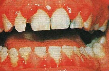
HEENT: PERRL, discordant horizontal eye movements, ptosis noted b/l, dry mucous membranes

CV: RRR no murmurs

Neuro: slowly able to raise both eyebrows b/l though requires significant effort, speech clear though girlfriend at bedside notes it to be “different,” 4/5 strength on shoulder shrug, normal facial sensation, 5/5 strength b/l UE and LE, sensation intact b/l UE and LE, normal cerebellar exam, normal gait

Skin: warm, dry, no diaphoresis, skin popping sites visualized, scar tissue present without any evidence of induration or fluctuance

Labs:

CBC: WBC 5.7, Hgb/Hct 13.7/39.1, Platelets 236

Chem: 136/4.0/98/28/10/0.71/89

LFTs wnl

CXR wnl

APAP/Salicylate/EtOH undetedtable

Applicable case questions:

1. What is this patient’s differential diagnosis?

  • Wound botulism
  • Myasthenia gravis
  • Eaton-Lambert syndrome
  • Miller-Fisher variant of Guillain-Barre
  • Paralytic shellfish poisoning
  • Tick paralysis
  • Lyme disease
  • Endocarditis with septic emboli

2. What are the symptoms of wound botulism?

  • Dry or sore throat
  • +/- nausea and vomiting (more common with food-borne botulism)
  • +/- fever
  • Descending cranial nerve palsies
    • Ptosis
    • Diplopia
    • Sluggishly reactive pupils
    • Dysphagia
    • Dysarthria
    • Dysphonia
    • Progressive symmetric descending paralysis
    • Clear mentation
    • No sensory loss
    • Constipation and ileus from decreased motility
    • Ultimately culminates in profound weakness involving respiratory muscles, can cause respiratory failure and death
    • History of exposure:
      • Usually skin popping, black tar heroin
      • Wound may not be present – manifestations may occur 1-3 weeks after onset of skin infection

3. What are the different causes of botulism?

  • Food-borne botulism: ingestion of preformed toxin in contaminated food
    • GI symptoms followed by onset of neurologic symptoms usually delayed 12-36 hours
    • Home canning – classic culprit
    • Infant botulism: most commonly reported type
      • Caused by ingestion of spores (not preformed toxin) in immature infant gut
      • Risks: ingestion of corn syrup or honey
      • Hypotonia, constipation, tachycardia, feeding difficulty, poor head control, diminished reflexes
      • Wound botulism
        • Classic culprit: black tar heroin
        • Spores contaminate wound, germinate in the anaerobic environment, and produce toxin that is systemically absorbed
        • Usually reported with skin popping – rare reports with open fractures, dental abscesses, lacerations, puncture wounds, GSW, sinusitis
        • Adult intestinal colonization botulism: very rare – adult ingestion of spores (similar to infant botulism)
        • Iatrogenic botulism: occurs following ingestion of botulinum toxin type A
          • Botox (cosmetic purposes)
          • Also used to treat spasticity, axillary hyperhidrosis, strabismus
          • Symptoms occur 1-2 days after exposure
          • Inhalational botulism: usually thought to be an act of bioterrorism, toxin is aerosolized, symptoms visible 1-3 days after exposure with longer onset times for low levels of intoxication
botulism.jpg

4. How is wound botulism diagnosed?

  • Usually a clinical diagnosis made by symptoms, history of exposure, and attempts to rule out other causes of symptoms – need to intervene before confirmatory tests available
    • Interestingly – many patients with botulism end up presenting to the Emergency Department or primary care doctor at least 3 times prior to the diagnosis being considered, due to nonspecific symptoms (i.e. dysphagia)
    • Need to have a high index of suspicion
    • Support for diagnosis can be found in EMG findings that differentiate from other causes, normal neuroimaging, laboratory work to rule out other causes
    • Confirmatory testing done by determination of toxin in serum, stool, gastric aspirate, or a wound – usually via mouse bioassay (contaminated body fluid injected into mouse and observed for development of symptoms)
      • Coordinated through the CDC
      • Testing may be negative due to toxin levels below the level of detection
      • Higher false negative rate in wound botulism than other causes

5. What is the management of wound botulism?

  • Botulinum antitoxin
    • Antibodies directed against toxins produced by C. botulinum
    • Equine derived
    • Binds circulating free toxin, prevents progression of illness
    • Does not reverse established neurologic manifestations
    • Most effective when given within 24 hours of onset of symptoms
    • Obtained by contacting state or local health department or CDC
    • ICU admission with close monitoring of respiratory parameters (NIF, Vital capacity)
      • Respiratory failure can develop rapidly
      • Often require prolonged intubation/tracheostomy due to paralysis of respiratory muscles
      • BabyBIG (human derived antitoxin) used for infant botulism
      • Antibiotics can be considered if a wound is present, along with wound debridement and irrigation
        • Penicillin
        • Avoid aminoglycosides – can exacerbate neuromuscular blockade
        • Not usually recommended for other causes of botulism

CASE CLOSURE:

  • Patient was admitted to the ICU and intubated within 6 hours of admission
  • Antitoxin flown in and administered to patient as soon as possible – approximately 8 hours after initial ED presentation
  • Neurology team followed closely and evaluated for other causes
  • Prolonged ICU course requiring tracheostomy placement

TAKE-HOME PEARLS:

  • Consider wound botulism in a patient with descending motor weakness or bulbar symptoms (diplopia, dysarthria, dysphagia) + history of exposure (skin popping)
  • See New England Journal Article from December 2010 - images in clinical medicine
    • http://www.nejm.org/doi/full/10.1056/NEJMicm1003352
    • Need a high index of suspicion to make the diagnosis: frequently missed by Emergency Physicians
    • Consider food-borne in patients involved in home canning, iatrogenic botulism in patients treated with Botox, infant botulism in infants with poor tone
    • Admit to ICU and closely monitor respiratory parameters
    • Antitoxin is obtained from the local health department or CDC: 404-639-2999
    • Antitoxin should be administered as soon as the diagnosis is seriously considered, as it only prevents further paralysis – it does not reverse established paralysis once it occurs
Botox_cartoon_1.jpg

What's the level?

TOXICOLOGY CASE OF THE MONTH

FEBRUARY 2014

55yoM h/o seizure disorder, Hepatitis C, HTN, DM, CKD, meningioma s/p resection and hepatic encephalopathy presents from assisted living for mental status changes. It is his second visit in the last 48 hours for similar symptoms. He has been difficult to arouse and was noted to have an unsteady gait. He denies fall or intentional ingestion. He is unsure whether he has had any recent medication changes. He complains of generalized weakness and unsteadiness.

Medications: Albuterol, Atorvastatin, Clonidine, Gabapentin, Hydralazine, Hydroxyzine, Insulin, Lactulose, Nifedipine, Phenytoin, Terazosin

Physical exam:

VS 97.2 142/84 69 18 100% RA

General: Awake, alert and oriented x3 though unsure of purpose of hospitalization, no acute distress, cooperative

HEENT: PERRL - 4mm to 3mm b/l, EOMI, + b/l horizontal nystagmus

Neck: supple no asymmetry

CV: RRR no m/r/g

Lungs: CTA b/l

Abdomen: + BS, soft, nontender,nondistended

Extremities: WWP, 2+ pulses in b/l radial, PT, DP, no clonus, no asterixis

Neuro: CN II-XII grossly intact, 5/5 strength in b/l UE and LE, sensation intact b/l UE and LE, wide-based ataxic gait with b/l dysmetria on finger to nose

Skin: warm, dry, no diaphoresis, no rash or lesions

1. What is this patient’s differential diagnosis?

  • Non-tox causes: hepatic encephalopathy, infection, incranial mass/hemorrhage, CVA
  • Toxicology differential:
    • Most likely antiepileptic given presentation - Phenytoin or Gabapentin
    • Hydroxyzine can cause somnolence but would expect anticholinergic presentation (elevated temperature and HR, dry skin and mucous membranes, delirium, urinary retention, etc)
    • Clonidine can cause somnolence but would expect different toxidrome (opioid-like - MS depression with respiratory depression, + VS abnormalities - hypotension & bradycardia)

2. What is the differential diagnosis of nystagmus in toxicology?

  • Many drugs and toxins cause nystagmus - usually horizontal. Here are a few examples:
  • Anti-epileptics
    • Barbiturates
    • Carbamazepine
    • Phenytoin
    • Gabapentin
    • Ethanol
    • Scorpion Envenomation
    • Isoniazid
    • PCP (horizontal, vertical, rotatory)

Horizontal Nystagmus

Labs:

Initial presentation:

  • 140/4.4/109/25/44/2.21/214
  • AST 30 ALT 41 Bili 0.2 Albumin 2.5
  • Phenytoin 14 mcg/mL (ref range 10-20)
  • Ammonia 25

Second presentation:

  • 136/4.8/107/22/49/2.0/200
  • AST 41 ALT 31 Bili 0.1 Albumin 2.5
  • Phenytoin 20 mcg/mL (ref range 10-20)
  • Ammonia 17

 (Creatinine is at baseline)

CT head with no acute change

3. How do these labs change your differential diagnosis? What is the culprit agent?

  • The patient's signs and symptoms are consistent with Phenytoin toxicity
  • Phenytoin is >90% Protein-bound - level needs to be corrected for serum albumin
  • This patient is at risk - hypoalbuminemia due to liver disease

4. What are the clinical characteristics of phenytoin toxicity?

  • Mild to moderate: horizontal nystagmus, ataxia, dysarthria
    • Less common: nausea, vomiting, diplopia, hyperglycemia, agitation, irritability
  • Severe: stupor, coma, respiratory arrest
    • Rare seizures have been reported (though should always consider an alternative cause of seizures in a phenytoin-poisoned patient)
    • Death extremely rare
    • Gingival hyperplasia is indicative of chronic use - may clue physician into diagnosis
    • Reported cause of Stevens-Johnson Syndrome and Toxic Epidermal Necrolysis
Dilantin-induced-gingival-hyperplasia.JPG
  • Propylene glycol component in IV preparations can cause myocardial depression - hypotension, bradycardia
    • Cardiac toxicity does NOT occur with oral overdose or with fosphenytoin

5. What are the applicable pharmacokinetics?

  • Absorption can be slow and unpredictable and in acute ingestion peak effects may be delayed
  • 90% protein bound
  • Hepatic elimination – zero order kinetics near therapeutic range – half-life increases as levels rise

6. What is the formula to correct the serum phenytoin level?

  • Corrected Phenytoin = Measured phenytoin level/(0.2 x Albumin + 0.1)
  • Available on most medical calculators
  • This patient's correct levels are 23 and 33 respectively using this formula

7. How is phenytoin toxicity managed?

  • Hold offending medication
  • Most importantly: protect the patient from self-injury caused by ataxia
    • This may require the patient to be admitted to the hospital for fall prevention
      • For PO toxicity - do not need to be admitted to a tele floor (common misconception)
      • Can also d/c home with reliable family member if close follow up with neurologist or PMD can be arranged, if circumstances are appropriate
      • If seizures occur - consider alternative cause, treat with other anticonvulsants (benzodiazepines generally recommended for toxicology-related seizures)
      • If hypotension occurs with IV administration - stop infusion, administed IV fluids/vasopressors as necessary
      • No antidote, no role for dialysis. May consider activated charcoal if appropriate though most often toxicity is chronic.

Case Conclusion:

  • Send-out free phenytoin level was 4 ug/mL (ref range 1-2), % free 21.6% (ref range 8-14%)
  • Culprit medication was held and patient recovered clinically after several days
  • Was discharged home with a lower medication dose

Pearls to remember:

  • Phenytoin toxicity si/sx: ataxia, nystagmus, dysarthria
  • High protein binding - level should be corrected for serum albumin
  • Treatment is supportive - fall prevention, hold medication, dose adjustment
  • Cardiac monitoring not necessary for toxicity due to oral preparations

"I think he took his gout medicine..."

JANUARY 2014

TOXICOLOGY CASE OF THE MONTH:

CASE:

58-year-old male presents to the hospital complaining of abdominal cramping, nausea, and vomiting.  He has a history of depression with suicide attempts in the past. He told his daughter he “may have taken some of his gout medication” earlier that day. His family says one of his bottles of medication, recently prescribed as needed for gout flares, is now empty. They are unsure how many pills were in the bottle.

XD141137.jpg

PMH: Gout, depression, hypertension

Meds: Hydrochlorothiazide, Colchicine, Allopurinol, Buproprion

All: NKDA

VS: 105/60 110 20 99% RA 36.6

Exam remarkable for tachypnea and dry mucous membranes. Lungs are clear. No focal abdominal tenderness though the patient does complain of cramping. Rectal exam with brown stool, guaiac positive.

Labs:

CBC: WBC 3.5, Hgb/Hct 11.3/34.1, Platelets 210

Chem: 132/3.2/95/14/45/1.2/110

LFTs wnl

Lactate 4.3

APAP/Salicylate/EtOH undetectable

Applicable case questions:

1. What medications are used to treat gout flares?

  • Colchicine
  • NSAIDs
  • Glucocorticoids (i.e. Prednisone)
  • Anakinra, an interleukin-1 antagonist, is used off-label
  • Note: Allopurinol is used for gout prevention, not in acute flares

2. What is the mechanism of action of colchicine? How is it prescribed?

  • Colchicine acts at a cellular level by arresting dividing cells during mitosis
  • It is used for treatment of gout and familial Mediterranean fever
  • It is also found in certain plants (Colchicum autumnale – meadow saffron)
  • It is rapidly absorbed into body tissues
  • FDA-approved therapeutic dose: 1.2mg orally, followed by 0.6mg after 1 hour
  • Should not be prescribed long term
colchicine.jpg

3. Why should I be worried about it?

  • Significant toxicity with no antidote and no effective means of elimination
  • One case series described the following:
    • 0.5 mg/kg: diarrhea and vomiting
    • 0.5-0.8 mg/kg: bone marrow aplasia and 10% mortality
    • >0.8 mg/kg: uniformly resulted in death
    • Fatalities have been reported with ingestions as little as 7mg (though some have survived with ingestions >60mg)

4. What are the clinical effects of an acute colchicine overdose?

  • Many organ systems effected from hours to days after exposure
  • 2-12 hours after presentation: GI symptoms
    • Nausea, vomiting, abdominal pain, diarrhea (often bloody)
    • Diarrhea: common side effect of therapeutic use
    • Progresses to shock: fluid loss and depressed cardiac contractility
    • Delirium, seizures, and coma
    • Lactic acidosis due to shock and inhibition of cellular metabolism
    • Myocardial injury
    • Rhabdomyolysis
    • DIC
    • Renal failure
    • Bone marrow suppression, alopecia, polyneuropathy (late effects)
    • Death after 8-36 hours by respiratory failure, intractable shock, and cardiac arrhythmias

5. How is colchicine toxicity diagnosed?

  • Clinical diagnosis
  • History of gout and familial Mediaterranean fever
  • Severe gastroenteritis closely followed by leukocytosis, shock, rhabdomyolysis, and acute renal failure followed by leukopenia and thrombocytopenia and general decompensation
  • Levels not readily available (forensic use primarily)
  • Other useful labs: CBC, CMP, CPK, UA, Troponin, EKG, Lactate

6. What is the management?

  • Aggressive supportive care and shock treatment
  • Anticipate respiratory and cardiac collapse
  • Highly fatal overdose with no antidote and no effective means of removal (cannot use hemodialysis)
    • Colchicine-specific antibodies (Fab fragments) tested in France, not commercially available
    • Rifampin is a CYP3A4 induced – may enhance elimination in theory but no good data
  • One of the only times in toxicology where aggressive decontamination is highly recommended
    • Charcoal, gastric lavage, whole bowel irrigation are considerations
    • Undergoes enterohepatic recirculation: may need multi dose activated charcoal
    • Some recommend intubation for aggressive decontamination
    • If survive initial insult may need treatment for bone marrow depression

7. What are the acute toxicities of other agents used to treat acute gout flares?

  • NSAIDs
    • Pharmacologic and toxicologic effects occur via COX inhibition
    • Usually asymptomatic or GI upset
    • May develop significant CNS depression, seizures, renal failure, acidosis, hepatic dysfunction with large ingestions
    • Usual treatment is IV fluids and H2 blockers
    • Hemodialysis not effective
    • Glucocorticoids
      • Small ingestions: usually GI upset
      • Anxiety, agitation
      • Fluid retention
      • Majority of toxic effects due to chronic use (i.e. Cushing’s syndrome)

CASE CLOSURE:

  • The patient developed hypovolemic shock
  • Intubated and aggressive decontamination with charcoal via NG
  • Ultimately death with v-fib arrest

TAKE-HOME PEARLS:

  • Colchicine is a highly toxic medication used to treat gout and familial Mediterranean fever
  • Presentation: gastroenteritis followed by shock, renal failure, and cardiorespiratory collapse, late bone marrow suppression
  • Aggressive supportive care and decontamination recommended
Gout-Cartoon_Beer-Consumption_2_Bar_CLR.jpg

Everything is toxic: the dose determines the (rat) poison.

DECEMBER 2013

TOXICOLOGY CASE OF THE MONTH:

CASE:

63yoF presents to the Emergency Department for her second time in a week complaining of epistaxis. On the first visit, she had packing placed. Bleeding was attributed to dry environment and changing weather. She was discharged home with Cephalexin. However, she returned to the ED 2 days later complaining not only of persistent epistaxis, but of hematuria and rectal bleeding. On further questioning, she admits to taking a “handful” of d-CON daily over the past 2 weeks as a suicide attempt.

PMH: Depression, HTN, no hematalogic disorders

Meds: Prozac, Amlodipine; no blood-thinning medications

All: NKDA

VS: 101/59125 16 99% RA 36.6

Exam remarkable for persistent epistaxis from L nare without a clear site of bleeding. Rectal exam notable for gross positive blood. No skin lesions (petechiae, purpura, ecchymosis).

Labs:

CBC: WBC 9.5, Hgb/Hct 9.2/27.9, Platelets 210

Chem: 132/4.2/95/26/55/0.8/110

LFTs wnl

INR: >15

Acetaminophen, Salicylate, EtOH undetectable

Applicable case questions:

  1. What is the Toxicology differential diagnosis of bleeding?
  2. What is d-CON?
  3. Why should I be worried about it?
  4. What is the toxic dose?
  5. What are the clinical effects? What labs should I order?
  6. What is the most appropriate management of rodenticide ingestions?
d-con

Answers:

1. What is the Toxicology differential diagnosis of bleeding?

  • Note: this differential dose not include other causes that should be considered such as bleeding disorders, hepatic failure, DIC, ITP, TTP
  • A lot of pharmacology but an important review – we see patients on may of these
  • Warfarins and suprawarfarins (i.e. rodenticide)
  • Direct thrombin inhibitors (ie Dabigatran/Pradaxa)
  • Factor Xa inhibitors (ie Rivaroxaban (Xarelto)
  • Anti-platelet agents
    • COX inhibitors (Aspirin)
    • ADP receptor inhibitors (Clopidogrel/Plavix, Prasugrel/Effient, Ticagrelor/Brilinta, Ticlopidine/Ticlid)
    • Glycoprotein IIB/IIA inhibitors: mostly IV use (Abciximab/Reopro, Eptifibatide/Integrilin, Tirofiban/Aggrastat)
    • Adenosine reuptake inhibitors (Dipyridamole/Persantine)
    • PDE inhibitors (Cilostazol/Pletal)
  • Rattlesnake envenomation
  • Arsenic toxicity (may have pancytopenia 1-2 weeks after acute ingestion, usually more leukopenia and anemia)

2. What is d-CON?

  • Anticoagulant rodenticide – contains long-acting suprawarfarins
    • Brodifacoum is most common: this is what is in d-CON brand
    • Others: Diphacinone, bromadiolone, chlorophacinone, difenacoum, pindone, valone
    • Interesting fact: warfarin initially used for rat poison, however rats and mice became resistant

3. Why should I be worried about it?

  • Like warfarin, suprawarfarins inhibit conversion of vitamin K to its active form: necessary to synthesize coagulation factors II, VII, IX, X, proteins C & S
  • The duration of anticoagulation from a single dose of warfarin is usually 2 to 5 days
  • Suprawarfarins may produce significant anticoagulation for weeks to months after a single ingestion

4. What is the toxic dose?

  • Warfarin
    • Single small dose (10-20mg) will not cause serious toxicity
    • Chronic or repeated ingestion (i.e. 2mg/day) can cause significant anticoagulation
  • Suprawarfarins:
    • Potential to have prolonged effects even after a single small ingestion
    • Large study of accidental suprawarfarin ingestion in children: no serious cases of ingestions less than 1 box
      • Ingels M, Lai C, et al. A prospective study of acute, unintentional, pediatric superwarfarin ingestions managed without decontamination.. Ann Emerg Med 2002;40(1):73-8.

5. What are the clinical effects? What labs should I order?

  • Excessive anticoagulation
    • Warfarin: anticoagulation effects within 15 hours, lasts up to 5 days
      • Suprawarfarin: Anticoagulant effects usually delayed up to 2 days and may persist weeks to months
      • CBC, PT/INR, Type & Screen (if bleeding),
        • Toxicologists will order Brodifacoum levels to determine duration of therapy
        • If you ordered warfarin-dependent clotting factors, these will be decreased
  • PTT, fibrinogen, liver enzymes, D dimer may help evaluatuation of other causes of bleeding if cause is unknown
  • Acetaminophen, Salicylate levels – rule out coingestants

6. What is the appropriate management of d-CON ingestions?

  • Most ingestions: accidental, by children
  • Poison center recommends: send child to the hospital if they ingest greater than a box of d-CON or other rodenticide, otherwise can be observed at home
    • Parents can observe for bleeding clinically
    • Poison center recommends that these patients can have a follow up at PMD office – a normal PT/INR 48 hours after exposure rules out significant ingestion (Studies suggest that if patients have no clinically evident bleeding that this measure may be unnecessary)
  • If patient comes to the ED just after this ingestion:
    • Activated charcoal
      • Appropriate conditions (recent exposure, normal mental status, not vomiting)
      • Not indicated if patient ingests less than 1 box
      • Labs will not give any information about toxicity but may establish baseline if there is concern for any underlying hematologic disorders
      • Do not give prophylactic Vitamin K (see below)
  • Significant bleeding
    • Treat shock – pRBC transfusion
    • Vitamin K
      • Give if evidence of clinically significant anticoagulation
      • Do not give prophylactically for any ingestion: if this is the case, then the 48 hour PT/INR cannot be used to determine severity -  will need at least 5 days of monitoring
      • Be aware: patients with significant bleeding due to suprawarfarin toxicity need weeks to months of treatment with Vitamin K – poison center can help admitting physicians with dosing but large doses may be required (up to 800mg per day!)
      • FFP for non-life threatening bleeding
      • PCC’s for life-threatening bleeding if available, otherwise FFP
      • Management can be difficult if patient requires long-term anticoagulation (ie prosthetic valve) – can consider heparin for maintenance anticoagulation

CASE CLOSURE:

  • Patient ultimately developed hemorrhagic shock in ED
  • Was treated with Vitamin K and PCCs – INR decreased to 1.3 in ED
  • Up to 5 the next day – still being treated with Vitamin K
  • No further significant hemorrhage initial reversal

TAKE-HOME PEARLS:

  • Classic toxicology teaching: “All things are poison and nothing is without poison: only the dose makes a thing not a poison.” –Paracelsus
  • One box of d-CON or less: patients can be observed at home for signs of bleeding and follow up with PMD in 48H
    • +/- follow up INR in 2 days – recommended if bleeding
    • Activated charcoal if appropriate conditions and ingestion of at least 1 box
    • No prophylactic Vitamin K
    • FFP, PCC, Vitamin K for significant bleeding
    • Will require long term vitamin K treatment for suprawarfarin ingestions – poison center can help with this management

Lipid Rescue Therapy in Overdose

Journal Club Synopsis November 2013

Lipid Rescue Therapy in Overdose

Many thanks to Abhi and Mala Katiyar for hosting and for the delicious south of the border feast!

Outstanding synopses, analysis, and additional review of the topic by Natalie K, Theresa, John P, Nick, Brian F, and Katie I.

Introduction:

Journal Club at the home of a toxicologist calls for a tox topic...in this case the use of lipid emulsion therapy to treat overdose.  IV lipid emulsions are usually thought of as part of nutritional support (TPN), but case reports of lipid emulsions used as rescue therapy for acute cardiotoxicity from lipophilic drug overdose in humans have been published since 2006.  We’ll call it LRT, or lipid resuscitation therapy moving forward, after the American College of Medical Toxicology guidelines.

Which overdoses?  Most compelling literature in overdose from local anesthetics, (LA) especially bupivacaine.  Also used in other lipophilic drug overdoses including calcium channel and beta blockers, TCAs and other antidepressants.

Mechanism?  3 are proposed:  enhancement of myocardial fatty acid transport  (preferred energy substrate of the heart), expanded intravascular lipid phase or “lipid sink” that alters the volume of distribution of the lipophilic toxin, and restoring cellular calcium transport/function.   Nobody knows for sure.

Risk?  Lipemia (who cares, except temporarily renders some lab results invalid), also possible acute lung injury although low incidence of this in patients receiving nutritional lipids, and critically ill patients after overdose already predisposed to acute lung injury. Need more experience to define scope and degree of risk.

When to use?   Great question.  American College of Medical Toxicology has a guideline that states “there are no standard of care requirements to use, or to choose not to use, LRT.  However, in circumstances where there is serious hemodynamic or other instability from a xenobiotic with a high degree of lipid solubility, LRT is viewed as a reasonable consideration for therapy, even if the patient is not in cardiac arrest.”  This actually is a protective document, allowing clinicians the choice of using/not using without declaring a medicolegally binding standard of care.  AHA 2010 ECC guidelines describe the use of LRT for specifically for LA toxicity.  There are reports of patients responding even after prolonged (up to 52 minutes) of cardiac arrest with LA toxicity.

Dose?  All over the place in the case reports...boluses, drips, repeat boluses.  The American College of Med Tox guidelines recommend 1.5 ml/kg bolus of 20% lipid emulsion over 2-3 minutes IV followed by infusion at a rate of 0.25 ml/kg/minute, with shortest duration of treatment possible until hemodynamic stability restored.  The room at JC agreed that using in unstable patients (not just those in overt cardiac arrest) seems reasonable, and LRT should be initiated immediately in LA toxicity.  There was some support for earlier administration in other overdoses, but concern that early LRT might delay treatment with more accepted therapies, and giving in anticipation of instability may result in unnecessary treatment and possible unnecessary complications.  It is reported that in bupivicaine toxicity, epinephrine administration is associated with a negative outcome, and reduces the efficacy of LRT (maybe because epi irritating to the heart?).  Therefore, as epinephrine hasn’t been shown to help in cardiac arrest anyway, consider avoiding epi or giving a smaller dose in the patient with a LA overdose.

Article 1:  Bologa C, et al.  Lipid Emulsion Therapy in Cardiodepressive syndrome after Diltiazem Overdose-a case report.  Am J Emerg Med. 2013;31:1154e3-1154e4.

Case report of 81 yo female who ingested 5.7 gm of diltiazem in a suicide attempt.  Even after gastric lavage, charcoal, calcium, hyperinsulin/euglycemia, epi drip and aggressive IV fluids patient remained hemodynamically unstable with a Mobitz II second degree AV block.   She received intralipid infusion and become hemodynamically and metabolically stable within 24 hours, and was ultimately discharged home neurologically intact.  Lots of missing information from this case report, but this patient appeared to have a positive outcome temporally related to the intralipid infusion.

Article 2:  Geib AJ, Liebelt E, Manini AF for the Toxicology Investigators’ Consortium (ToxIC). Clinical Experience with Intravenous Lipid Emulsion for Drug-Induced Cardiovascular Collapse.  J Med Toxicol 2012;8:10-14.

This retrospective chart review described the presentation and management of 9 cases of drug-induced cardiovascular collapse  (cardiac arrest or refractory shock) treated with LRT from 45 centers in 2.5 years.  Five patients (55%) met the main outcome of survival to hospital discharge.  Interestingly there was no significant increase in MAP immediately after LRT infusion (would expect increase in MAP if LRT helps contractility).  A number of possible adverse effects were described, but the scoring system used to assign causality to these events has been faulted as an inappropriate instrument for this purpose.  Again, lots of missing information regarding additional treatments and LRT dosing, no comparison group, and a very small retrospective study.  Also comment that this is likely an incomplete picture of the experience for this number of centers.

Article 3:  Presley JD, Chyka PA.  Intravenous Lipid Emulsion to Reverse Acute Drug Toxicity in Pediatric Patients.  Ann Pharmacother.  2013;47:735-743.

Moving to kids, this article is a brief description of 14 case reports of pediatric patients who received LRT to treat acute drug toxicity (7 cases of LA, 7 from other drugs (Ca channel blockers and other psych drugs).  Thirteen patients did well, one died.  One patient developed hypertriglyceridemia and pancreatitis.  Again, different dosing regimens.

Impressive how well LRT worked in the local anesthetic cases.  Patients received usual ACLS care with successful resuscitation from cardiac instability/cardiac arrest, and in one case resolution of V Tach using only LRT.  As anticipated, the nonanesthetic medication cases were more complicated, and information regarding additional treatment was incomplete.

Sobering that 20 cc of misplaced local anesthetics in 2 teenagers undergoing routine surgeries led to V tach and cardiac arrest.   A plea to be calculate maximum safe doses of local anesthetics, especially in small kids.

Also, point raised that for all of these articles, there is likely significant reporting bias.  Case reports are more likely to be written up and published when they are positive, especially with dramatic outcomes.  A skewed picture results.

EBM teaching point:  You’re not going to find many RCTs in the tox literature.  There are challenges in coordinating trials across multiple centers for what are often sporadic/rare ingestions, and currently there isn’t a good system to organize these research efforts.  Therefore, the level of evidence for many tox questions is low.  A generally accepted hierarchy of levels of evidence is below, and case reports are close to the bottom, but this is what we have to work with for this topic.

ebm pyramid.jpg

Interesting discussion at the end of JC about whether or not we’ve reached clinical equipoise on the question of the use of LRT for LA and other overdoses.  The problem with a drug such as LRT being used without controlled study is that by the time many case reports are published, the genie is out of the bottle.  Consensus in the room was that IRBs would not approve a RCT for LRT in LA use (eg bupivacaine), but that given the weak evidence for LRT in other ingestions (eg calcium channel blockers), there should be further study, ideally through a RCT.  Simply hoping that it might work as a last ditch isn’t a good reason to use it, and medical history is littered with examples of initially popular therapies ultimately shown to have no benefit (or to cause harm).   Even if this is beyond the point of IRB approval for a RCT, it was suggested that a more comprehensive review of national Tox center data would provide a less biased data set than continued publication of case reports.  A plug to call and report all tox cases to Poison Control, even if you are comfortable with management!