Conference Notes 10-5-2016

Conference Notes   10-5-2016

Chastain/West   Oral Boards

Case 1.   Child with anaphylaxis.   Patient has angioedema of lips/pharynx and signs of shock.  Initial treatment with IM epinephrine,  IV steroids, IV Benadryl, IV Pepcid.  Epinephrine drip was started after initial IM epinepherine.    It is ok to give repeated doses of IM epinepherine before moving to IV epinephrine.   Steroids/Benadryl/Pepcid/ nebulizer treatments are second line therapies in anaphylaxis.   First line/life-saving treatment for anaphylaxis is epinephrine. Epinepherine dosing in kids is 0.01mg/kg.  This can be difficult to measure out appropriately.  Easy dosing for kids is to use the EPIpen junior (0.15mg of 1:1000) IM for kids less than 30kg.

Delay in epinephrine for anaphylaxis increases mortality.   The rapidity of onset of anaphylaxis predicts the severity.  Most fatalities occur in the first hour.  

Elise comment: EPIpens are the way to treat anaphylaxis.  It greatly simplifies the dosing.  Patients weighing less than 30kg get EPIpen junior.  You can redose the patient every 15 minutes if needed. 

Case 2.  55 yo male brought in by EMS after being rescued from a house fire.  Patient has soot on his face and body.  Patient has cough and sore throat.   Exam of the oro-pharynx demonstrates soot and carbonaceous sputum.   Patient was tested for CO poisoning.  Labs did not indicated severe acidosis making cyanide poisonin unlikely. During ED course, patient developed increased dyspnea, throat pain, and stridor.  Patient was intubated early to avoid airway obstruction.  Patient was also treated with 100% FIO2 for CO poisoning. 

Harwood and Elise both felt this patient needed to be treated with hyperbaric O2 even though he was intubated and receiving 100% FIO2.

There were some questions about the mechanism of how hyperbaric oxygen works.  This is an exerpt from a Medscape article:

Most oxygen carried in the blood is bound to hemoglobin, which is 97% saturated at standard pressure. Some oxygen, however, is carried in solution, and this portion is increased under hyperbaric conditions due to Henry's law. Tissues at rest extract 5-6 mL of oxygen per deciliter of blood, assuming normal perfusion. Administering 100% oxygen at normobaric pressure increases the amount of oxygen dissolved in the blood to 1.5 mL/dL; at 3 atmospheres, the dissolved-oxygen content is approximately 6 mL/dL, which is more than enough to meet resting cellular requirements without any contribution from hemoglobin. Because the oxygen is in solution, it can reach areas where red blood cells may not be able to pass and can also provide tissue oxygenation in the setting of impaired hemoglobin concentration or function.

Basically hyperbaric oxygen increases the amount of oxygen dissolved in the serum. This increased oxygen can then diffuse further into tissue than normobaric oxygen.

Michelle comment: Be aggressive with prophylactic intubation in patients with signs ofinhalation injury.  Inhlation injury is the main cause of fire-related deaths.  It is associated with patients being in a closed space fire.


Case 3. 19yo male with difficulty breathing.  Patient developed left chest pain while lifting weights. Chest x-ray showed pneumothorax.  Patient was treated with chest tube.   Spontaneous pneumothoraces can be treated with mini-chest tube (pneumothorax kit).   

*Pneumothorax   Look for the pleural reflection.  Dr. Asokan’s tip: look in the spaces between the ribs for lung markings.


Muhammad     Neonatal Resuscitation

Normals

1) Periodic breathing is normal-looks like rapid breathing followed by pause of about 10 s. Apnea requires 20s pause and should be associated with cyanosis or bradycardia

2) HR 100-220 normal. BP MAP > gestational age. Temp should be between 36.5 and 38 or sepsis eval required.

3) Weight loss of up to 10%V in first week normal. Should be back at birth weight at 2 wk. Double breath weight at 6 months. Should be fed about 1 oz/hr. Constipation is defined by consistency of stools not frequency. About 8 wet diapers or more in 24 hrs.

Case 1- g1p0 at 25 y/o presents in labor. How do you prepare?

1) History- term (plastic wrap for pre-term)? Maternal risk factors? How many babies? Fluid clear?

2) Basics- Dry, stimulate, warm (radiant warmer). Three Questions- Term? Good tone? Breathing or crying? If yes, no resuscitation. Apgars at 1 and 5 min


One minute to treat if ventilation compromised.


ABCs

A- suction only if needed (changed from previous). If you do suction, mouth before nose. Avoid deep suctioning

B- assess effort, apnea, bradypnea, gasping

C- auscultate for HR (vs palpate base of umbilicus)

Use PPV if apnea after 30s, gasping, or HR < 100 (usually secondary to respiratory problem)

Use either flow inflating bag (control pressure manually, set at 10-20, more for pre-term) or T-piece resuscitator (set pressures)

Sniffing position with shoulder roll.

Start at room air O2 (21%), and titrate up if needed.

Chest compressions if no response to 30 s of PPV and HR < 60. Use 2 thumb technique. Compress 1.3 diameter of chest. 1 & 2 & 3 & breathe & 1 & 2 & 3 & breathe. Intubate or LMA at this point.


Term babies ETT 3.5 with Miller size 1. Pre-term 3, Miller 0. Insert ETT to depth of 3x tube size.

Should place UVC if no IV access, low lying UVC- only until you get blood flash. You can catheterize UVC up to 1 wk post-partum.

Umbilical line technique-

Pick the big, floppy one. 3.5 F catheter. Sterilize, stabilize with umbilical tape. Cut to 1-2 cm above skin surface. Clear thrombus if present. Place catheter about 4 cm, until blood return


Doses-

Epi- 0.1 mg/ml

Naloxone 0.1 mg/kg

dextrose 5 ml/kg of D10

NS 10 ml/kg

pRBCs 10 ml/kg


Special Considerations- Meconium

1) If vigorous, do nothing.

2) If not vigorous, its complicated. No longer emphasize intubation and aspiration (no proven benefit) although it is still IIB recommendation by AAP

Discontinuing resuscitation- Justified in stopping at 10 min if no signs of life.


Neonatal HPI-

Pregnancy and delivery- infection, GBS status.

Gestational age, d/c with mom? Birth weight?

Meconium passed, feeding, eliminating, issues with feeds?

Case 2.  15 do infant presents with seizure.    Any infant less than 60 days with a seizure needs a big time work up including CT and labs.  If CT and labs don’t give you a cause of the seizure, you need to proceed with LP.   Test CSF for bacterial infection and HSV. 

Child has another seizure in the ED.  Check blood glucose.  Treat hypoglycemia with 5ml/kg of D10.   Place an IO for access to give benzo’s.   Alternatively, you can give rectal valium, IM midazolam, IN midazolam. 


* Elise Reference on IM Midazolam:  Shorter duration of seizure, fewer patients seizing on arrival to the ED


Check the sodium level.  This is a common electrolyte abnormality in neonates.

If child is still seizing, give phosphenytoin or keppra IV.

Sodium comes back 120.  You need to treat with 3%hypertonic saline (3ml/kg).  Remember 3 and 3= 3ml/kg of 3%hypertonic saline.

Think inborn errors of metabolism if hypoglycemia,metabolic acidosis, or elevated ammonia level is present.

You unfortunately have to consider non-accidental trauma in the seizing infant.

Case 3.  5 do infant presents with lethargy and poor peripheral pulses/delayed capillary refill.   Heart rate is 200.  Order prostaglandin immediately. 

Get access with UVC or IO.   Start IV fluid bolus and antibiotics.

You have to consider coarctation of aorta.   These kids can get shocky after ductal closure.   PGE (0.1mcg/kg/min) will open the ductus.  PGE however can cause apnea.  So you need to be prepared to intubate if necessary.  If you need to transfer an infant who is receiving PGE you should consider intubating prophylactically prior to transport.


Case 4.  4 do male vomiting.  Emesis is green.   You have to think malrotation with volvulus in any infant that has bilious emesis.   Consult surgery emergently.  Start IV fluids and antibiotics.    Start with plain abdominal x-ray but patient will need upper GI study.  


*Malrotation with volvulus


*Another malrotation with volvulus

Plain xrays may be non-specific, you will need to consult surgery and get upper GI study.

Hart/Regan     Ortho Cases of the Week

*SCFE   More commonly affects boys 13-16 yo.  Patients may have hip or knee pain. Get AP pelvis to compare both hips.   Treat with non-weight bearing and operative repair.


*Jones fractureIs a fracture at the metaphyseal/diaphyseal junction of the 5th MT. It will be at the level of the inter-metatarsal joint.


*Pseudo-Jones fracture.  

Jones fractures are prone to non-union.   Pseudo-Jones or dancer’s fracture is due to avulsion injury.   Jones fracture needs short leg post-mold and crutches.    Avulsion injuries can be treated with ace wrap and post op shoe.


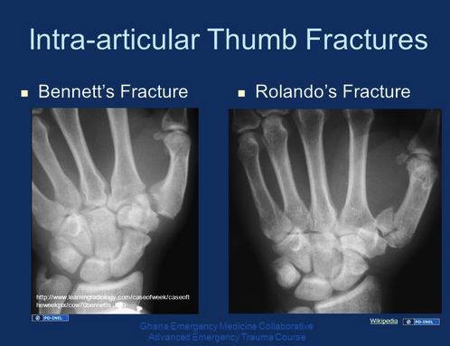
*Bennett fracture vs Rolando Fracture   Treatment is thumb spica splint and operative repair. Benett has 2 syllables in the name and parts to the fracture.  Rolando has 3 syllables in the name and 3 or more parts to the fracture.


*Maisonneuve fracture   If you see a medial malleolus fracture, check the proximal fibula for tenderness.  Keep your guard up for a maisonneuve fracture or a syndesmosis tear.


Ortho Lab   


Conference Notes 9-22-2016

Windy City EM Joint Conference of all the Chicago Area EM Programs   Held at the UIC Forum

Pick Your PoisonToxicology Topics

AKS    Overview of Tox

The “Mickey Finn” started in Chicago.   A bartender named Mickey Finn would slip a purgative in a patron’s drink causing vomiting/diarrhea. His buddies would then hustle the “sick” patron out into the alley and beat them up and rob them.

The first Poison Control Centerin the nation was in Chicago.

CheemaUrine Drug screens

A positive urine drug screen for amphetamines has about a 1 in 4 false positive rate.  There are many meds that can cross react with the urine screen for amphetamines.

Surprisingly, Xanax, Ativan, andKlonopin won’t give a positive urine drug screen for benzos.  That is because the screen looks for the metabolite oxazepam and these three benzos don’t get metabolized to oxazepam.  It’s unfortunate that the drug screen won’t pick up these common benzos.

Second-hand marijuana smoke is very unlikely to trip a urine drug screen.  A patient will not get a positive drug screen for marijuana by being at a concert near others smoking marijuana.

Fentanyl and methadone are not picked up on a urine drug screen.  Hydrocodone and hydromorphone are picked up <60% of the time.

Research has shown that taking a history from the patient is superior to the drug screen for identifying illicit drug use.  Drug screens rarely change how we manage patients.

Carlson       Marajuana and THC Concentrates

 50% of people will use marajuana at some point in their lifetime.

Alaska, Oregon, Washington, Colorado and Washington DC allow legal recreational use of marijuana.

In Colorado, there has been a large increase in the number of pediatric patients accidentally exposed to edible forms of marijuana (candy, brownies)

Concentrated marijuana (hash) can cause psychosis.  Stopping use of marijuana products for 1-2 weeks can resolve psychosis.

Synthetic cannabinoids can also cause psychosis.  There have also been reports of MI’s and strokes in young patients using synthetic cannabinoids.  The cause is uncertain but it gets to the point that synthetic cannabinoids are metabolically complex molecules.

THC increases appetite.  This is one of the medical benefits of marijuana.

Marajuana can result in amotivational syndrome and attention deficit symptoms.

In a young patient with new onset psychosis you really have to consider concentrated marijuana use as a cause.

Marajuana is stored in adipose tissue.  Habitiual users can have a positive screen for up to 12 weeks.  Synthetic cannabinoids do not trip a urine drug screen for marijuana.   Single use of marijuana will cause a positive drug screen for 2-3 days.


Lank      Psychiatric Medications

Bupropion, Citalopram, and Venlafaxine are the 3 SSRI’s that can cause seizures with overdose.  Admit these overdoses for 23 hour OBS with telemetry.  Citalopram can cause torsades.  2 patients with bupropion overdose have been reported to require ECMO to survive.  So these drugs are not totally benign in overdose.

Patients on lithium can have a significant drug interaction with a prolonged course of NSAID’s.  A patient on lithium can have a single or few doses of ibuprofen but not 600mg Q8 for 7 days.  They can get lithium toxic.

Treat lithium toxicity with fluid and electrolyte management and if the patient appears ill, dialyze them.   Dialysis may decrease the incidence of neuro-cognitive sequelae of lithium toxicity.


Bryant     Hyperbaric O2for CO

Consider CO poisoning in patients presenting with headache, vomiting, vision changes, altered level of consciousness, and chest pain. CO poisoning is a chameleon and can masquerade as many other diagnoses.

VBG is adequate to measure the CO level.

Treat CO toxicity with HBO if the patient has neurologic symptoms, loss of consciousness, EKG changes, is pregnant, or has a CO level over 25% (some centers say 40%)

It is totally unclear from current data whether HBO is effective for CO poisoning.  It may have a role in decreasing delayed neurologic sequelae.

The EP’s role is to identify CO poisoning and discuss  management with a HBO center.


Jordan    Body Packers , Body Pushers, and Body Stuffers

Packers swallow large amounts of well-packaged narcotics to smuggle thru customs

Body pushers conceal narcotics by placing them retrograde into the rectum or vagina

Body stuffers rapidly swallow unpacked drugs when they fear imminent arrest by police.

All these patients are initially unreliable because they are hiding narcotics. 

As an emergency physcian caring for these patients you cannot force them to have imaging.  If they refuse evaluation, they will need to remain in police custody and under observation in the ED until they have a bowel movement.

Sensitivity of x-rays for identifying body-packed narcotics ranges between 40-90%.  Specificity is 93%.

Plain CT has a sensitivity and specificity of 97% for drug packets in GI tract.

Admit all confirmed body packers

Give them WBI or even just a laxative or cathartic to get the packets out.

Get confirmatory imaging (plain xray or CT?) to assure all packets are removed

Symptomatic patients should get exploratory lap to remove packets.


Chhabra      Toxidromes


*Basically know your toxidromes.  The big 4=Opioid, Sympathomimetic, Anticholinergic and Cholinergic.


Visual diagnosis

*Red cap mushrooms are amamita muscaria.  These are hallucinogenic.   Red top=hallucinogenic


*Death Cap mushrooms are aminita phylloides.  These cause symptoms >6 hours after ingestion.   These can kill you.


*Gyrometra mushrooms look like a brain (think gyri of the brain) but they can kill you as well.  They get metabolized to the compound in rocket fuel.


The TOX EKG

A wide QRS is a marker for badness in toxicology.  It is due to a  depolarization/sodium channel dysfunction.  Treat with 2 amps of NaHCO3.

Sodium channel problems get sodium bicarb.

Long QT interval (500ms) is a potassium channel problem.  Treat with magnesium.  Also give potassium up to the upper limits of normal.

Potassium channel problems get magnesium and potassium.

Fast atrial rhythms/Fast ventricular rhythms with AV block,  bradycardia, afib with slow ventricular rate, and bidirectional ventricular tachycardia are all potential signs of digoxin poisoning.  Treat tox patients with combined fast/slow problems on EKG with digibind.

Devgun                  Antidotes

Hydroxocobalamine has a deep purple color and can affect the colorimetric assays for creatinine and bilirubin.  You can see a high creatinine and bilirubin in patients who have been treated with hydroxycobalamine.

Panel Discussion of Toxic Alcohol Management     

Excellent discussion of management points of toxic alcohols

Use serum osmolality and osmolal gap as well as serum bicarb to raise your suspicion for toxic alcohol ingestion.  You need to be aware that these labs are tricky with wide variation in normal values for patients and moving targets on both the osmolality and bicarb depending on time since ingestion.

Fomepizole basically prevents metabolism of toxic alcohols by competitively inhibiting alcohol dehydrogenase.  It serves as a time bridge to allow you to set up dialysis to remove the toxic alcohol.   A ph of 7.25 was recommended as a cut-off number indicating need for dialysis.  This cut-off number is associated with increased incidence of renal failure and other morbidities.    Another panel member made the point that if the patient still has an elevated osmolal gap that means they still have unmetabolized toxic alcohol on board and would benefit from dialysis.

There have been 50 episodes of mass methanol toxicity events in the last 15 years.  In less developed countries or if fomepizole is not available, IV or oral ETOH is very effective to block alcohol dehydrogenase.  You need to keep the blood alcohol level above 100. 


Conference Notes 9-7-2016

Alexander   M&M   

 For M&M’s,   I will focus on a few of the take home learning points.

Kelly:  The decision-making when dealing head with injured patients who are combative is very tricky.  Elective intubation is always risky but sometimes you have to do it to protect the patient and move on to safely image and workup the patient.

Harwood: Every combative, head-injured patient deserves an initial attempt at sedation to calm them before attempting intubation.  Also check the glucose in every combative patient.  Hypoglycemia is a common cause of altered behavior.

Elise: Chemical restraint should either precede or be given at the same time as physical restraint.

Elise and Andrea: Ketamine and/or Precedex would both be great options in this situation.  Ketamine is now considered neuro-protective instead of previous thinking that it was dangerous in head injured patients.

EAST Guidelines state that intubation is indicated in combative patients that are not responsive to attempts at sedation.

Elise:  It is so crucial to perform an airway assessment prior to performing an intubation.   If  despite your best efforts, the airway attempt is going south, your bridge to cricothyrotomy is LMA.

 Girzadas: If the airway is at all expected to be difficult, strongly consider performing intubation with ketamine and topical anesthesia and no paralytic.   Also the McGrath Video laryngoscope has an angulated optic device to allow you to see a very anterior airway.

Be sure you have suction, oxygen, back up devices (bougie, LMA, Cric tray) prior to starting intubation.


*EMCrit Intubation Checklist.  You can download or print out checklists for intubation at emcrit.org.


Elise and Kari: When using the glidescope, only use the glidescope stylet.  Using another more malleable stylet has much potential for failure in securing the airway.


Kelly: If the intubation looks to be difficult and it is not immediately emergent, you should consider calling anesthesia as back up.   It is not a sign of weakness to call anesthesia, it is called proper preparation for a difficult airway.


Barounis Tips concerning Glidescopes and Extubation: Links to a few articles on glidescopes and traumatic intubations:

http://link.springer.com/article/10.1007/s12630-012-9824-3

http://www.oapublishinglondon.com/article/341


1. Glidescopes can be dangerous see above

2. Tips

1. The stylet is basically a harpoon. when you can't see the tube on the screen be very careful when inserting it into the oropharynx and mindful of the location. (can cause perforation and trauma to tonsillar pillar as you often have the stylet pointed towards the cheek to avoid blocking your point of view).

2. Point head and neck in neutral position for glide (unlike sniffing with MAC).

3. Do not insert ett too posterior in the pharynx, otherwise the ett is trying to go into trachea but bumps into the arytenoids and it is hard to disengage. 

4. When engaging the arytenoids, have a friend pull out the stylet (which I know most people do), but another advantage is to ROTATE the ett clockwise as you enter through the cords and the friend or assistant is pulling out the stylet. this prevents the ett hanging up on the right posterior arytenoid. 

5. LUBE the ett, it will go in smoother over a VERY DRY mucosa and arytenoid. 

6. LUBE the stylet it makes pulling it out much easier

3. Extubating patients with laryngeal edema

One major problem with edema that is above the cuff is that a cuff leak is meaningless. A cuff leak is the stand-alone idiot test to tell someone if there is edema. Problem is the cuff is below the cords and if deflated there might be air movement around the deflated cuff, but above the ett is still a splint around swollen vocal cords or an edematous soft palate. 

if one suspects trauma or edema best to perform laryngoscopy and fiberoptic evaluation prior to extubation.


If one anticipates a difficult extubation, for any reason can place cook airway exchange catheter in place. 

I do this.

1. Patient is sedated with precedex

2. Patient gets lidocaine down ett to numb cords

3. Patient gets topical 4% lido in mouth

4. Patients get lidocaine atomizer around cords and posterior pharynx

5. I meausre how deep to insert cook exchange catheter

6. I remove ett over cook exchange catheter

7. I sit patient up with catheter in place and monitor 

8. I have all aiway equipment and personel available if they crash (happens usually immediately)

9. I have an RT bring a racemic epi neb to bedside BEFORE extubation, i do not wait until an emergency arises and someone needs to find and start a racemic epi neb. when you need it, you need it NOW

-Dave


Schmitz        Financial Issues for Residents

You should plan your finances to prepare for a life expectancy of 90 years old.

Most financial experts state that you will need about 88% of your pre-retirement income to maintain your lifestyle in retirement. You may be able to get by with less but shooting for the 88% mark will give you the best chance of not having to severely change your lifestyle.

Starting the habit of regular saving early in your career is the key to a lifetime habit of saving.  A lifetime of saving is the best way to hit that 88% mark.  Saving also teaches you to have a lifestyle that doesn’t use 100% of your income.  Thus when you retire, you actually can get by on less than 88% of your pre-retirement income.


*Compound interest is the most effective way to grow your savings to meet your retirement needs.  To take advantage of compound interest, you need to start saving as early as possible.

You absolutely need to save enough of your yearly income to get the full employer match in your retirement account.  The match amounts to a 3% raise.  Basically, it’s free money if you have the discipline to get it.


*The power of starting early coupled with compound interest.  Blue line is saving started early and continued during working years. Green line is starting somewhat late in working years. Dashed line is starting early but then not continuing beyond age 35. You can see how the dashed line falls off the trajectory of the solid blue line when saving stops.

Excellent websites for financial guidance:

WhiteCoatInvestor.com

Investor.gov

Bogleheads.com


Alexander     Bradyarrythmias


*Third degree heart block.   Compare 3rd degree heart block with the two types of second degree heart block pictured below.  3rd degree heart block has regular R-R intervals and regular P-P intervals and the P-P and R-R intervals are asynchronous with eachother.   Both types of 2nd degree heart block have grouped beats with intermittent pauses in the R-R intervals.


*Second degree heart block Type 2


*Second degree heart block Type 1 Wenckebach


Muelleman       Rural Injuries


The most common fatal injury mechanism in rural areas is motor vehicle crashes.

47 million Americans live more than 1 hour from a trauma center

6 million Americans live more than 1 hour from an ED

There are 5,000ED’s in the US


Firearm associated homicide is less common in rural areas

Firearm associated suicide and unintentional injury is more common in rural areas.

MVC death rates drop as population density goes up.   Many factors that could explain this relationship.    Research has shown that age of patient and crash characteristics predict 50% of the added mortality.   High speed crashes account for increased mortality.  No seatbelt also increases mortality.  ETOH use increases mortality.   Distance of the crash from a board certified EP increases your mortality.  Most rural trauma deaths occur in the ED.   These deaths mostly are due to hemorrhage, lack of airway, or pneumothorax.

The ATLS course was actually developed by an emergency physician.  ACEP turned down sponsorship of ATLS.  So the emergency physician took the course to the American College of Surgeons and they made it the world-wide course it is today.

Muelleman      Your Path to ABEM Certification

 The ABEM mission is to ensure the highest quality of Emergency Medicine care.

Board certification allows you to be recognized as the specialist you have trained to become.

The In-training exam is designed to predict your probability of passing the ABEM Qualifying exam.   The In-Training exam will be moving to an electronic format probably 2018. 

ABEM Certification requirements:

Successful completion of an accredited EM Residency training program  

Valid and current Medical License

Pass the written and oral tests within 5 years of completion of residency training

Total cost for initial certification is right around $2500.


Delay taking the exam for any reason, makes you less likely to pass the exam. 

Passing score is 76 for the Qualifying exam.  Pass rate is around 90% nation-wide

Oral exam pass rate is around 98% nation-wide

There are about 34,000 ABEM diplomats.

Conference Notes 8-31-2016

Barounis      Engineering a better Cardiac Resuscitation

 Organized practice on a regular basis can improve the function of the team during resuscitation.  Dave spoke about optimal function of a resuscitation team based on practicing resuscitations, defining specific roles and locations for each member of the resuscitation team, and having a very organized and specific location for drugs and devices you may need in a resuscitation.  It takes a lot of pre-planning and practice to have a great resuscitation team.  It is very difficult to change hospital culture to accomplish this.

Physiology of Cardiac Arrest:  There is a massive SIRS response when a patient has a cardiac arrest.


*Coronary Perfusion pressure is a key to getting ROSC.  If CPP is <15 no arrested patient gets ROSC.  IF CPP is >25 the patient usually will get ROSC.    Pauses in chest compressions cause a drop in CPP.  You have to really work on decreasing the number and length of pauses in compressions during ACLS care.   Longer peri-defibrillation pauses of chest compressions have been show to result in higher mortality.


Mechanical chest compression devices have not shown a benefit over human chest compressions.   Dave andEM faculty present at this lecture all felt the mechanical devices should be better than human compressions due to more consistent compressions and shorter pauses.  Dave felt that all the studies using mechanical devices had relatively prolonged delays to defibrillation initially while medical personnel were applying the device.  Erik also noted that centers that do these studies have outstanding quality of human CPR at baseline in the comparison group making it harder to demonstrate a difference.  This level of quality of human chest compressions is really not reproducible in other medical centers.  


Epinepherine is a double edged sword.  It increases MAP and the likelihood of ROSCbut it also increases the risk of arrhythmia.  Epi has alpha effect but also beta 1 effect. The beta 1 effect increases the arrhythmic risk.  Electrophysiologists use esmolol to manage arrhythmias in the lab.  There is a growing body of evidence for giving esmolol in patients in cardiac arrest who have failed multiple doses of epinephrine.  Dave felt that if you have a patient who has failed defibrillation, amidarone, and 3 doses of epinephrine, it is reasonable to try esmolol.    Elise asked if there is enough data on this concept to support an emergency physician giving esmolol during prolonged cardiac arrest.  Dave felt it was reasonable to try 50 mg of esmolol in the patient who has failed the entire standard ACLS algorithm.  Esmolol is used all the time in the electrophysiology lab and there are some papers supporting esmolol in cardiac arrest that has failed shock/epi x3/amiodarone.   If you don’t get ROSC in 5 minutes after esmolol you can call the code.  If you get ROSC, start an esmolol drip.

Bicarb doesn’t really help in cardiac arrest.   It doesn’t change pH effectively.

Dave places an A-line in cardiac arrest patients to monitor their pulse and blood pressure.  He feels it is way better than pulse checks.  It picks up a pulse much better than palpation of the carotid and eliminates the pause in CPR for pulse checks.  It allows you to better titrate pressors to elevate blood pressure to get CPP above 25.  It also takes away “pseudo-PEA” where a pulse is present but you just can’t feel it.  If you see an arterial wave form on the arterial line tracing you know you need added pressor effect to raise blood pressure.

Every cardiac arrest patient needs ETCO2 monitoring.  If the ETCO2 jumps above 20 you have ROSC.  Some people don’t even do pulse checks, they just watch for a jump in ETCO2.

Every cardiac arrest patient needs transthoracic echo to look for tamponade, pneumothorax or other causes of cardiac arrest.   Dave says TEE in cardiac arrest is coming.  Emergency medicine studies are already looking at this.

High dose epi did not have improved neurologic outcomes despite more frequent ROSC.

ECMO can save a patient if the patient had CPR started within 5 minutes of arrest.  Patients with massive PE are the most optimal candidates.  Dave advised consulting with CV surgery prior to giving TPA for massive PE to see if the patient can go to OR to be cannulated for ECMO.


*Double defibrillation with 2 defibrillator machines is something that can be tried if prolonged ACLS care with multiple defibrillations has failed.   You applythe pads from two machines and defibrillate with both at the same time.


Regan/Kustad     Oral Boards

 Case 1. 76yo male with malaise, vomiting, and confusion.  BP 104/76, febrile, other vitals OK.   Exam demonstrated RUQ tenderness and mild jaundice.   Imaging identified inflammatory changes around gall bladder.    Diagnosis was ascending cholangitis. Treat with IV fluids and IV antibiotics. Consult GI for ERCP.  Admit to ICU.   Consult surgery. 


Snip20160902_1.png

*Acute Cholangitis   (Slide from Dr. Ruby Wang)


Case 2.  40 yo female with severe pain in hand after exposure to rust remover containing hydrofluoric acid.  


*Management options for HF acid exposure.  If you are going to give intravascular calcium gluconate, give intra-arterial thru an A-line.


Case 3.  50 yo male presents with garlic odor.  Patient has a lot of oral secretions.  Lungs sound full of fluid.   Patient had diarrhea in the bed.  Patient ingested insecticide. 


*Cholinergic Toxidrome


Critical actions:  Hi dose of atropine.   2-Pam also should be given.   The patient required intubation.


Kennedy        

 Ketamine is a very useful drug for multiple intubation scenarios.  It doesn’t cause respiratory or CV depression.  It is a bronchodilator.  It also provides pain relief.    It was once thought to be contra-indicated in patients with increased  intracranial pressure.  Now it is thought to be neuro-protective and indicated in patients with normal or increased intracranial pressure.


Etomidate works fast and is fast offset as well.  It does cause respiratory depression.  There can be some hypotension.

For procedural sedationKetofol (0.5mg/kg Ketamine mixed with 0.5mg/kg Propofol)   This combination has fast onset, adequate length of sedation for most procedures and has less emergence phenomenon, less vomiting than ketamine alone, and less respiratory depression, and less hypotension than propofol alone.

Case scenarios: medication choice

Cardioversion: ketamine or propofol or ketofol.

Hip dislocation: ketofol

Pediatric forehead lac: Intranasal versed or intranasal fentanyl or both.  There were conflicting views among the faculty on this one.  Also give local anesthetic and po Tylenol.

Pediatric oral laceration: Ketamine.   Harwood preferred Ketamine solo versus ketafol in this scenario.

70yo pneumonia who needs intubation: Etomidate and rocuronium

35 yo asthmatic requiring intubation: Ketamine for induction

22yo with status epilepticus: Ketamine and succinylcholine, however this scenario was also controversial


EMcrit reference:  Although usually Rocuronium is the preferred paralytic, in status epilepticus succinylcholine may be preferable to allow determination of whether the seizure has been controlled.    If Rocuronium is used, there is a risk that the patient may have ongoing seizure activity which is not observable, but which is nonetheless causing brain damage.   Hyperkalemia secondary to rhabdomyolysis takes time to develop, so status epilepticus of short duration (<15-20 min) itself is not a contraindication to succinylcholine.   For a patient who presents to the ED with seizure of unknown duration, Rocuronium is safer.   Alternatively, this may be one situation in which sedative-only intubation may be a reasonable approach, as high-dose propofol will typically provide good intubation conditions provided that it breaks the seizure [more discussion about this below – see Addendum #2].


18yo altered trauma patient:  Etomidate or ketamine.    Ketamine these days may be preferred for neuro-protective effect.

50yo with FB in airway:  Ketamine sedation with topical anesthetic.  Pull out FB with a mcgill forceps.

Angioedema:  Ketamine, topical anesthetic, no paralysis.


Marshalla/Ohl/Okubanjo     Head & Neck Trauma


3 goals of management: Prevent secondary neurologic injury, identify treatable intracranial findings, identify other injuries.


*Epidural vs Subdural Hematomas


*DAI is due to axonal shearing caused by rapid deceleration injury.


*PCARN Head injury guideline for pediatric patients


*Gradual “return to play” recommendations following concussion.  The patient should not move to the next stage until they have been asymptomatic for at least one day at the current stage.


Lowering Intracranial Pressure: Either mannitol or hypertonic saline can be used.  Dr. Lee (Trauma) prefers hypertonic because there is less risk of hypotension compared to mannitol.


*Neck triangles


*Zones of the neck.   Girzadas mnemonic: Zone 3 is close to the third ventricle and zone 1 is close to Big Red 1 (aorta).


*Management guideline for penetrating neck trauma.   Esophogeal injury is the leading cause of delayed death.


*Hard and soft signs of penetrating neck trauma


*NEXUS Criteria for C-spine Injury   Harwood comment: NEXUS does not perform well in patients over age 65.


*Canadian C-spine Rule    Elise comment: The Canadian C-spine rule scans all patients over age 65.  It will also scan all patients with dangerous mechanism.


Conference Notes 8-24-2016

Carlson       Toxicology


Case 1 .   18yo female presented with strange behavior (grimacing, not speaking).   Patient had history of depression, pituitary adenoma,  and pseudoseizures. Patient was on multiple meds: Soma, Effexor, Parlodel and Norco. Patient was agitated, hypertensive, tachycardic and febrile.


Diagnosis:  Serotonin syndrome


*Clinical Characteristics of Serotonin Syndrome.   There is no objective diagnostic test.   There are 3 diagnostic criteria


*Hunter Criteria for Serotonin Syndrome.  Andrea said that this was the most up to date and accurate diagnostic criteria.


Treatment for Serotonin Syndrome is stopping all serotonergic medications and giving cyproheptadine.


*Serotonin Syndrome vs Neuroleptic Malignant Syndrome.   If you see the term “lead pipe rigidity” on the boards, that is NMS.  Treat NMS with bromocriptine.


Core temperature is the most important vital sign to correct in the setting of overdose. Andrea said the fastest way to cool a hyperthermic patient is to pack them in ice.   Girzadas comment:  We had amazing success with packing a patient in ice and using a high powered fan blowing air over the ice/patient.


Staley      Approaching the Febrile Infant


Fever is  defined as 100.4F or 38C.  Also beware of hypothermia (<36.5) in infants as a marker of infection.  If the parents take the temp by axillary or ear methods, don’t add or subtract anything to the reported number.  In the infant under 60 days old, even reported fevers at home that are not substantiated in the ED need to be strongly considered for work up.


Around 10% of infants under three months of age with fever have serious bacterial illness (SBI).  Physical exam in these infants is not able to distinguish kids with/without SBI. 


Hi Flow O2 in septic infants is useful to prevent intubation.

If you can’t get an IV in 2-3 sticks go right to IO.  IO has actually been shown to be less painful than multiple IV sticks.


If the child is less than 28 days old and they have fever, they get a full sepsis work up including LP.  Give ampicillin and cefotaxime and admit.  Give vancomycin and acyclovir if child appears critically ill.


If the child is 29-60 days old with fever,  all get CBC, blood cultures and urinalysis/urine culture.  If the CBC, urine, vitals are all OK, you can consider discharging the patient without antibiotics.   If you decide to give antibiotics for any reason you have to do an LP.  If you identify a UTI in infants this age and you decide to give antibiotics you still need to do an LP prior to giving antibiotics. 


In infants older than 60 days, say 2-4 months old, with fever 39.5 or above, there is about a 6% risk of serious bacterial illness and you need to consider doing CBC, blood culture and UA/urine culture.


Tips for LP success in infants:  Use EMLA prior to giving lidocaine.   Use a pacifier dipped in sweetese.   Sugar on a pacifier has been shown to be a very effective pain reducer in infants.   Using a firm surface under the child (chest compression board covered in a blanket) this board keeps them from sinking into the mattress and it may line up landmarks better.   Advancing your needle with the stylet removed after you have gotten past the skin helps you identify CSF more readily.    If you get any flow of CSF, don’t try to adjust the needle to get better flow.  Needle adjustments increase the risk of a bloody tap or moving the needle out of the CSF containing space.


Pecha Kucha


Nejak          Managing Shoulder Dystocia


The infant’s shoulder gets hung up on the pubic bone.   You need to calm mother and try to limit contractions/pushing.  Call OB for help.


Step 1. Put mom in extreme lithotomy position and apply suprapubic pressure to move the shoulder under the pubic bone.


Step 2Try turning the infant’s shoulder in the vaginal canal


Step 3.  Deliver the posterior arm to free up some space


Step 4.  Get mom/infant to OR.


*Management of Shoulder Dystocia


West          Bedside Ultrasound for Shock


Evaluate the Pump:   Evaluate the Heart for pericardial effusion/tamponade, contractility,  abnormally large RV


Evaluate the Tank:  Evaluate the IVC for flattening/collapse, do a FAST exam, and check the lungs for CHF or pneumothorax.


Evaluate the Pipes:  Check for AAA,  check for DVT.


DeStefani          Brain MRI for Dummies


The DWI and ADC sequences of an MRI imaging set will identify acute strokes.

The DWI sequence will show all strokes acute and chronic.

ADC sequence differentiates acute vs chronic stroke.

Acute stroke region will be Bright on DWI and Dark on ADC MRI sequences.


Holland        EMS VooDoo


There is no evidence to demonstrate benefit of routine use of long spinal immobilization boards.


There is no evidence to demonstrate benefit of routine use of cervical collars.

We as the EM community should advocate for a more rational and evidence based use of long boards and C-collars.  

Suggested Indications for long boards: Transferring patients to an EMS cart or ED cart.  After that, get the patient off the board.

Suggested Indications for Cervical Collars:  Positive NEXUS criteria, high risk mechanism,  or known cervical spine fracture.  

Kennedy      Intro to Ventilator Management

Always start with Assist Control

Lung Protective Strategy: RR=18, TV 6ml/kg, FIO2 100%, PEEP 5.

Obstructive Strategy (Asthma/COPD): RR=10, I:E Ratio should be 1:5, TV 6 ml /kg.  FIO2 50%.  Let pCO2 ride high.  Watch for autopeep.


*Managing Problems with the Intubated Patient


Hart/Regan     Ortho Cases


Boxer’s Fracture.  Requires ulnar gutter splint.  Elise comment: For all splinted patients, be sure to document that you re-evaluated the patient after splinting and splint was applied correctly and N/V status intact. 


*Boxer’s Fracture


 Tibial Plateau Fracture.   High energy trauma in young patients.  Plateau failure in older patients.  Patients will have significant pain and won’t be able to bear weight.  Management is commonly surgical.


*Tibial Plateau fracture types.  There can be either or both depression and fracture fragments. 


Conference Notes 8-10-2016

Thanks to Elise for writing last weeks Conference Notes.     

Shorter, summer version of the Notes today

Regan    STEMIConference

 Case 1.  Middle age male with some chest pain during the night.  Pain resolved but wife made him come to the hospital.  Further history revealed that patient has been having chest pain with exertion over the last few weeks.

EKG showed anterior ST elevation with Q waves and no reciprocal changes.   Patient went to the cath lab and had a 99% LAD occlusion.

This case was interesting because the patient had a STEMI without pain in the ED.

About 5-10% of STEMI’s are painless.  Painless STEMI’s are more common in female patients, diabetic patients, and patients with EKG’s that have ST elevation with Q waves.   There is no difference between painless and painful STEMI’s regarding culprit lesions and maximal troponins.  Painless STEMI’s in general have worse outcomes compared to painful STEMI’s.

There was some discussion among the cardiologists about whether they would take a patient with history of intermittent pain but no pain in the ED to the Cath Lab acutely.  In the end they agreed that the ST elevation pretty much pushes you to do a cath.


Harwood comments:  Patient may say they don’t have pain but if you ask them if they have pressure or tightness or discomfort they will say yes.   Also, reciprocal ST depression is only present in about 80% of STEMI’s.


Case 2.   Middle age male with sudden onset central chest pain.   Initial EKG shows subtle anterior STEMI.  ST elevation was at most a box but inferior leads show down-sloping ST depression.   Emergent cath showed total occlusion of the LAD and 80% occlusion of obtuse marginal.


Interestingly, patient had a normal stress test a few weeks prior to this episode. 

As part of the nuclear stress test,  a little known aspect of the report (at least to me)  is a TID score (Transient Ischemic Dilation score).  It is usually listed in the body of the nuclear stress test report.  If elevated the TID is a reliable marker of multi-vessel disease.

TID is an abnormal finding in stress myocardial perfusion imaging that suggests severe and extensive CAD and signifies a worse prognosis. TID has been reported with exercise and pharmacologic stress testing, planar and SPECT imaging, and Tl-201, Tc-99m, and dual-isotope protocols. Underlying mechanisms include a combination of stress-induced subendocardial hypoperfusion, ischemic systolic dysfunction, and less likely physical LV dilation with severe ischemia. TID appears to represent a significant ischemic burden and, compared with increased pulmonary Tl-201 uptake, suggests less permanent LV dysfunction and more myocardium at risk. Stress-to-rest LV volume ratios of 1.12 (epicardial) and 1.22 (endocardial) have been consistently shown to be highly specific for severe and extensive CAD. Reference: McLaughlin, M.G. & Danias, P.G. J Nucl Cardiol (2002) 9: 663. doi:10.1067/mnc.2002.124979

Last teaching point on this case: Subtle ST elevation does NOT have a better prognosis compared to marked ST elevation.

Lovell/Cirone       Oral Boards

 Case 1.  Middle age patient presents with seizure.  Patient travelled from Mexico.  Diagnosis was neuro-cysticercosis.   Manage the airway. Treat seizure. Order a CT scan to identify ring lesions.  Get an ID consult to discuss management.   Albendazole and steroids are the first line treatment.


*Neurocysticercosis on CT

If a person eats infected pork they get intestinal worms.   If a person eats food contaminated by feces with tenia eggs they get cysts in the brain.  This disease is a common cause of epilepsy world-wide.  Be alert to this illness in patients travelling from endemic areas.


Case 2.   Female patient with vaginal bleeding and abdominal cramping.   Patient has a positive pregnancy test.   History reveals patient had recent in-vitro fertilization.  U/S shows an IUP and a right ovarian mass.   Diagnosis is heterotopic pregnancy.  Patient had severe bleeding.  Treatment is blood product replacement and emergent surgery.  Patient was RH negative so Rhogam was administered.   1/3 of intrauterine pregnancies will be lost when patient goes to the OR for ruptured heterotopic pregnancies.    Fertility therapy markedly increases the risk of heterotopic pregnancy.  


When a patient has a history of recent in-vitro fertilization/fertility therapy, think of 2 diagnoses:  heterotopic pregnancy and ovarian hyper-stimulation.

Case 3.  Pediatric patient with itchy red rash of right foot after waking on the beach in Puerto Rico.


*Diagnosis is cutaneous larva migrans.   Hookworm larva get into the skin.  Treatment is ivermectin or albendazole.  Manage itching with Benadryl and topical steroids.


Einstein/Ohl     Patient Communication & Satisfaction

Noah and Sean discussed a new printed form that protocolizes how we keep patients informed about their ED course and discharge plans.

Kennedy          Pressors


*Pressor Chart


*EMCrit   Pressor decision-making


*EMCrit    Push dose Epi


Ohl             Bullous Skin Disorders


*Porphyria cutanea tarda


*Porphyria cutanea tarda


*SJS vsTEN


*SJSvs TEN


*Orf disease     Shock and awe among the conference attendees when Elise knew this one immediately.

(Wikipedia Reference)    Orf is a zoonotic disease, meaning humans can contract this disorder through direct contact with infected sheep and goats or with fomites carrying the orf virus. It causes a purulent-appearing papule locally and generally no systemic symptoms. Infected locations can include the finger, hand, arm, face and even the penis (either caused by infection from the hand during urination and/or bestiality). Consequently, it is important to observe good personal hygiene and to wear gloves when treating infected animals.[1] The papule may persist for 7 to 10 weeks and spontaneously resolves. It is an uncommon condition and may be difficult to diagnose. There have been no reported cases of human to human infection.

While orf is usually a benign self-limiting illness, it can be very progressive and even life-threatening in the immune-compromised host. One percent topical cidofovir has been successfully used in a few patients with progressive disease. Serious damage may be inflicted on the eye if it is infected by orf, even among healthy individuals. The virus can survive in the soil for at least six months.[2]  


Hart/Regan     Visual Diagnoses

 There were so many great images in this lecture.  I only can show a few.


*Gingival hyperplasia50% caused by phenytoin, 20% caused by cyclosporine


*Ranula


*Plunging ranula


*Vitreous hemorrhage on ultrasound


*Retinal detachment on ultrasound


Stanek        Safety Lecture


Rebuilding a better suture cart.


Holland        Administrative Update

 Patrick updated the residents and faculty on ongoing administrative and process improvement initiatives.


Conference Notes 8-3-16

Conference Notes Wednesday August 3

8 am: Cardiovascular Study Guide #3; Christine Kulstad: I was keeping Jeopardy score, so unable to provide notes on this outstanding S.G.

9 am: Ethics-Forms/End of Life; Dr. Shayla Garrett-Hauser

Advanced Directives

•Advanced directives are only operative if patient is unable to make and communicate the decision.

 Illinois POLST Form : took the place of the old Illinois Portable DNR form

•Has multiple sections with more options; is still a portable valid physician order It is illegal to generalize DNR designation beyond the coding condition. You cannot withhold other treatments based on DNR status

POA

•POA designates who will decide for the patient if they are unable to decide for

themselves

•This person can make any decision-from band-aids to withdrawing care, palliation

and hospice

•They are the legal equivalent of asking the patient

LET

•An internal form at Advocate hospitals

•It is a physician order consented to by the patient or their surrogate

•Gives additional guidance for very sick patients beyond the DNR

•It can be revoked by the patient, the agent or the surrogate

Withdrawal Withholding Care Form

•Life sustaining medical treatment health care surrogate act physician certification “double doc form”

•Asks 2 physicians to certify that pt has a qualifying condition: Terminal, Permanent unconsciousness, Incurable or irreversible condition

•Patient must lack decisional capacity

•Certifies a surrogate: in descending order of power: Guardian of person=POA, Patient’s spouse, Adult son or daughter, Either parent, Adult brother or sister, Adult grandchild, Close friend, Patient’s guardian of the estate The surrogate, who must sign the form, can withdraw or withhold treatment as specified

End of life facts:

•30% of Medicare dollars are spent in the last 1 year of life

•When they die: 75% of people are nonambulatory, 40% are cognitively impaired, 80% die in an institution, 20-30% die in an ICU

Principle of “Double Effect”:

•A legal/philosophical principle first proposed by Thomas Aquinas

•An action that has 2 effects, one is desired and the other is accepted-eg ok to give morphine to relieve pain at end of life understanding it may also cause respiratory depression.

Hospice

Can occur anywhere-it’s not a specific facility. It’s an insurance designation, and controls insurance dollars. There are specific designated hospice providers (eg Dr. Mien at ACMC)-most docs cannot admit to hospice. Palliative care is NOT an insurance designation, and any physician at ACMC can admit to any bed for palliative care. Palliative care is medical care focused on symptom control, enhanced quality of life, and maximizing functional capacity.

Hospice failing to control symptoms is one of the most likely reasons hospice patients are seen in the ED. Use the “ED comfort care” power plan to help with management.

10 am: Endocarditis; Dr. Logan Traylor

Risks: male, older, IVDU (repetitive bombardment with particulate matter/talc, ischemia from vasospasm), valvular disease (pressure gradients/turbulent flow), prosthetic heart valves, IV devices, Hemodialysis, HIV. Outside of USA, rheumatic heart disease #1 risk.

Epidemiology:

-- Native valves-usually left sided, initially marantic (nonbacterial vegetations) endocarditis. No big deal until transient bacteremia.....Strep viridans (slow) and Staph (fast). Fun facts: Strep bovis is assoc. with colon cancer. Also Enterococcus after manipulation of GU/GI tract. HIV: listeria, salmonella. 5 % will be culture negative (HACEK). IVDU=Staph.

-- Prosthetic valves: Staph epidermidis, Aspergillus, Candida albicans.

Valves: overall most common Mitral, second Aortic, third Tricuspid. IVDU more right sided, rheumatic more left sided.

Janeway Lesions-painless plaques “Jane is nice”, microabscesses

Osler nodes-“O is for ouch”

Clinical features:  Fever, chills, N/V, fatigue, malaise.  80% febrile on presentation (20% not febrile!).  Murmur 85% usually regurgitant.  Acute or progressive CHF in up to 70% of cases.   Have to think about this diagnosis to make it-frequently missed-ask about risk factors, listen for murmurs. 

MCA embolic stroke is the most common neurologic complications.

Can have many other embolic phenomena-retinal artery, mycotic aneurysms, pulmonary osteo.

If you see different infectious presentations/sites and can’t tie them together, think ENDOCARDITIS!

Most important test-blood cultures:  >/= 3 blood cultures, first and last at least one hour apart, 3 different sites.  Allows you to start antibiotics more quickly.  Unless HACEK, this should nail the diagnosis.  Also Echo, ECG, look for PR prolongation/other conduction abnormalities.   Also CXR, +/- CT, procalcitonin. 

Duke’s Criteria:  2 Major, 1 Major and 3 Minor, or 0 Major and 5 Minor...

Treatment:  Strong suspicion/acutely ill:  Vancomycin +/- gentamycin.

If stable may wait until blood cultures return.

Prosthetic valves get rifampin too, increased penetration of biofilm.

Prophylaxis:  previous endocarditis, unrepaired cyanotic congential heart diease, prosthetic valve/tissue in heart.  Do this if invasive dental procedures maybe tattoos.  Use Amox.

10:30 am:  M&M; Dr. Mark Bamman

“7 Deadly Sins”....lessons learned.  AKA the Bamman confessional.

Pride: 

First chest tube-supervision offered, deferred.  Next day with new hemothorax, likely from intercostal vessel damange.  

Afib/RVR-didn’t sync for cardioversion. 

If not comfortable with procedure, get help.

“Humility is not thinking less of yourself, it is thinking of yourself less” C.S. Lewis

Envy:

“Speed envy” when self-comparingto more senior residents.  Risk=cutting corners, incomplete history, will miss things.

Gluttony:

Diet and exercise-prioritize.

Anger:

“Anger is an acid that can do more harm to the vessel in which it is stored than to anything on which it is poured.”  Mark Twain

 “It isn’t the mountain ahead to climb that wears you out, it’s the pebble in your shoe.”  Muhammad Ali

Many daily system challenges in our ED

Anger towards patients.

Must learn to let it go, in order to preserve your humanity and longevity.

Lust:

Lust for “Dr. Done”.  Elderly woman with abdominal pain, CT read said SBO, missed incarcerated inguinal hernia...look at your own CT scans, careful/repeat physical exams.  Consider why diagnoses occur (look for underlying primary problem).

Greed: 

Greedy with Time.  

Consider service for others, beyond what brings you secondary gain.

“We make a living by what we get, but we make a life by what we give.”  Winston Churchhill

Sloth:

PICU extubation:  patient extubated, then received usual push dose sedation which led to oversedation and re-intubation.  Could have been avoided with clinical reassessment before giving meds?

Re-assess your patients, especially prior to interventions, going upstairs, going home, or if nurse says change in condition.

Procastination of reading/studying-you are cheating yourself; of administrative requirements-just makes it harder to complete.

“Diligence is the mother of good luck.”  Benjamin Franklin

11:30 am:  Health Care Disparities-Social Determinants of Health; Dr. Oyinkansola Okubanjo

WHO:  Social Determinants of health:  “conditions in the environments in which people are born, live, learn, work, play, worship and age that affect a wide range of health, functioning, and quality-of-life outcomes and risks”

EG:  childhood asthma rates in children on the south side, access to public transportation, food deserts

Categories of Social Determinants of Health:

Economic Stability:  employment (access to insurance, PMD).  Employment associated with better health.  Blacks, Hispanics less likely to be in management positions.

Neighborhood, physical environment:  housing-time for EMS, smoke/CO detectors, mold, safety

Education:  language, literacy-tied into health, nutrition, employment

Food/Nutrition:  hunger in Chicago children, access to healthy options.  Food deserts correlate to areas where African Americans live.

Community and Social Context:  Diversity, Race (which is only a social construct) and discrimination, incarceration rates (normalized in certain communities)

Health Care system: insurance, provider availability, provider linguistic and cultural competency, access to health care, health literacy-pay attention to discharge instruction comprehension.

How Determinants Interact:  individual life style factors + social and community networks + socioeconomic, cultural and environmental conditions

Take home points:

-Determinants affect individual and community health directly and indirectly

-Different determinants affect different social groups

-Inequitable distribution contributes to health care disparities

-Increased knowledge of these determinants leads to providing better care for YOUR patients!

Next time:  Patient and Provider factors

12:00 pm:  Pulmonary Hypertension (PH); outside speaker Valerie Laroy, APN

Pulmonary artery hypertension-can be idiopathic or due to multiple underlying disease processes. Seen more in women, starts in small arterioles

Diagnosis with Right heart catheterization.

Pulmonary circulation:  low pressure system, low resistance, high capacitance, dynamic vascular bed. Pulmonary circulation has one fifth the pressures of systemic circulation despite the same CO as systemic circulation.

Usually several year delay from symptoms to diagnosis.  Presents with shortness of breath without hypoxia, tachycardia, fatigue, peripheral edema.  Late symptoms syncope, JVD, CP, hypotension, hepatomegaly, ascites, SOB

Patients asymptomatic until RV is affected.  How RV reacts to preload and afterload predicts outcome.

Treatment:  endothelin receptor antagonist (eg Opsumit), prostacyclin analog (eg Remodulin) and nitric oxide enhancement (PDE5 inhibitor-Viagra, Adempas).  PA pressures diagnose the disease, but goal of treatment is to remodel RV to maintain cardiac output.  Terminal treatment is therefore lung transplant, rather than heart transplant.  If you transplant a heart into patient with PH, heart will rapidly dilate/fail, death.

From Valerie Laroy (NP for PH team).  If a patient presents with PH diagnosis on PH meds, please call PH team on arrival.  They need to be on consult for the admission.   Type in “Pulmonary Hypertension” to perfect serve to identify who is on call.  If our PharmDs see the med list and identify a PH med, they will alert us.  PH meds are life saving-there are oral meds that must be continued in the ED, and pumps must stay on

Why do they present?  Fluid overload-need for diuretic assessment.  Site pain at site of infusion-possible infection.  These patients are usually baseline hypotensive.  If BP support needed, usually the preferred pressor is Neosynephine (avoid tachycardia).  Usually will go to MICU (preferred ICU), possibly 7W.  For admission, there is not a preferred admitting physician.  On the back of the patient’s IV or SQ pump (SQ pump is actually an insulin pump) will be a phone number for their specialty pharmacy-call this number for dose/rate.  NEVER SHUT OFF THE PUMP.  Inhaled meds have unique inhaler-it’s NOT our usually MDI.  Initial skin pain may not be infection-look for infectious sx beyond pain.

Testing:  Order BNP, need stat Echo

If respiratory failure, try CPAP first.  Intubation dangerous-PA pressures very high, anticipate code.   Anticipate need for pressors and inotropes.

Conference Notes 7-27-2016

Carlson/Kennedy   Oral Boards

Case 1. 67yo male with back pain and lethargy.  He also has lower extremity weakness.  ESR was elevated. Imaging showed multiple lytic lesions in the spine.   Patient had hypercalcemia secondary to multiple myeloma.  Critical actions were to diagnose multiple myeloma with hypercalcemia and treat with IV saline.   


*Hypercalcemia symptoms


*Hypercalemia management


*Multiple myeloma


Case 2.  22 month old female presents with parent saying she cannot wake the child up.  BP 52/26   HR 162,  shallow respirations.  Accuchek is 28.   History reveals that older sibling is taking phenobarbital.   CXR shows non-cardiogenic pulmonary edema.  Patient required glucose supplementation and intubation.  Hypothermia was also treated. Urine was alkalinized.  Multi-dose activated charcoal can be useful with phenobarbital ingestions.  Diagnosis was child abuse.  Mom was giving phenobarbital to keep child calm and to sleep.  Child was placed in protective custody.


*Multi-dose Activate Charcoal MnemonicsABCD for the toxins it works for.   PHAILS for the toxins that charcoal does not work for.


Case 3.  30mo male who is vomiting after taking his vitamins.  The vitamins contained ferrous fumarate.  However, calculations showed the amount the child ingested was not toxic.  If child takes less than 60mg/kg the ingestion should be non-toxic.  A serum iron level < 500 is non-toxic.   You can get vomiting with a non-toxic dose of iron.   A toxic dose typically will cause an anion gap acidosis.  This patient did not have an elevated anion gap.  Andrea made the point to get the numbers 60mg/kg and an iron level of 500 in your head regarding iron poisoning.


C & EKulstad    CV Study Guide


*Age adjusted d-dimer EBM


The test to evaluate for pelvic thrombosis in pregnant patients is MRI.  It may be the best study as well for identifying pelvic thrombosis in non-pregnant patients.


*phlegmasia cerulean dolens


*phlegmasia alba dolens


Wikipedia: The disease presumably begins with a deep vein thrombosis that progresses to total occlusion of the deep venous system. It is at this stage that it is called phlegmasia alba dolens. It is a sudden (acute) process. The leg, then, must rely on the superficial venous system for drainage. The superficial system is not

adequate to handle the large volume of blood being delivered to the leg via the arterial system. The result is edema, pain and a white appearance (alba) of the leg.

The next step in the disease progression is occlusion of the superficial venous system, thereby preventing all venous outflow from the extremity. At this stage it is called phlegmasia cerulea dolens (PAD). The leg becomes more swollen and increasingly more painful. Additionally, the edema and loss of venous outflow impedes the arterial inflow. Ischemia with progression to gangrene are potential consequence.  ePhlegmasia alba dolens is distinguished, clinically, from phlegmasia cerulea dolens (PCD) in that there is no ischemia.[1]

Emedicine/Medscape: In PAD, the thrombosis involves only major deep venous channels of the extremity, thus sparing collateral veins. The venous drainage is decreased but still present; the lack of cyanosis differentiates this entity from PCD. In PCD, the thrombosis extends to collateral veins, resulting in severe venous congestion with massive fluid sequestration and more significant edema. Without established gangrene, these phases are reversible if proper measures are taken.

Of PCD cases, 40-60% also have capillary involvement, which results in irreversible venous gangrene that involves the skin, subcutaneous tissue, or muscle.[3] Under these conditions, the hydrostatic pressure in arterial and venous capillaries exceeds the oncotic pressure, causing fluid sequestration in the interstitium. Venous pressure may increase rapidly, as much as 16- to 17-fold within 6 hours.[4


Elise comment: You now need to treat superficial thrombophlebitis with heparin or LMWH or one of the new oral anticoagulants if the thrombophlebitis is large or proximal. 

From the ACMCEM website: Thrombosis of superficial veins has long been considered benign, and deemed a separate entity from venous thromboemolism (VTE)

-- However, multiple studies illustrate a significant association with VTE (DVT and PE).

-- When patient with ST (diagnosed clinically, no ultrasound) are thoroughly evaluated, the degree and the extent of clot are underestimated 75% of the time.  Further, such patients are found to have co-existent DVT or PE 25% of the time and/or rapidly progress to DVT 10% of the time.  

---- Teaching point: get ultrasounds on all clinical superficial thrombophlebitis

-- The risk factors for ST and VTE are the same, and many argue that ST should be treated the same as VTE.  

-- In general,

anticoagulate (as you would for DVT) the patient if they have known clotting risk factors, greater than 5 cm of clot, or clot < 5 cm within the sapheno-femoral or sapheno-popliteal junction.

-- Another way to think about it is that a superficial vein thrombosis is a manifestation of a systemic clotting cascade gone awry.  To even further simplify things, seems pretty pathologic to have any blood vessel clot; ahh hello, you are clotting off blood vessels.

*Treatment algorithm for superficial thrombophlebitis


The treatment of pulmonary edema is high dose IV NTG or sublingual NTG as the mainstay of therapy.   Aggressive use of NTG allows us to avoid intubating pulmonary edema patients.

Morphine has no efficacy in the management of CHF.


*OESIL Rule for syncope is considered better than San Francisco Syncope Rule


*Rose Rule    Elise likes the Rose Rule for syncope.  She made the point that an elevated BNP is a marker for badness in syncope and can be a surrogate for CHF.


*Hypertrophic Cardiomyopathy    Patients have LVH QRS complexes with lateral inverted T waves.


Treat pericarditis with colchicine.


Hart/Regan 2015 ACLS Updates


Social media can be used to summon rescuers.

Christine, Erik and Elise spoke about an app called Pulse Point that will notify you of a nearby cardiac arrest.


*Pulse Point App


Do Chest compressions 100-120 per minute.   Compressions at a pace over 120 has a worse outcome.

Avoid leaning on chest to allow full recoil.

Vasopressin is out.  It has no advantage over epinephrine.

We discussed automated CPR machines.  Faculty present at the meeting have found them more reliable with less interruptions anecdotally, but the research does not show any benefit over human CPR.

Routine use of lidocaine is not recommended.

If ETCO2 is <10 after 20 min of downtime ROSC is unlikely

ECMO can be used in cardiac arrest with a potentially reversible cause (hypothermia, myocarditis, transplant candidate)

Any post-arrest patient not responding to verbal stimuli should be cooled to 32-36C.

Avoid and treat hypotension in a patient who had ROSC.


ACLS Workshop


McGinnis   MIDAS Reporting Update

Please place any patient safety concerns in the MIDAS system.  We really want you to make a report of any safety issues.

When placing a bed request, please check the special request drop down menu to be sure you make note of suicidal patient needing a sitter, peritoneal dialysis, LVAD, chronic trach, c-diff, andother isolation needs.  These requestswill limit the patient to certain floors that can provide that specialized care.


Conference Notes 7-20-2016

Barounis     Management of the Hypotensive Patient

Recent study on HelmetNon-Invasive Ventilation

Results  Eighty-three patients (45% women; median age, 59 years; median Acute Physiology and Chronic Health Evaluation [APACHE] II score, 26) were included in the analysis after the trial was stopped early based on predefined criteria for efficacy. The intubation rate was 61.5% (n = 24) for the face mask group and 18.2% (n = 8) for the helmet group (absolute difference, −43.3%; 95% CI, −62.4% to −24.3%; P < .001). The number of ventilator-free days was significantly higher in the helmet group (28 vs 12.5, P  < .001). At 90 days, 15 patients (34.1%) in the helmet group died compared with 22 patients (56.4%) in the face mask group (absolute difference, −22.3%; 95% CI, −43.3 to −1.4; P = .02). Adverse events included 3 interface-related skin ulcers for each group (ie, 7.6% in the face mask group had nose ulcers and 6.8% in the helmet group had neck ulcers).

Conclusions and Relevance  Among patients with ARDS, treatment with helmet NIV resulted in a significant reduction of intubation rates. There was also a statistically significant reduction in 90-day mortality with helmet NIV. Multicenter studies are needed to replicate these findings.

*Helmet NIV


*AVAPScan be used in patients who are not getting optimaltidal volumes with NIV.  It gradually increases IPAP.  Usually used in COPDer’s.

Management strategy with BIPAP: For COPDer’s move up on the IPAP.   For CHF, go up on the EPAP.   For ARDS go up on EPAP.

Dave’s first two steps when evaluating hypotensive patients are: 1. Touch the patient to evaluate their peripheral perfusion.  2. Put an ultrasound probe on the patient to evaluate their cardiac pump function.   Mitral valve movement directly correlates with LV function.  The other method he likes is calculating fractional shortening.

*Fractional shortening.  You drop an M mode line through the left ventrical and the machine will calculate the percentage change in width of the left ventrical.   Less than 50% is c/w heart failure.

Dave brought up the situation of unilateral R side CHF.  This can be caused by severe mitral valve disease causing an eccentric regurgitant jet into the left atrium forcing backflow preferentially into the right pulmonary vein.   If you see a patient with a right unilateral big infiltrate that looks like unilateral CHF, throw an ultrasound probe on the heart.  If the mitral valve looks abnormal, get a formal echo to evaluate for an eccentric reurgitant jet. 


*Unilateral Pulmonary Edema   Editorial comment: Unilateral right side pulmonary edema is a Zebra diagnosis but you could look amazingly good if you pick this one up. 

Snip20160720_5.png

 *Eccentric mitral regurgitant jets.


*Sonographic B lines go all the way to the bottom of the screen.  These indicate most commonly CHF but could also be due to other fluid in the alveoli.

In hypotensive patients with Afib and RVR don’t use diltiazem.   Give magnesium 2-4 grams.   Don’t worry to much about the magnesium level.  Patients can tolerate mag levels up to 4.9.  2nd line drug is Amiodarone 150mg bolus followed by 1mg /min.

E. Kulstad       CV Study Guide

*Indications for Transcutaneous Pacing

Compared to placebo, Heparin and LMWH do not provide morality benefit in non-STEMI acute coronary syndromes.

Compared to placebo, Heparin and LMWH demonstrate a trend toward mortality benefit in STEMI’s.


*Sgarbossa Criteria


*Heart Score Components

*HEART Score Outcomes MACE (Major Adverse Coronary Events)


Carlson       Toxicology Cases

*Acetaminophen Overdose Decision-making

*Rumack Matthews Nomogram

*Acetaminophen Toxicity Mechanism.  The amount of acetaminophen overwhelms the conjugation pathways and the excess acetaminophen instead gets metabolized to NAPQI which is toxic.

*NAC Dosing

Dextromethorphan can give you a positive toxicology screen for PCP.  It is a dissociative drug similar to ketamine.  It is commonly mixed with marijuana.  Patients can have bad trips.  Treat patients with benzo’sto manage the agitation.

Drugs that cause a positive toxicology screen for PCP: PCP, dextromethorphan, and ketamine.

Caffeine is a methyl xanthine that binds the adenosine receptor. If you are treating SVT for caffeine overdose you will need to use higher dosing of adenosine.  You should start at 12 mg of adenosine.

Caffeine will cause vomiting and hypokalemia.  Caffeine acts at the kidney to increase potassium diuresis.   Hypokalemia is variable in caffeine overdose and the level of hypokalemia should not be used to risk stratify or rule in/out the diagnosis.

Toxidrome: Anxiety/agitation, tachycardia, vomiting and hypokalemia think caffeine toxicity.

Iron toxicity causes an anion gap acidosis, hyperglycemia, and leukocytosis.  Serum iron levels >500 are dangerous.


*5 Stages of Iron poisoning.   Treat with deferoxamine.

Vitamin A is really the only dangerous vitamin overdose.  It can cause cerebral edema.

If you identify a patient with an anion gap metabolic acidosis and a respiratory alkalosis they have ASA toxicity.  Death from salicylate is a CNS death, not a pulmonary or cardiovascular death. Alkalinize serum to keep salicylate out of CNS.  Alkalinize urine to enhance elimination. Replace potassium.  Finally, hemodialysis can be life-saving.

Tinnitus is common with a salicylate level above 20. 

*Indications for dialysis in ASA toxicity

Hart    Code STEMI

Goal: Get the patient to the Cath Lab in less than 60 minutes for a STEMI.  Identify alternative critical diagnoses such as PE, Aortic dissection, perforated ulcer, GI bleed,  hyperkalemia, pericardial effusion, valve rupture.

No need for O2 if a Code STEMI patient is sating at or above 94% on room air. 

Air Versus Oxygen in ST-Elevation Myocardial Infarction (AVOID) trial compared supplemental oxygen vs no oxygen unless O2 fell below 94%.

"The AVOID study found that in patients with ST-elevation myocardial infarction who were not hypoxic, there was this suggestion that, potentially, oxygen is increasing myocardial injury, recurrent myocardial infarction, and major cardiac arrhythmia and may be associated with greater infarct size at 6 months," lead author Dr Dion Stub (St Paul's Hospital, Vancouver, BC, and the Baker IDI Heart and Diabetes Institute, Melbourne, Australia) concluded.

Methods and Results—We conducted a multicenter, prospective, randomized, controlled trial comparing oxygen (8 L/min) with no supplemental oxygen in patients with STEMI diagnosed on paramedic 12-lead electrocardiogram. Of 638 patients randomized, 441 were confirmed STEMI patients who underwent primary endpoint analysis. The primary endpoint was myocardial infarct size as assessed by cardiac enzymes, troponin (cTnI) and creatine kinase (CK). Secondary endpoints included recurrent myocardial infarction, cardiac arrhythmia and myocardial infarct size assessed by cardiac magnetic resonance (CMR) imaging at 6 months. Mean peak troponin was similar in the oxygen and no oxygen groups (57.4 mcg/L vs. 48.0 mcg/L; ratio, 1.20; 95% confidence interval [CI], 0.92 to 1.56; P=0.18). There was a significant increase in mean peak CK in the oxygen group compared to the no oxygen group (1948 U/L vs. 1543 U/L; means ratio, 1.27; 95% CI, 1.04 to 1.52; P= 0.01). There was an increase in the rate of recurrent myocardial infarction in the oxygen group compared to the no oxygen group (5.5%vs.0.9%, P=0.006) and an increase in frequency of cardiac arrhythmia (40.4% vs. 31.4%; P=0.05). At 6-months the oxygen group had an increase in myocardial infarct size on CMR (n=139; 20.3 grams vs. 13.1 grams; P=0.04).

Conclusions—Supplemental oxygen therapy in patients with STEMI but without hypoxia may increase early myocardial injury and was associated with larger myocardial infarct size assessed at six months.


 For STEMI’s make sure all patients get 4 chewable baby ASA.  If the patient received ASA from EMS or took it at home, document that.  Start IV Heparin (max dose 4000u).  Discuss anti-platelet agents with your cardiology consultant.  Some cardiologists may choose not to use an anti-platelet agent to avoid bleeding complications if the patient requires CABG.

If you have a patient with an inferior STEMI with ST elevation in lead 3>lead 2 get a right-sided EKG to identify a possible Right ventricular infarction.

*DeWinter’s Syndrome

*Dewinter Teaching Points

Lee    

Unfortunately I missed this excellent lecture. 

Nand    Observation Service and Care Management

Basically if you are admitting for a symptom, the patient should be brought in as observation.  For the most part, if you have a diagnosis for the patient then  bring them in as a full admit.  Exceptions would be TIA, syncope, and cellulitis which can be observation status.  If you have a doubt, start as an observation. Social issues should be brought in as an observation.

When patients are brought in as an observation patient, theyhave more personal financial exposure than they would as a regular admission.  If the patient is expressing concerns about the financials of an OBS admission, consult the ED Care Manager to help with this discussion.

If you have to change the admit or observation order, cancel and reorder the bed request. This action will re-generate a new level of care order.

If the patient will be staying for 2 midnights that is a patient who should be a full admit.

Don’t use the word “observation” as a verb.  Use “monitor” or “watch” instead.   For example don’t write “we will observe the patient in the ICU”.  Instead write, “we will monitor in the ICU”  Using the word “observation” as a verb in your note causes confusion for reviewers regarding whether the patient is admitted or Observation status.


Conference Notes 7-13-2016

LambertUltrasound Basics

The basis of ultrasound imaging is that every tissue in the body has a different acoustic impedance.   Sound travels at different speeds thru different tissues.   When sound waves reflect back to the probe at varying speeds, the ultrasound machine can generate an image based on these differences.


*Acoustic Impedance

Low frequency waves penetrate deeper into the tissue but provide less resolution.   High frequency waves have less penetration but better resolution.


*High Frequencyvs Low Frequency ultrasound waves

Terminology synonyms:

Longitudinal = sagittal=long axis

Transverse=axial=short axis

Coronal is basically a longitudinal view taken from the side of the body


* Sagital,  Axial, and Coronal Planes


Knobology

Mike discussed the importance of controlling the depth and gain of the image you are looking at.   You want to adjust the depth to optimize to proportion of the body structure being visualized in the image.  You want the structure of interest to take up the majority of the screen.   You need to adjust the gain to have a uniform appearing image both in the near and far fields. 


*Near field is top half of image,   far field is bottom half of image.  Gain is uniform throughout image.


Lambert      Bedside Echocardiography

Bedside echo is a game changer for identifying life-threatening illness in patients with chest pain or shortness of breath.   You can identify PE, pericardial effusion, CHF, problems with contractility/wall motion.   It is useful during cardiac arrest as well.


There are two main views of the heart when doing Bedside echo


*Parasternal Long axis


*4 Sub costal


*Pericardial effusion*


*Pulmonary embolism Note the large RV in comparison with the LV.


Mike classifies LV dysfunction as either OK or Bad.   Bad is usually obvious on bedside echo and helps you identify CHF or cardiogenic shock.   


Elise’s Journal Club Summary:

Article 1:  Flato UA, et al:  Echocardiography for prognostication during the resuscitation of intensive care unit patients with non-shockable rhythm cardiac arrest. Resuscitation  2015  Jul; 92:1-6. 

This was a Brazilian prospective, observational cohort study of 49 ICU patients with in hospital asystole or PEA cardiac arrest. Of 88 eligible patients, 39 were excluded. PEA without contractility was classified as electromechanical dissociation (EMD), and PEA with contractility as pseudo-EMD.  Two echo trained intensivists performed transthoracic echos. Rates of ROSC were 70% for pseudo-EMD, 20% EMD, and 24% for asystole.  Good ROSC percentages for all groups, but survival to hospital discharge was only seen in pseudo-EMD patients, and discharge is what counts.  Four patients survived to 180 days, all in the pseudo-EMD group, with CPC of 1, 1, 1, 2.  Echo was feasible, with maximum duration of 10 seconds, so non-disruptive to the ongoing code.  Echo also helped identify underlying etiology of arrest in selected patients, eg unexpected tamponade.  This was a very small study, and conducted in an ICU with a large number of DNR patients who were never entered into study, so different from our ED population.   

This study reinforces our usual practice of using echo to verify presence/absence of cardiac contractility and guide futility of resuscitation.  Blaivas and Fox (go ACMC!!) published a larger study in 2001 demonstrating 100% mortality in patients with asystole or PEA cardiac arrest and no cardiac contractility on bedside echo. This modality helps conserve resources (time and personnel), and may identify the underlying reason for code.  In the future, in young otherwise healthy patients with arrest, an echo demonstrating contractility may help risk stratify for ECMO.

Bottom Line:  Rather than pulse check, consider echo as the more reliable marker for viability.  Also remember end tidal CO2 to guide prognostication.  

(AHA 2015 ACLS guidelines:  “In intubated patients, failure to achieve an ETCO2 of greater than 10 mm Hg by waveform capnography after 20 minutes of CPR may be considered as one component of a multimodal approach to decide when to end resuscitative efforts but should not be used in isolation.”)


Lambert    IVC and Aorta

*Aortic Aneurysm

*IVC with hypotension. 

*The proper landmarks for observing the IVC for hypovolemia include the heart, portal vessels and IVC in the same image.

Mike feels a collapsing IVC with inspiration is the most useful sign of hypovolemia on ultrasound.  He has doubts that IVC ultrasound is the holy grail of identifying low central venous pressure.

Lambert and Team Ultrasound        U/S Lab

Conference Notes 7-6-2016

Hart/Girzadas   Oral Boards

Case 1.   15yo female suffered blast injuries in an explosion.   Patient required intubation fordyspnea, respiratory distress , and bloody sputum secondary to blast lung injury.  Patient also had a traumatic amputation of the right upper extremity that was hemorrhaging.  A tourniquet was applied to stop bleeding.  Patient was resuscitated with PRBC’s and IV LR.  Patient also had 2nd degree burns on right side of body of about 20%.   Parkland formula was started.


*Blast Injury Categories


> Mortality increases from 8% to 49% when blast occurs in an enclosed space

>  Blast Lung Injury is the most common primary blast injury causing death

>  Traumatic Amputations portend a much higher mortality from blast injury

>  Military data demonstrate that tourniquets decrease mortality from a hemorrhagingextremity from 90% to 10%.


Case 2. 5 month old child presents with bloody stool, shock, and metabolic acidosis.  Patient had an anion gap acidosis and markedly elevated LFT’s and LDH.   History revealed that grandma was giving excessive amount of ferrous sulfate supplementation.   Serum Iron level was 600.  Patient was treated with IV fluids and IV Deferoxamine.  


*CAT MUDPILES


Case 3. 35yo male presents with left hand puncture wound on the palmar surface.  A paint gun fired into his palm when he was cleaning the nozzle.  The patient has severe pain.  Treatment for high-pressure injection injury included pain control, IV antibiotics, TDAP updated, emergent surgical debridement in the OR.


*High pressure injection injuries can look innocuous on first look.


*Xrays can show the extent of subcutaneous spread of pain or grease.


Regan    M&M

Tension pneumothorax is treated initially by a needle thoracostomy followed by a chest tube.  A common complication of chest tube placement is the tube can slide up into the subcutaneous tissue and not actually enter the pleural space.   You have to make sure the tube passes thru the ribs.


*Chest tube that never passed thru ribs and is the in subcutaneous tissue. After tube placement you need to feel all the way around the tube and be sure it passes thru the ribs.  It is amazing how easily the tube can pass up thru the subcutaneous tissue and feel like it is in the pleural space.


Harwood comment:  Any time the paramedics place a needle thoracostomy in the field, the patient should get a chest tube when they arrive in the ED.


If you are managing a bloody airway, you may need to use direct laryngoscopy rather than video laryngoscopy.  Blood can obscure video laryngoscopy.

When breaking bad news to families in heartbreaking situations, don’t hesitate to get support for yourself.  Some days our job can be terribly sad and emotionally disturbing.  Reach out to our faculty, chaplain, a crisis worker or co-workers for support.


Alexander      EKG Basics


*Basic Step-wise approach to EKG interpretation


Ari suggested the Rule of 4 for initial EKG interpretation

4 Features: Clinical context (patient age, chest pain), rate, rhythm, axis

4 Waves: P, QRS, T, U

4 Intervals: PR, QRS, ST, QT


Ari then demonstrated this approach on a number of EKG’s.   If you would like further EKG training, Ari has an excellent EKG teaching blog (Christ-ECG.com) linked to the ACMC EM website (click on Enlightenment)


Regan/Hart       Intro to Codes

Code 30:  The onset time of stroke is the “last known normal.”  If a patient wakes up with stroke symptoms, the onset of symptoms is not the time of waking but rather the last time the day prior that the patient felt normal or a family member noted them to be normal. 


Check a blood sugar in anyone who you can’t have a conversation with, anyone with a seizure, and anyone with neurologic findings.

There are phone apps and paper copies of the NIH stroke scale to make it easier to perform in patients you have a concern for stroke.


Girzadas comment: You may want to use the NIH stroke scale as the new standardized neurologic exam.  If you start doing it on all your patients with neurologic complaints you can get pretty fast at performing the exam.  Some non-stroke patients may need some additional exam components such as halpike or gait testing.


In the setting of acute stroke within 4.5 hours, if you get a stat CT head and the scan shows no hemorrhage, you should consider and discuss with neurology team about moving next to getting a CTA.  CTA will evaluate for the opportunity for interventional thrombectomy up to 6 hours out from onset of stroke symptoms. CTA is used to identify large proximal clots in the MCA.  These types of clots are the ones amenable to thrombectomy.


TPA outcomes: 1 in 3 patients will have some degree of improvement.  6 in 100 will have bleeding (some studies show higher rates of bleeding).   1 in 100 will have death or serious disability secondary to bleeding.

As of 2015, all contraindications to TPA are relative.  However, you need to weigh risks carefully.  If the patient has had prior ICH or is anti-coagulated you will probably evaluate the risk of TPA as outweighing the benefit.

Code 44:  Basic preparation: Assemble your team ASAP.   Get IV access.  Start O2.  Get them on a monitor/pulse ox and get an EKG.  Check the blood sugar.  Be sure you have airway tools in the room.


Holland       Hypertensive Emergencies

Deweert      5 Slide Follow Up

Holland       Admin Update

Unfortunately I missed these 3 excellent lectures. 


Conference Notes 6-8-2016

Ede/Herron   Oral Boards

Case 1.  Middle age woman with acute weakness and diarrhea.  Electrolytes showed hyponatremia and hyperkalemia.  Further history revealed that patient was not taking her daily steroids for several days.  Diagnosis was acute adrenal crisis.  Treatment required IV hydrocortisone or dexamethasone, IV fluids and correction of electrolyte abnormalities.

Elise comment:  When dealing with an endocrine emergency always look for an inciting cause such as infection or AMI. 

*Adrenal Crisis


Case2. Firefighter trapped in a house fire suffered burns and had altered mental status and metabolic acidosis.  Treatment required Intubation for airway protection, applying Parkland Formula to manage fluids. Patient had a profound metabolic acidosis so he also required presumptive treatment for cyanide toxicity. Finally, the patient required transfer for hyperbaric O2 for elevated CO Level.   


*Parkland Formula


Elise comment:  If you get a burn victim on the boards always look for other traumatic injuries.   Look for circumferential burns on extremities that require escharotomy.  Always consider CO and Cyanide toxicity. 


Andrea comment:  A “poor man’s” rapid cyanide level in a person trapped in a house fire is a lactate.  Cyanide poisoning should give you a lactate of 10 or higher.  Hydroxycobalamine is a safe drug and there really isn’t much downside to giving it presumptively.


Case 3.   50 yo male fell from ladder and injured his wrist.  Pt has wrist

swelling and tenderness on exam.   No other injury.


*Xray is c/w perilunate dislocation.  


Girzadas comment: Lunate and perilunate dislocations can be confusing.  To me the easiest way to remember it is if the lunate is out of line with the capitate and the radius, it is a lunate dislocation.  If the lunate is in line with the radius but the capitate is out of line with the radius and the lunate it is a perilunate dislocation.  “Peri” means around, so perilunate is not a dislocation of the lunate but rather of the bone around the lunate which is the capitate.  As an aside, any time you see overlapping bones on the AP view of the wrist, you have a dislocation of some sort.  Both the lunate and perilunate dislocations require operative repair. 


Carlson   Summer Toxicology Cases


Case 1. 16 yo male ate seeds from a “loco pod”  (black seeds)  and develops anticholinergic syndrome.  The seeds were jimson weed, which is basically, plant-based Benadryl.


*Anticholinergic symptoms


Treat antichoinergic syndromes with benzos and supportive therapy.  Most cases of anticholinergic ingestions are short lived and can be observed in the ED until symptoms improve.


Case 2.  Little kids frequently ingest caustic materials.   Caustics include swimming pool products, rust removers, toilet bowel cleaners, hair treatments, detergents, bleaches, and denture cleaners, etc.   Detergent pods for dishwashers are a common caustic ingestion because kids see them as candy and try to eat them.


Never induce emesis in a patient with a caustic ingestion.   You don’t want to bring the caustic material back up the esophagus and cause further injury.

Alkali ingestions cause deeper injury by liquefaction necrosis.

Acid ingestions cause coagulation necrosis and less local injury but cause more systemic effects due to acidosis.

If the patient is drooling, has stridor, has painful swallowing, vomiting, or chest pain they have a significant caustic ingestion.

Plain films of the chest and abdomen can be useful to identify mediastinal or intra-abdominal free air. 

Endoscopy does not need to be performed emergently.  It is best done at 12-24 hours after ingestion.

For ED management of symptomatic patients, have a low threshold for starting antibiotics such as Zosyn or Unasyn.  

Steroids are really not an Emergency Physician decision.  Your GI consultant should direct steroid administration.

Andrea said the only kids she would let go home after a caustic ingestion would have to be running around the exam room, happy, eating and drinking, no tachycardia.  They have to look fantastic. Also the history has to suggest a minimal exposure.


Case 3.   Poison ivy exposure.


*Poison ivy exposure

ED treatment options:  If the exposure is recent you can advise the patient to purchase Tecnu to remove the resin.   Benadryl can help with the pruritus.    Prednisone 60mg/day for 5 days then 50mg for 2 days, then 40mg for 2 days, then 30mg for 2 days, then 20 mg for 2 days, then 10 mg for 2 days.  Steroidslessen the hypersensitivity reaction.  Finally Andrea said astringents such as domeboro solution can help dry the weeping lesions.


*Tecnu Poison Ivy treatment product


John Meyers comment:  Topical Benadryl gel is very effective for localized itching from poison ivy.  Topical steroids are also very effective for small areas of poison ivy exposure.


Case 4.  There are actually venomous snakes in Illinois.  It is rare that a patient will get a significant envenomation from a snakebite in Illinois.  Treat with local wound care, update tetanus, give antibiotics and give antivenin based on usual recommendations


*Antivenin indications.  I would also add local progression of edema/pain/skin changes at the site of envenomation


Case 5.  4 yo child ate red berries growing in the yard.  It is climbing nightshade.  Climbing nightshade causes nausea and vomiting and in general is self-limited.   Deadly nightshade is rare in the US but can cause fatal anticholinergic syndrome. 


*Climbing nightshade


*Deadly night shade


Case 6.  Sitting in the grass can result in chiggers due to a trombiculid mite.  They are very itchy but self-limited.  Treat with antihistamines and topical steroids. 


*chiggers


*trombiculid mite


Case 6.  Tick paralysis can cause weakness of bilateral feet and knees.  It is rare and all reports are in kids under age 10.  Dog tick is most common. It usually requires 5-7 days of tick attachment.  When the tick is removed the symptoms resolve rapidly. 


Case 7.  Lyme disease is cuased by borrelia burgorferi vectored by the ixodes tick .  The tick needs to be attached at least for 36 hours to cause Lyme disease.


*Erythema migrans

Patients can also have heart block and facial palsy caused by Lyme disease.


*Tick removal

Harwood comment: While pulling a tick out with forceps, you can use an 11 blade to excise the skin in which the tick is embedded.  Andrea said if you hold traction for 1-3 minutes the tick will usually release on it’s own.


Holland     

Unfortunately, I missed this excellent lecture.


Parker     TPA for CVA EBM

Dr. Parker went through all the evidence regarding TPA for acute CVA.  His conclusion was that:

If you have a patient with an acute CVA and a Rankin score of 4 or 5 (moderate to severe disability) the benefit of TPA outweighs the risks.  For patients with less severe disability the risk benefit analysis is closer to equipoise and is a tougher call. Shared decision making with the patient and their family is critical when deciding to administer TPA for acute CVA.


Okubanjo    5 Slide F/U

The management of patients with incarcerated hernias is manual reduction and out patient follow up for surgery.   Manual reduction frequently requires procedural sedation and placing patient in trandelenburg position.

Harwood comment:  To reduce a hernia, apply circumferential pressure to the hernia.  The reduction attempt may take up to 5 minutes of pressure.  After reduction, you need to observe patient for an hour or two to be sure they don’t develop peritonitis.   If you can’t reduce the hernia, consult surgery for possible emergent surgery.


Ohl    5 slide F/U

Doxylamine ingestion can cause an anticholinergic toxidrome.   The drug blocks acetylcholine at muscarinic receptors.   Patients with anticholinergic toxidromes will be grabbing at imaginary objects.  It is a peculiar aspect of altered mental status specific to anticholinergic overdoses.

Treat with IV Ativan.  This will calm the agitation and may help decrease the risk of seizures.

Physostigmine is indicated for anticholinergic toxidromes with seizures and/or severe mental status changes.

Check a CK in all these patients for possible rhabdomyolysis.

Charcoal is not indicated for anticholinergic overdoses. 

Andrea:  It is always OK to not give charcoal.


Jamieson/Walchuk    The EM-3 Final Lecture

A funny, yet sweet reminiscing of 3 years of training that went by so quickly. 


Conference Notes 5-25-2016 & 6-1-2016

Today’s Conference Notes contains 2 weeks of Learning!  Both May 25th and June 1st are included in this post.  


Meyers and Faculty        Wilderness Medicine Conference


Top things I learned without taking notes:

1.     Top causes of death in the wilderness are: 1. sudden cardiac death/ACS, 2. drowning, and 3. exposure (hypo/hyperthermia)

2.     Most common injuries in the wilderness are musculoskeletal and soft tissue injuries such as fracture, sprain, dislocation, contusions, lacerations.

3.     Prior planning can significantly decrease your risk of death, illness, injury in the wilderness. Be sure to have enough water, some food, and a rain jacket/insulating layer in case weather conditions change suddenly.

4.     We all agreed that probably the best strategy tomake a fire in the wilderness would be to pack water/wind proof matches and take some cotton balls with Vaseline smeared on them in a plastic sandwich bag.  Thewater/wind proof matches are not that expensive and the Vaseline coated cotton balls are very light and compact.  When you use the match to light the cotton balls they flame very quickly providing a great fire starter.

5.     Have a first aid kit with medications/supplies to treat anaphylaxis, clean and repair wounds, treat pain and ACS with aspirin, and have a broad spectrum antibiotic with multiple indications.  Elise recommended Levofloxacin as a once a day antibiotic with a wide spectrum of indications (pulmonary, urine, skin, bite wounds).

6.     Tourniquets are a good thing for life-threatening limb bleeding.  Recent military data demonstrates that if you apply a tourniquet proximal to life-threatening bleeding in an extremity before shock develops, the survival rate is around 95%.  If you apply the tourniquet after shock develops, the survival rate drops to around 35%.  Tourniquets can remain in place for 2 hours without much negative effect.  The tourniquet articles that John Meyers sent out are an excellent read and worth your time.

7.     Treat jelly fish stings by irrigating/rinsing with sea water.  For patients having severe pain try immersing the limb under hot water.  Some toxins are heat labile and pain may improve.  Most jelly fish stings resolve in 15-30 minutes.

8.     Moving injured people in the wilderness is energy intensive and difficult.  Splinting a patient so that they can walk or even partially mobilize them can sometimes be life-saving.

9.  Humans don't have as much fear of fast moving water as they do of cliffs but more people die of drowning or going over rapids/water falls than falling of cliffs.  Be cautious of fast moving water.

IMG_0060.JPG


Htet   STEMI Conference

Case 1.  Patient presents with chest pain, altered mental status, and hyperglycemia.  EKG shows diffuse ST elevation. 

*Example of similar EKG. 

Troponin was elevated above 8.   Second EKG shows similar diffuse ST elevation.   For the most part, the ST segments were concave upward making myopericarditis more likely.   ABG and anion gap shows DKA.  Troponin continued to trend up. Echo was performed in the ED which showed 35% EF and hypo-kinesis of the baso-inferolateral wall. Patient was managed medically for DKA.


The next day patient went to cath lab and had clean coronary arteries.   Diagnosis was myopericarditis.

Patient was transferred to ICU.   At about 6 hours in the ICU, patient suffered a PEA arrest.

A recent retrospective review of myopericarditis shows that the prognosis of myopericarditis is for the most part excellent.  3.5% of patients will have residual CHF.    A handful of patients had sudden cardiac death or tamponade.


Case 2.   Elderly patient presents with syncope. 

EKG shows   Sinus rhythm, slightly long PR interval, some inferior Q waves and poor R wave progression. 

Initial work up with labs and CT was non-specific.

3 hours later in the ER the patient had another episode of syncope.

Cardiac Catheterization did not demonstrate acute coronary occlusion but did demonstratechronic coronary disease.   Patient had episodes of V-tach in the lab and was treated with amiodarone and lidocaine. 

Treatment for Recurrent V-tach

Amiodarone

Lidocaine

Magnesium

Correct hypokalemia

Atropine

Transvenous overdrive pacing

Beta-blocker

AICD placement

Monomorphic V-tach is usually due to scarring of the ventricle.  Polymorphic V-tach is usually due to ischemia.   Cardiologist consensus was that amiodarone was the go-to drug for any form of Ventricular tachycardia.

Cardiologists felt that beta-blockers should be given for recurrent V-tach if the patient is not hypotensive, in shock, or has asthma.   I asked cardiology how it works that we are beta blocking the patient while at the same time overdrive pacing them.  They said at a cellular level it makes some sense.  You want to block ventricular beta receptors and use the pacing to shorten the refractory period.

Case3.   Patient with diffuse ST depression and ST elevation in AVR can mean proximal LAD occlusion.


*1 Proximal LAD Occlusion EKG.


Marshalla     5 slide F/U


Patient presents with sore throat and fatigue.  On exam the patient has malaise.  Initial clinical picture appeared more like a viral syndrome.   Patient requested water from the doctor.  

Lab work up shows hyperglycemia, anion gap acidosis consistent with DKA.   Patient did turn out to have positive mono test.


*Diagnostic Criteria for DKA and HHS


Treatment of DKA= VIP=Cautious Volume replacement, Insulin, Potassium replacement. 

Be cautious of the patient with malaise, weakness, and fatigue.  Keep DKA in your differential.

Einstein     5 Slide F/U

Young adult male presents with chest pain.  Exam was unremarkable.  EKG showed diffuse ST elevation consistent with pericarditis.   Troponin was elevated.  CRP was elevated.  ECHO and MRI showed acute myocarditis.   MRI of the heart can show ventricular dysfunction in myocarditis.   Diagnosis was myopericarditis.


*There is a spectrum of myo-pericarditis depending on the involvement of the pericardium and myocardium.


Kennedy        Neutropenic Fever Bundle and A-Lines


I missed this excellent lecture but with the Neutropenic Fever Bundle, nurses can now access the porta-cath to draw blood and give fluids and meds without getting a CXR prior to usage. 


Lee     Ectopic Pregnancy


The discriminatory zone for transvaginal U/S is 1500-2000 hCG units.   Viable IUP’s have an hCG increase of at least 66% over 48 hours.  15% of normal pregnancies can have a lower than expected (<66%) increase over 48 hours.  72 hour re-test may be more practical for patient convenience and getting a significant rise in hCG.   If the hCG is not rising normally over three measurements then the pregnancy is considered abnormal.    


Okubanjo  Healthcare Disparities

 Health disparities= Certain populations have different health problems due to genetic predisposition (type 2 dm, sickle cell disease, kidney stones)


Healthcare disparities= Certain populations have different healthcare access and quality due to racism, economics, or other discriminatory social factors.


Pain management has been shown repeatedly to take longer and have lower dosing in minority populations. 


Girzadas question:  How does a doctor monitor this issue in their own practice.  How do we know we are not giving disparate care?   Answer: Work to consistently treat all patients the same way.


Hayward       Heme-Onc Study Guide


*Tumor Lysis Sydrome


Petechiae and mucosal bleeding are associated with low platelets or dysfunctional platelets.  Thrombocytopenia does not usually result in deep tissue bleeding such as retroperitoneal bleeding or hemarthrosis.


*Transfusion strategies in GI Bleeding

The most recent recommended threshold for PRBC transfusion in a patient with GI bleeding is HGB of 7.  This more restrictive strategy demonstrated a better outcome than the liberal strategy of a cutoff of HGB of 9.  The faculty all felt that if the patient is actively bleeding from the GI tract and the HGB is 8 or 7.5 they are going to start a PRBC transfusion rather than wait until the HGB gets to 7.   Also patients with coronary ischemia associated with a GI bleed should be transfused at a HGB of 9.


Ted Toerne rule: In any test question or in real life, if the O2 sat is 85% and the patient is cyanotic consider strongly methemoglobinemia.    The light wavelength of methemoglobin sets the pulse oximeter to 85%.

The youngest age a sickle cell patient can receive hydroxyurea is 9 months of age.


Acute hemolytic transfusion reactions will demonstrate on lab testing: shistocytes, low haptoglobin, free HGB in the blood and urine.  Patients may develop hyperbilirubinemia over several hours.


*Cryoprecipitate components.  


*Discuss with cardiology before giving Plavix or Brilinta for acute MI patients.  If cardiac cath shows the need for CABG, use of these agents may delay or complicate surgery.


Conference Notes 5-18-2016

Urumov            Study Guide   Environmental

Harwood comment:  If a patient is shivering, they will survive hypothermia.  You just need to passively rewarm them.  If they are hypothermic and not shivering, you need to initiate active rewarming.


Treat heat cramps by removing the patient from the hot environment andgiving IV saline or oral fluids with electrolyte replacement.


Prickly heat is due to plugging of glands.


*1 Prickly Heat


Heat Stroke defined by elevated core temperature with mental status changes due to environmental and exertional heat. It should be treated by either evaporative cooling or immersion in an ice bath.


Prognosis for heat stroke is best determined by duration of hyperthermia and the patient’s comorbidities.


We had a discussion about the definition of drowning: The ILCOR recommends that the terms dry and wet drowning, active and passive drowning, near drowning, and secondary drowning no longer be used as they are confusing and not clinically relevant.

In accordance with the ILCOR guidelines, patients should be referred to as drowning victims if they have suffered a suspected respiratory injury following submersion in a liquid medium, regardless of their clinical status, which may vary from essentially asymptomatic to severely ill at time of presentation. Additional descriptors such as whether there was a precipitating event that led to drowning or whether the drowning was witnessed may be used as necessary. The primary outcome of a drowning episode is either death or survival. Adopting this clinical nomenclature will allow future studies to better characterize, study, and risk stratify drowning victims.  (Trauma Reports)


*2High Altitude Illness

Indications for antivenin: Progression of swelling. Low platelets/low fibrinogen. Hand bites.  Unstable vitals/shock.  Rhabdomyolysis.

48 hour absolute lymphocyte count is the best predictor of outcome after radiation exposure.  If the absolute lymphocyte count is <300 the prognosis is dismal.


*3Gamma rays are the most penetrating radiation and can pass through all layers of the body.  Alpha rays can be blocked by paper.  Beta rays can be blocked by aluminum.

Treatment of jelly fish stings/burningincludes using vinegar as the first choice of irrigation solution.  Ocean water is the second choice of irrigation solution.   After irrigation use a razor or credit card edge to mechanically remove the nematocysts.

Scorpion stings cause: nystagmus, tongue fasiculations, swallowing difficulty, tachycardia/hypertension, in addition to burning pain and paresthesias at the site of sting.  Treatment is pain management, wound care, tetanus prophylaxis, benzo’s, and supportive care.

Cirone/Tekwani     Oral Boards

Case 1.  Neonatal chlamydia pneumonia and conjunctivitis.   Treat with oral erythromycin.  Diagnosis is with conjunctival/nasopharyngeal culture.  You need to swab the epithelial cells in the conjunctiva.   Even in a well-appearing child, treat with oral antibiotics.

*7 Neonatal chlamydial conjunctivitis

Case 2.  Torison of appendix testes.  Rule out torsion and reassure patient’s parent about the benign nature of the disease.

Case 3.   5yo child with history of sickle cell disease. Patient has cough and fever.  CXR shows a large right side infiltrate.    Diagnosis is acute chest syndrome.  Treatment is: IV Fluids,  IV antibiotics, exchange transfusion.  This patient also required intubation for respiratory failure.

Elise comment:  In sickle cell patients, if you are considering exchange transfusion, it will depend on the HGB level.  If the HGB level is very low, the patient just needs regular PRBC transfusion.  If the HGB level is not particularly low then you do need to do an exchange transfusion.

Faculty consensus and AAP statement:  For neonatal conjunctivitis and or pneumonia, you need oral erythromycin or azithromycin for 14 days.

Girzadas   Study Guide Neurology

Faculty and Pharmacist discussion highlighting a recent change in acute ischemic stroke management: There are no longer any additional age, comorbidity restrictions for administering TPA in the 3-4.5 hours time window other than the usual contraindications  to TPA for the 0-3 hour window.


We discussed the work up of vertigo at length.  There was faculty consensus that it was difficult to do the HINTs testing in dizzy patients.  HINTs has been shown to be highly accurate when compared to ABCD2 score and MRI for identifying central causes of vertigo, but all faculty said they were not using this physical exam method due to difficulty.

*10 HINTs vs ABCD2 score

*11 Some suggested Red Flags in the setting of Dizziness/Vertigo to consider Brain Imaging.  These are red flags from my reading on this topic. This is not a validated list.   It should be noted that CT scan is much less sensitive than MRI for posterior circulation strokes.  Harwood commented that if he is going to image a patient with dizziness or vertigo he goes right to MRI.


Bamman/Ryan    Oral Boards

Case 1. Rocky Mountain Spotted Fever.  Adult male presented with rash and fever.

*4RMSF rash

Treatment is doxycycline 100mg q 12hours

Case 2. 24 yo male presents after being “Tased.”  Vital are normal.   Patient still has a Taser prong in his back.  Taser prong was removed by making an incision in the skin to allow the barb to be removed.  TDAP was updated.  Wound was irrigated.  Antibiotic ointment and bandage applied.  

Dennis made the point that most taser prongs can be removed with simple traction on the prong.

Otherwise asymptomatic patients don’t need any cardiac work up .  There was faculty consensus that if you have a young, healthy, asymptomatic patient who was “tased” and now is fine, you don’t need to do an EKG or labs.

Harwood comment: You could use a needle to cover the barb of the taser prong and remove the prong similar to the fish hook removal method.

*8 Needle over barb technique


Girzadas comment: If the taser prong resists minimal to moderate traction to remove, I think local anesthetic and making an incision to aid removal makes sense.

Case 3. 24yo female presents with depression, somnolent, and tearful. Patient is tachycardic and hypotensive.  History reveals that patient is taking amitriptyline.


*5EKG c/w TCA overdose, note tachycardia, wide QRS complex and tall wide terminal R wave in AVR.

Treatment includes: IV Bicarb bolus and drip.  IV fluids.  You can use norepinepherine if the patient is hypotensive and unresponsive to IV fluids.

Pharmacist asked when would we use intralipids for TCA overdose.  Faculty consensus wasthat they would use intralipids if no improvement with bicarbonate (still with wide complex QRS and still tachycardic).


Schmitz    5 Slide F/U

Pediatric patient presented with unilateral facial weakness, nystagmus and limited eye movements.

Diagnosis was Acute Disseminated Encephalomyelitis (ADEM).  Typically presents with fever, headache, nausea and vomiting.  Patients can have ataxia, cranial nerve palsies, altered mental status and seizures.  Treatment is steroids and IVIG.

*7 ADEM

*8 Treatment for ADEM


Denk    5 Slide F/U

Adult male presents with loss of memory.  No focal neuro findings on physical exam. CT head was normal.  Labs were all normal.

*6Transient Global Amnesia

Cause is unknown.  Leading theory is cerebral venous congestion.  Heavy lifting or valsalva prior to episode can increase venous congestion in the cerebral veins.  It is thought that possibly patients prone to TGA have incompetent head and neck venous valves.

Long term there may be possible mild cognitive impairment. No increased risk of stroke.   Harwood comment: There is an association long term with dementia.


Conference Notes 4-27-2016

Joint EM-Peds Conference   Physcian Wellness and Resilience

Misuse of Adderall is a dangerous and under-estimated risk for students and residents who use this drug for test and work performance reasons. There is a serious risk of arrhythmia in persons using this drug. The risk is especially high in persons with structural heart disease. Dr. Bunney discussed a tragic case of fatal arrhythmia in a resident using Adderall as an alertness aid to work night shifts.


Most students and residents who misuse Adderrall get the drug from friends or it is prescribed by fellow residents.  Do not use this drug unless prescribed for an accepted indication by a treating physician who is not a friend or family member and who is documenting the prescription and indication in a medical record.


Physicians make bad patients because we fear the loss of our identity as a healthy healer.  There is stigma to being ill.  We feel weak if we are sick.  We don’t like being seen as a  vulnerable person.  We also have medical skepticism.  We know the problems of the healthcare system and the errors that can occur.  We are uncomfortable putting ourselves at risk in the healthcare system.


As physicians and healthcare workers, we need to look out for each other and care for each other.


Wellness=Longevity

Sleep hygiene matters for our health and nongevity

Take care of yourselves and each other.


*Casinos have learned that giving all their workers anchor sleep (sleep at some point during the hours of 1a to 6a) improves performance and longevity. Casino’s purposely schedule night shifts to change over at 4 am so that both night and day shift workers can get some anchor sleep during that 1a-6a time period. 


As ER docs, bad things are going to happen to our patients and us.  We need to have a spiritual anchor that’s get’s us through the tough times.  We all need to find our own spiritual anchors.


Residents are at risk for burnout due to social isolation due to work demands.  Residents tend to neglect their own emotional and health needs. Residents have limited control of their schedule.  Perfectionism is a common trait of residents that can lead to burnout.  Poor relationships with colleagues can increase burnout.  Some residents may feel regret over their career choice.   Anxiety over medical errors can lead to burnout.


As educators we need to teach residents and student how we have dealt with anger, anguish, grief, fear, failure, and other strong emotions that we have faced as physicians.


Dealing with stress: Anticipate your stressors. Interpret your feelings of stress as a sign or opportunity to take positive action to mitigate that stress. Believe in yourself that you can influence events and how you react to them. Talking about feelings and emotions can be very useful for lowering stress levels.


Dealing with stress in the moment:  Take some time to remove yourself from a situation.  Meditate with deep breathing.   Rethink your strategy. 

After the event practice self-compassion and think positive thoughts about yourself or do something nice for yourself or do something you enjoy. 


Munoz/Naik   Oral Boards


Case 1.  48 yo male with severe vomiting.  Patient developed chest and abdominal pain. HR=116.  BP=100/62.   Patient has a history of pancreatitis.   Patient was drinking the night before. 

Diagnosis was Boerhaave’s syndrome.  Patient’s pain was treated with IV morphine.   IV fluid resuscitation was started.  Broad spectrum IV antibiotics were also given.  Surgery was consulted.


*CXR of Boerhaave’s syndrome.  Look for mediastinal air.  CT is more sensitive than CXR of course.

Boerhaave’s is a life-threatening disease and mortality is time dependent.  Get patients to the OR emergently.


Case 2.  74 yo male with leg pain.  Vital signs are normal.   Patient has a history of CAD, DM, and vascular disease.  He is a smoker.  Exam reveals cool lower extremity with absent distal pulses.  EKG shows Afib.

Diagnosis was ischemic limb from embolus.   Treatment is with IV heparin.  Patient also needs vascular surgery consultation.   Be sure to consider other possibilities causing an ischemic limb such as dissection, thrombosis, and trauma.


Case 3.  3 yo male with temp of 38.9.  Parents also note that patient has had fevers for 5 days.  Child is well appearing on initial exam playing with toys.  Patient also has conjunctivitis and rash.


*Kawasaki’s Disease    Medium sized vessel vasculitis. (conjunctivitis, rash, palmar/plantar erythema, red/cracked lips/tongue, and lymphadenopathy).  Consider Kawasaki’s in any pediatric patient with fever for more than 4 days.

Treatment for Kawasaki’s includes IVIG and ASA.


*Aneurysms secondary to Kawasaki’s disease


Katiyar        Toxicology


TCA’s are the most common drug overdose responsible for ICU admissions.

TCA’s have a 3 ring chemical structure.


* Tricyclic Chemical Structure


*Tricyclic Overdose EKG.  Note prominent R wave in AVR.   Also EKG shows widened QRS.


Drugs with high volume of distribution have most of the drug in the tissue rather than in the plasma.  TCA’s have a high volume of distribution Drugs with a high volume of distribution are not amenable to dialysis because the drug is predominantly in the tissue rather than the plasma and dialysis really only works on molecules in the plasma.


4 C’s of TCA overdose: Cardiovascular collapse, Coma, Convulsions, Anti-Cholinergic effect.


* TCA effects on the QRS predict the clinical severity of the overdose.  Abhi also made the point that if the patient has persistent sinus tachycardia they may be more prone to arrhythmia and OBS/monitoring should be considered.


*Anti-Cholinergic toxidrome can be caused by TCA’s.


Treatment of TCA overdose:   The antidote is Sodium Bicarb.   Give 2 amps as a bolus then run a bicarb drip (1 liter of D5W with 3 amps of Bicarb added) at 250ml/hour.

Indications for bicarb are: QRS>120msec, Arrythmia, and hypotension.


Treat TCA-induced seizures with benzos and phenobarbital, third line is propofol. 


Optimize electrolytes (K, Mg, Ca) to reduce the risk of torsades.


Avoid amiodarone(class 3 antiarrythmic) and 1a’s (procainamide) and 1c’s.


*Anti-arrythmic classification


Intralipid rescue may be useful for patients who are crashing despite the above management options.  Finally ECMO is a last ditch move.


If patients who overdosed on a TCA are asymptomatic for 6 hours they are medically clear.  If they have any cardiac or neurologic findings they should be admitted.


EDE   Lightning Oral Presentation for SAEM


Hemorrhage after thrombolysis for acute ischemic stroke.

HAT score   Hemorrhage after thrombolysis.


*HAT Score


The HAT score performed moderately well predicting ICH after thrombolysis.  In patients with high HAT scores you may want to adjust your risk assessment of intracranial hemorrhage upward when discussing TPA for stroke with patients and their families.


Jeziorkowski      M&M


Know your equipment.  Be sure you have the correct specimen collection swab or container before you collect the specimen.   Make sure it is labeled properly before you go into the patient’s room.


With Code Strokes, consult everyone as early as possible.   The time cutoffs for TPA and Invasive strategies come quickly in the ED.   You have to push to get a timely CTA if indicated.


Possible ED Imaging decision making for stroke: Bad stroke (NIH stroke score>8) or devastating deficit (aphasia) get plain CT scan first. Consult neurology and interventional neuroradiology.  If no ICH and patient within TPA window, start TPA infusion.  Get CTA looking for large proximal clot.  If CTA is positive patient may be candidate for interventional procedure.


Beware of framing bias.   Just because patients are triaged as low acuity it doesn’t always mean they have a minor problem.


Conference Notes 4-20-2016

Holland/McKean       Oral Boards

Case1.   55 yo diabetic male with right lower leg pain after getting a fish hook embedded in his right lower leg.  Patient was fishing in the Gulf of Mexico.   Xrays show SubQ Air.


*1 SubQ Air

Diagnosis was necrotizing fasciitis.  Patient was treated with surgical debridement, and broad spectrum antibiotics.  Patient required IV fluid resuscitation as well.   Vibrio vulnificus should be considered when patients have a rapidly progressive soft tissue infection after exposure to salt water related injury. 


*3 Vibrio infection


*6 Management of Vibrio Infection


Case 2.  21yo male has a syncopal event.  Vitals are basically normal except HR of 101.


*4Patient Had Lown Ganong Levine Syndrome.


*5 LGL EKG


Case 3. 31 yo male with elbow pain following scuba diving. Patient was diagnosed with decompression sickness (bends).  Treatment is hyperbaric oxygen.


Decompression Illness (the bends)  Laying the patient flat is thought to reduce the chances that an air embolus will go to the brain.

Study Guide   Pediatrics


Treat ductal dependent lesions with Prostaglandin E1.  The main side effect of PGE1 is apnea.   Consider elective intubation for patients receiving PGE1 who need to be transferred.


Most common cyanotic heart defect is tetralogy of fallot.   Key pharse is “boot shaped heart.”


*7 Management of Tet Spell.    Christine Kulstad also made the point that intranasal fentanyl may be a good choice rather than morphine. 


SVT is the most common cardiac arrhythmia in kids.   To treat, first attempt vagal maneuvers.  Ifvagal maneuvers fail, try Adenosine 0.1mg/kg (can give 0.2mg/kg for second dose).  If you give adenosine 2 doses and still have SVT, consult cardiology for either synchronized cardioversion or amiodarone.  Of course if the patient is truly unstable go ahead and cardiovert emergently.


For mild dehydration, kids need 50ml/kg of oral rehydration.

For moderate dehydration kids need 100ml/kg of oral rehydration

Faculty recommended apple juice or Gatorade diluted with pedialyte as a rehydration solution.

For moderate to severe dehydration if you give IV fuids, give 20ml/kg bolus and consider a second bolus to give a total of 40ml/kg.


*8TTP vs HUS .   TTP has low ADAMTS13 activity.   Treat TTP with plasmaphoresis.


Elise commented on malrotation with midgut volvulus: If a neonate has bilious emesis, you have to get an upper GI.   Ultrasound will miss this diagnosis.


*9 Midgut volvulus.  Apologies for blurred image but it had the best content.


*10 NEC


*11Pneumatosis intestinalis in NEC(air in the bowel wall)


Treatment of status epilepticus in kids who you don’t have an IV: valium 0.5mg/kg PR, intranasal versed 0.2 mg/kg, IM versed 0.2mg/kg


Elise comment:  In seizing kids be sure to check the glucose, sodium, and calcium.  Those are the most common metabolic causes.


Bernard    5 Slide Follow Up

Patient with AIDS and Pneumocystis Pneumonia


Estimating the CD4 count with the total lymphocyte count is imperfect.   85% sensitivity and 45% specificity for the total lymphocyte count as an estimate of the  CD4 count.


LDH is sensitive for PCP pneumonia. Often the level is over 300.

Treatment for PCP is Bactrim first line.  Pentamadine second line.


*12 Pneumocystis Pneumonia

Dean    5 Slide F/U

Pediatric patient presents with vomiting and diarrhea.   Patient is listless and has dry mucosa.  HR=125, BP 78/45.  Labs showed metabolic acidosis and elevated lactate. Patient had hyperkalemia and elevated Bun.

Patient initially responded to IV fluids in the ED. 


Dr. Dean made the point that you always need to keep up your guard for sepsis and other diagnoses even though the clinical picture looks like dehydration.


Editorial comment: consider QSOFA criteria of tachypnea, altered mental status, and hypotension when considering sepsis.


*QSOFA Criteria.  These findings of course need to be considered in the clinical context.  Diagnoses like anaphylaxis and hemorrhagic shock can have hypotension with altered mental status and are not sepsis.


Patient was later diagnosed with likely sepsis and treated with IV fluids, IV pressors, IV antibiotics.  The patient improved with treatment.


There was discussion among the faculty that this was an unusual and difficult case.  But all agreed that a broad DDX is important in ill-appearing pediatric patients.


Dr. Sullivan (visiting professor)    Medical-Legal Issues in EM


A report needs to be made to the National Practitioner Data Bank any time there is a settlement, adverse action, or payment made on behalf of a physician.  Hospitals must query the data bank when you apply for privileges. 


States with the most reports are New York, California, Florida, and Pensylvania.

There are 308,723 reports due to malpractice payments.


Average time from alleged malpractice to settlement is 4.75 years.


Most common allegation of malpractice is diagnostic error.


Residents can and do get named in lawsuits.


The statute of limitations for medical lawsuits is 2 years from the time that the patient or family learned about the issue.  This can be extended in pediatric cases.


State Medical Licensure Actions have increased significantly since 1993.

There is a broad range of actions a Medical Licensing Board can take regarding a physicians license.


Judicially tough places for docs: California, New York, Florida, Cook County IL, Pennsylvania

In Cook County, 29% of trials result in plaintiff verdicts.  Median verdict $1.1mill.  Average Verdict $3 mill.    Plaintiffs get half of the award. Plaintiff attorney’s get a 1/3 of the verdict award.

If you ever have to go to trial, don’t tick off the judge.  The judge holds the keys to the trial.

There are 4 Aspects of Medical Malpractice: Duty, Breach of duty, Causation, and Damages.

Duty is created by the physician-patient relationship.  Phone advice also creates a duty.  You may have a duty to other people who may be at risk from a patient (homicidal ideation).  If you treat a co-worker who asks for some medical treatment as a curbside.  Probably don’t do it.  You are creating a duty.  The State Medical Regulations require that a chart is created for any medical evaluation/treatment even a curbside.

Breach of Duty examples: The physician failed to uphold the standard of care.  If you don’t follow a hospital policy that is a breach of duty.

During depositions be cautious about how you describe your actions.  Your statements can be used against you to show that you did not meet the standard of care.  Bad outcomes don’t necessarily equate with breach of the standard of care.


Causation means there is a direct causal relationship between the negligent act and the injury.  There also has to be a temporal relationship between the negligent act and the injury.


Damages require that there is compensable damages or injury to the plaintiff.  Lawyers won’t usually pursue a case for less than $250,000 in damages.


Any battery, or unwanted touching of patient can result in civil and criminal liability.   Your malpractice coverage does not cover battery-related liability.

Why do patients sue physicians?   Bad feelings toward the physician.   Bad outcomes plus bad feelings toward the physician=lawsuit.

Unsatisfactory explanations.

Families don’t want it to happen to anyone else.

Patient’s feelings were ignored.

The doctor made a terrible first impression

The doctor rushed thru the visit too fast

The doctor was rude and insensitive.

80% of malpractice claims are attributed to communication problems

People won’t remember your medical knowledge but they will remember how you made them feel.


Depositions are very important.  They are intended to gather further facts, lock the deponent into a certain fact pattern, get an idea of how the deponent will act in front of a jury.   You want to present yourself as a caring, responsible doctor.  If you can do that, it makes it less likely that you will go to trial. 


Conference Notes 4-13-2016

Htet/Myers     STEMI Conference

Case 1.  65yo female with Afib & RVR.


*EKG


*CHADS2Vasc Score for Stroke risk with Afib


* ACMC ED Afib Clinical Pathway


Dr. Silverman stated that chemical cardioversion of afib with ibutalide is preferred prior to electrical cardioversion because:  1. No sedation needed, 2. If Ibutilide fails, it does makes electrical cardioversion more likely to be successful.


Elise comment: Beware of hypomagnesemia when using ibutilide.  Low mag can result in torsades when using ibutilide.


Case 2.   60 yo male presents with chest pain.   Pt had prior coronary stent placement.


*EKG shows anterior q waves.   Cath showed irregularity of LAD with no acute occlusion of stent.


Dr. Silverman comment: There is a new change to our STEMI protocol: The ED doc should ask the interventional cardiologist if they want an anti-platelet drug given in the ED.   There are some new recommendations favoringBrilinta/Effient over Plavix.  If Brilinta is given however, cardiac surgery is recommended to be delayed for 5 days.   To keep it simple in the ED just give heparin and asa and ask the interventionalist whether they want Plavix, Brilinta, Effient, or no additional anti-platelet drug given .    The P2Y12 anti-platelets are not time-sensitive and can be given in the cath lab.

Dr. Avula comment: No benefit to IV beta blockers in the ED. 

Elise comment:  Just to be clear we are

not routinely giving beta blockers in the ED.  All the cardiologists agreed.

Risk of restenosis of a stent increases with 3 factors: DM, multiple stents, and small vessel lumen(<3mm).


Case3.   40 yo male with chest pain.


* EKG

Cath showed 100% LAD occlusion.

All cardiologists agreed this was a tough EKG to call a STEMI.  Everyone felt bedside echo was useful in this case to identify focal wall abnormality.  Also it was important to see that the EKG was evolving over minutes to an hour.  Pericarditis does not evolve over minutes to an hour.   Josh Eastvold/Jason Thomasello comment:  Benign early repol does not have focal ST depression and the QTc will be less than 380.   With STEMI’s, the  QTc is usually >380.   Early repol almost always has prominent R waves in V2-4.  Pericarditis never has ST elevation in V1.


Follow up email from Elise:   Dr. Silverman asked that we not give P2Y12 platelet inhibitors (Plavix, Brilinta, Effient) in the ED routinely for STEMI.  There is not a time sensitivity to giving these agents in the ED as opposed to the cath lab, and Brilinta/Effient have received a higher level of recommendation in the latest iteration of AHA recommendations, so some cardiologists will prefer a different agent than Plavix.  He will be discussing this with the interventionalists with the anticipation that these medications will routinely be given in the cath lab rather than in the ED.  For now, it's reasonable to ask the interventionalist if they want Plavix or not, and please document if given.

Also, a reminder that the new Atrial Fibrillation pathway is active and on the Advocate website. It includes the option for Flecainide for chemical cardioversion of stable patients with Afib for < 24 hours of duration and no structural heart disease.  (Flecainide + Structural heart disease = higher risk of bad dysrhythmias).  Another alternative discussed this morning although not on pathway is Ibutilide. If using this agent be sure Mag and K are normal (Ibutilide + hypomag = Torsades).

Navarette      M&M

60yo female with DM, HTN, CHF and smoking history.  Patient presents with SOB and increased O2 requirement at home.

Exam showed 97% O2 sat on 4l.  Patient has some lower extremity edema and scattered wheezing with diminished air movement bilaterally.

CXR shows infiltrate.

Initial treatment was nebs,steroids and antibiotics.  

Patient had increased respiratory distress so team moved to intubate.

After intubation, BP drops to 60 and heart rate drops to 60 as well.

Re-evaluation of patient determined that  auto-Peeping and air trapping in lung was the cause of patient’s rapid deterioration.


*Flow diagram of auto-Peep.


Strategies to counter Auto-Peep.

Increase the expiratory time by using an I:E ratio of 1:5

Decrease the respiratory rate

Use a tidal volume of 6ml/Kg

Sedate and Paralyze the patient

Use a large ET tube

Suction frequently

Use bronchodilators and steroids

Elevate the head of the bed


Chastain     Study GuideEndocrinology


* Management of Thyroid Storm


* Management of Myxedema Coma.   You also need to identify/treat the underlying cause such as infection or MI.   Be careful giving thyroxine. IV thyroxine can cause an MI. Use small doses and give slowly. 


* AKA can have normal or  only slightly elevated serum glucose levels. 


*Adrenal Crisis Identification


* Adrenal Crisis Management

DKA management: no insulin bolus, no bicarb unless ph<6.9, no fluid bolus in kids unless they are in shock.  Supplement potassium early on as long as patient is making urine.   Activate the DKA protocol as soon as possible.


*Rule of 50 Glucose replacement for kids.   Use D10 if child is <30 days old.  D25 for kids 30 days to 2 years.   D50 for kids 3 years and up.


Eastvold/Thomasello        Lessons from the Community ED

Make sure you sedate patients adequately if you are going to use a neuromuscular blocker.

No need to rate control Afib with Cardizem if the rapid rate is due to fever or sepsis.  Treat the sepsis and the rate will come down.

Don’t under-resuscitate the septic patient with a history of CHF or ESRD. Try to get as close to the 30ml/kg recommendation as possible. They can handle more fluid than you think. 

When using insulin/glucose for hyperkalemia consider giving 2 amps of glucose and checking blood sugar on an hourly basis.  There are many cases of hypoglycemia from this giving 10 units of insulin and 1 amp of glucose.

Kayexelate has no value for acute management of hyperkalemia.   It takes hours to work  and sorbitol by itself is similarly effective.   Kayexelate has significant risks (intestinal necrosis, bowel perforation and concretions)  Faculty in the room said they would still give it if nephrology advised it. 

Strategies toimprove Press-Ganey scores:  Look the patient in the eye.  Shake the patient’s and family member’s hand. Sit down. Listen. Try to make 2-3 contacts with patient during their ED stay.  The power of saying yes.   (Avoid saying No directly to patient. Try to lower their expectations without using the word no.  Say, “sure I will definitely try to do X but if we can’t, this will be our alternative plan.” )

Girzadas comment:  Try to make a conscious note of the patient’s eye color.  It is a mental strategy to spend enough time looking the patient in the eye. 

Ways to calm a patient: Sit down, use calmest voice, and clarify any confusion.  Don’t say "calm down", it doesn’t work.  Feed the patient.   Food is very effective in calming patients.   If patient or family is upset, do a more thorough or protective work up.  Ask the patient, “Did I do something to upset you?”    Ask the patient, “What are you most worried about?”

Christine comment: If patients don’t like you, a more cautious work up may counter your internal bias against that person. 

Nate West comment: Use the phrase, “We did extensive blood work today to evaluate your problem” (He learned this from Christian DenOuden)

If patients are very ill and you expect them to die, don’t sugar coat the prognosis to the family.  Giving false hope may lead to blame down the line.  Tell them,  “the next 48 hours could be very rocky and you may want to call family to the hospital.  Your family member is that sick.

Be alert for pyelonephritis with obstructing kidney stone.  These patients get very sick very fast.   Consider imaging the kidneys with bedside ultrasound in all urosepsis patients.  Patients with pyelonephritis and an obstructing stone need emergent ureteral stent or urostomy tube placement.

Kelly comment: I have recently changed my practice to do a bedside renal ultrasound on all patients with pyelonephritis or urosepsis.  I am looking for signs of hydronephrosis.

Eastvold comment: In any male with a UTI, you need to rule out 3 things: Ureteral stone, urinary retention, and prostatitis.

Be very concerned about pelvic fractures:  Wrap the pelvis as soon as possible.  Get blood/plasma started early.  Transfer the patients to a Trauma Center if you are not at a Level 1 facility.  If you are at the Trauma Center, look for free fluid in the belly with ultrasound.  If free intra peritoneal fluid is present go to OR, if absent go to IR.

When intubating sick kids, just use ketamine.  Don’t paralyze them.   The acidotic pediatric patient can deteriorate so quickly that neuromuscular blocking increases your risk greatly.

Patients with trauma or sepsis who have transient hypotension with etomidate or pain meds are under-resuscitated.     They need fluids/blood products and possibly pressors.     Josh has observed that Tylenol in febrile septic patients can result in hypotension as their fever resolves.   He won’t give Tylenol to febrile, septic patients until he has 2 liters of fluid on board.

PCARN guidelines do not apply to non-accidental trauma.


Conference Notes 4-6-2016

Kennedy/Walchuk       Oral Boards

Case 1. 71 yo diabetic male with fever and altered mental status.  Patient had seizures pre hospital and in ED.  Patient was given Ativan and propofol to halt seizures. Patient was intubated.  Dr. Kennedy ordered an EEG to determine if patient was still seizing while intubated/neuromuscular blocked.   Physical exam showed otitis media.  CT head showed extensive mastoiditis.  INR was supra-therapuetic so LP was contraindicated.  Patient required management of sepsis with IV antibiotics and IV fluids.  ENT consultation was also indicated.

Diagnosis was otitis media with severe mastoiditis resulting in sepsis, seizures, and encephalopathy .

Case 2.  65yo male who crashed his motorcycle and presented with neck pain and upper extremity weakness.  Mechanism of injury suggested hyperextension injury of the neck.

Diagnosis was Central Cord Syndrome.  Immobilize the neck, careful neuro exam, consult neurosurgery.  No steroids.  Decompressive surgery within 8 hours is optimal.


*2Central Cord Injury


Snip20160406_3.png

*3 Central Cord Injury


Case 3. 35 yo female with erythematous rash after taking Bactrim.  


*erythema multiforme

Patient had no blisters or mucosal lesions.

Diagnosis is erythema multiforme.   Stop the offending agent.  Get a thorough rash history including medications, travel, and sexual history.  Perform physical exam looking for blisters and mucosal lesions, which would indicate EM major/SJS.  Treat with antihistamines and topical sterooids for itching.  Oral steroids are controversial.  Consider testing for mycoplasma, HSV, TB.  However, testing is not usually indicated unless history suggests one of these diagnoses.


*4 Causes of Erythema Multiforme

Comments:

Elise: For my rash exam I document there are no mucosal lesions and no blisters.  I also note whether the rash blanches.

Trushar: Make the statement “I will put the patient in spinal precautions”


Lambert       Soft Tissue Ultrasound


Snip20160406_5.png

*5Cellulitis


*6 Abscess


*7 Necrotizing Fasciitis.  Look for StAF=Soft tissue thickening, Air, and Fluid.   Air shows up on ultrasound with a hyperechoic band with downward streak artifacts/shadowing.  Ultrasound sensitivity for necrotizing fasciitis is mid 80% range. Specificity though is upper 90’s%.


Mike showed multiple examples of using ultrasound to diagnose clavicle fractures, shoulder dislocations, and AC joint separations.


The supraspinatus is the most common muscle/tendon injured (97%) in a rotator cuff injury.


*8 Supraspinatus Injury .  You position the probe anterior/superior on the right shoulder.  Position the patients arm with their hand on their buttock like a “hand in a the back pocket position”  Aim the probe in the direction of the patient’s ipsilateral ear.


*9 Hip Effusion   Position the probe anteriorly with the hip slightly externally rotated.


US is very good to identify quadriceps tendon, patellar tendon, and Achilles tendon ruptures.


*10 Patellar tendon rupture


Lambert           US Guided Nerve Blocks

 Mike discussed multiple nerve block techniques.


Team Ultrasound                     Soft Tissue/MSK Ultrasound Lab


Conference Notes 3-30-16

8:00 am   Pediatric Appendicitis Update:  Drs. Raghavan and Slidell

Pathophysiology:

Luminal obstructionà Increased pressure with continued mucus/fluid secretion à growth of bacteria, recruitment of WBCs/purulent fluid à higher pressures à venous outflow obstruction leads to wall ischemia à bacterial invasion of the appendiceal wall and subsequent extravasation of bacteria à “perforation”

5X higher rate of perforated appendicitis if 48 hour time of symptoms c/w less than 24 hours of symptoms

Complex/Complicated appendicitis:

Phlegmon, abscess, perforation or gangrene.  +/- appendicolith.

Pediatric Appendicitis Score (as opposed to Alvarado more used in adults)

8 components, total possible score of 10.  Score does not include time; much less likely to be appendicitis if prolonged/intermittent pain.

Imaging Choices:  US (fast/cheap/safe but operator and patient dependent), CT (accurate, makes other diagnoses, but radiation) MRI (accurate, but cost, time, availability).  Each with pros/cons

Normal US:  appendix less than 6 mm, compressible, no free fluid, normal hypoechoic muscular layer and echogenic mucosa, no peri-appendiceal inflammatory changes

Appendicitis on US:  larger than 6 mm, non-compressible, hypervascular, appendicolith, associated findings of periappendiceal fat, free fluid, abscess, and point tenderness over appendix

Transverse and Lateral thickened noncompressible appendix on US= appendicitis

MRI gaining traction, accurate.  Unlike in US, MRI can be called “negative” even if can’t see appendix, as long as no secondary signs of appendicitis on MRI

So far at ACMC:  100 cases in new protocol, with 30 positive cases by MRI, 29/30 true positives, 1 false negative

In general, more of a push for antibiotics/IV hydration, both while waiting for OR, as well as the potential for antibiotics as definitive care.  Antibiotics are especially important in complicated appendicitis.

ACMC Pathway:

Start with Pediatric Appendicitis Score:  PAS

PAS less/equal to 2 unlikely appendicitis

PAS greater/equal to 3 possible consider imaging (MRI if daytime weekday)

PAS >7 probable appendicitis, consider imaging, consult surgery and discuss antibiotics

As soon as diagnosis of uncomplicated appendicitis (no phlegmon, perf, abscess), then start Cefoxitin in the ED).

If complicated appendicitis then start Ceftriaxone and Flagyl

Basically, as soon as diagnosis of appendicitis is made, please start antibiotics, NPO, 1.5 x maintenance IVF

The future???  There will likely be another arrow in the pathway for uncomplicated appendicitis, with medical management using IV antibiotics only, as is reflected in evolving adult literature.  Anticipated one-year success rate with IV antibiotics instead of surgery of 80%.


9:00 amM/M Dr. John Meyers

Case I:  17 yo female, MVC trauma patient 3 days prior seen at OSH, unremarkable initial eval, returned to ED with back/neck pain and vomiting.  In ED with hypotension/tachycardia, developed fever, renal failure, ultimately with gram negative sepsis (EColi) due to UTI.  Ultimately did well, renal function returned after treatment of sepsis.  Received IVF, pressors, antibiotics, admission PICU.

Bias due to trauma history, one set of normal VS in ED (disregarded other multiple sets of abnormal VS).  Patient given ketorolac (Toradol) in ED, which in retrospect not a good choice with the renal failure, hypotension.  Was a good opportunity to switch from System I (intuitive) to System II (deliberate, reflective) thinking when initial evaluation and reaction the presenting symptoms don’t make sense.  If stepped back, may have considered the differential of shock and reached diagnosis of sepsis more quickly. 


Case II:  Septic patient from NH. 

Early central line placement, inadvertent arterial placement in femoral artery.  Picked up by MICU nurse who read entire report of CT (mentioned “arterial line” in body of report).  Teaching point-pay attention to your gut and any concerns about possible arterial placement.  Confirm placement!  Can use blood-column monometer (http://emcrit.org/central-lines/), or bubble test for IJ/subclavian (https://www.youtube.com/watch?v=XBNQw0BFJLI), or just US the wire to verify that wire is in the vein (both transverse and longitudinal views of the wire).  Look at your imaging studies, and read the whole report!

Case III:  Busy signout, in hurry to get to conference after overnight shift.  Pending BMP on a patient that was signed out as “doctor done, nothing to do”, missed K of 7. Teaching point:  signout is a dangerous time.  Although signing out tasks for others to complete has negative juju, all outstanding labs and testing must be accounted for with a physician taking responsibility.  Before hitting  “doctor done”, look at all the data again.  Remember, a new set of eyes can be very helpful-both for having coordinated signout of data, and to re-consider complicated/sick/undifferentiated patients.


10:00 am Fast Track Pitfalls-beware the snakes in the grass!  Dr. Steve Anneken

Worry about these common/minor presenting complaints that are often missed on initial ED eval, that actually require urgent specialist followup, where outcomes may result in serious morbidiy for patient and medico legal exposure for the doc.  Use dynamic stress testing with exam.  Special xray views can improve sensitivity of diagnosis, CT when in doubt, and always look at your own images!  If in doubt immobilize and refer, and carefully document your concern and plan of care/referral plan.

Top 12 MSK “snakes”

1.  Game keeper’s or skier’s thumb-don’t worry about stress testing in ED, just splint and send to ortho!

2.  Infectious Flexor Tenosynovitis-remember Kanavel’s cardinal signs.  IV abx, early OR.  Evil dorsal cousin....Human fight bite.

3.  Recurrent branch of Median nerve “million dollar nerve” laceration.  Think about it with laceration to palm at the base of the thumb/thenar eminence.  Need to test opponens (opposition) strength.  If concern, contact Hand.  Needs repair within 2 weeks.

4.  Snuff box tenderness, FOOSH, negative Xray = occult scaphoid fracture.  Try axial load scaphoid pressure, and get scaphoid view xray, splint and send to ortho if unsure. Scapholunate dissociation another “miss” in that area.  Use the “clenched fist view” xray to diagnose.

5.  Elbow effusion (large anterior/”sail sign” or any posterior fat pad) without fracture on xray = occult fracture.  75% of fracture, long arm splint and ortho followup.  Kids typically have occult supracondylar fx, adults usually have occult radial head fx.

6.  Shoulder injury with lateral impact mechanism: posterior sternoclavicular dislocation.  Rare, but can be clinically subtle, usually need CT to make diagnosis and look for associated injuries.  Huge risk for mediastinal injury.  Needs OR for reduction.

7.  Quadriceps tendon rupture-sudden violent contraction of quadriceps with a slightly flexed knee-doesn’t require much force.  Many not have a lot of pain if complete and may not have obvious deformity (swelling fills in the defect).  Can’t lift heel off the cart!  (extensor mechanism injury; other 2 possibilties are patella fracture and patellar tendon rupture). Xray with patella baja (low riding) in quads tendon rupture.  Knee immobilizer, urgent ortho referral for OR, best result if OR in 72 hours.  Could also use US to help with diagnosis.  Tibial plateau fracture also a potential low impact fracture, esp. in elderly and the obese.  Obese also with higher rate of occult knee dislocation!  Patella fracture most common knee fracture, usually from fall onto flexed knee; consider sunrise and oblique xray or CT.

8.  “Twisted ankle” with widened mortise/medial tenderness and tenderness over syndesmosis (squeeze tib and fib together about 6 inches proximal to ankle = squeeze test) concern for Maisonneuve fracture.  Need to get tib/fib xray to look for associated proximal fibula fracture!  Usually need operation. 

9.  “Twisted ankle” with lateral tenderness...think of peroneal tendon dislocation.  More common in past with low ski boots.  Other mechanism when walking down stairs, feels “snap.”  Posteriorly located peroneal tendon ruptures, will have tendernessposterior to the posterior mallelous rather than anterior to malleolus as typical for simple sprain.  Can do a physical exam stress test of the tendon.

10.  “Snowboarder’s fracture”, when lands after jump, fracture of the lateral process of talus.  Looks like a little chip, missed as an ankle sprain, tenderness is in the same spot as sprain.  Look carefully at mortise view.  May need OR.

11.  Jones vs. Avulsion (Dancer’s) fracture of based of fifth metacarpal .  Distinction if fracture goes into cuboid space (Avulsion) vs intointer-tarsal space (Jones). Danger of Jones = non-union, needs immobilization, non-weight bearing, and possible OR.  Avulsion fracture can walk in a cast shoe.  Also look for anterior process of calcaneus avulsion fracture.

12.  Lisfrance:  Can do weight bearing stress view xrays to help with diagnosis.  5% will also have compartment syndrome.


11:00 am Safety Lecture Dr. Nathan West:  Morphine, Dilaudid, Fentanyl Oh My!  Opioid safety.

--Remember different potencies of opioids, and mg vs mic dosing for morphine/dilaudid (hydromorphone) vs fentanyl. 

--Morphine:Dilaudid 7:1 potency ratio.  Assess your patients within minutes of medication dosing to determine effect/need for more meds. 

--Duration of action 3 hours for morphine/dilaudid, one hour for fentanyl. 

--Higher risk patients for adverse effects:  extremely of age, obesity with risk of sleep apnea, opioid naïve, concomitant use of other sedation drugs, preexisting cardiopulmonary disease/major organ failure, thoracic trauma/incision/disease that may impair breathing.

--safety story of delayed apnea after ketamine and dilaudid administration

--Joint Commission recs:  identify tolerance, find hidden fentanyl patches, opioid pumps, consider starting non-narcotic, goal of tolerable pain, if opioid naïve, start low and go slow.  Take extra care when dosing patients who are being discharged. Avoid using opioids to meet an arbitrary pain rating.

11:30 am Dr. John Meyers Wilderness Medicine Elective/Avalanche Awareness course

Great stories, great pictures, thanks!


Noon:  5 slide FollowupMatt DeStefani

64 yo female, healthy, usually completely independent, now confused, found at home, generalized weakness.  H/o one month of abdominal pain, h/o kidney stones.

Exam:  unkempt, slow to respond, obese, dry mucous membranes, diffuse abdominal tenderness no guarding.

Workup:  Hypercalcemia, normal TSH, CT abdomen/pelvis with gyn tumor, metastatic disease.

Hypercalcemia:  90% due to malignancy or hyperPTH.

ECG short QT, J waves, arrthymia

Treatment IVF, correct K, Mg, bisphosphonates, calcitonin, hemodialysis.  NO loop diuretics-will worsen dehydration/electrolyte disturbances

Mnemonic:

·        Stones (renal or biliary)

·        Bones (bone pain)

·        Groans (abdominal pain, nausea and vomiting, constipation)

·        Thrones (polyuria) resulting in dehydration

·        Psychiatric overtones (depression, anxiety, cognitive dysfunction, insomnia, coma)


Patient received 3 liters NS, 150 cc/hr, IV zoledronate, Calcium normalized by HD#3, poorly differentiated adenoCA, started chemotherapty, d/c HD #20


Conference Notes 3-2-2016

We had our first Wellness Retreat. 

I did not take notes at this retreat but a few key take home points:

1.  Andrea spoke about burn out in EM physicians.   Across the country, 70% of EM physicians have burn-out as measured by the validated Maslach assessment tool! Burn out includes 3 components: Loss of enthusiasm,  cynicism/depersonalization, and low sense of personal accomplishment.   We have a great career/calling but we have to be very aware of the toll our work can take on us. 

2.  Andrej introduced the Maslach burn-out survey.  All present at the retreat took the survey and were able to see their own level of burn-out.

3. We practiced yoga,focused breathing, and meditation with yogi Danny B.   These practices are all useful tools to maintain our mental health and compassion.  Danny taught us his Triad: Daily physical movement/breathing practice/meditation.  He recommended a practice of 5 minutes of each component daily to keep ourselves mentally healthy.  

Yogi Danny B   comment: 

Leo Tolstoy on practice and slow growth..........

“The greatest changes in the world are made slowly and gradually, not with eruptions

and revolutions. The same things happen in one’s spiritual life.”

“To be good at any activity requires practice: no matter how hard you try, you cannot do

naturally what you have not done repeatedly.”

“A person uses the wisdom of those who lived before him. The education of mankind

reminds me of the creation of the ancient pyramids, in that everyone who lives puts

another stone in the foundation.”

Hugs and high fives!!

ASANA (5min)

In general, move your body for 5min in a mindful way. Below is an example of what you

could do.

-establish your breath seated

-move into table top and find some movement of the spine, neck, hips

-down dog

-ragdoll

-standing-intention, breath

SUN SALUTE A

-raise your arms

-gentle back bend

-forward fold

-half way lift

-forward fold

-rise up to stand up with arms raised

-a breath of rest (arms at sides or at heart)

(do 3 times if you have the time)

SUN SALUTE B

-down dog

-lunge

-stand up to high crescent lunge

-open up to warrior 2

-extended side angle (tick tock your torso forward keeping side bodies long)

-reverse warrior (tick tock your torso backwards keeping side bodies long)

-table top or plank

-lower to belly or half way down

-cobra or upward facing dog (lift your torso up with your legs down, toes untucked)

-simple twist; standing, seated or on back

-bridge or wheel pose (a back bend)

-svasana for at least 30sec (laying flat on back, arms at sides, be the witness to the

energy you’ve created.)

-----------------------------------------------------------------------------------------------

PRANAYAMA (5min)

Two essentials for pranayama: a stable (achala) spine and a still (sthira) but alert mind.

Find a tall, comfortable seat and an alert spine.

Shoulders comfortably pulled back to feel the chest expanded.

Don’t over exert.

Soften your skin.

Close your eyes. Gaze downward.

Set alarm for a 5min alert. DO NOT look at the timer!! Trust it is working.

The most simple breath is to simply BREATH. Take your breath off of auto-pilot, use

your ears and control the sound of the breath in and out so it sounds the same. Soft,

smooth, eased. Tension creates dis-ease. If the breath becomes “work”, stop. Take a

few normal breaths and start over. We don’t want to fight with the breath. We create

more harm than good.

More challenging “beginner” breath technique:

Samavrtti Pranayama (4 part equal breath)

Ideal ratio is equal. 1:1:1:1

If INHALE is 4 counts, HOLD breath at top for 4 counts, EXHALE for 4 counts, HOLD

breath out at bottom for 4 counts.

Don't stress yourself with holding breath out after exhale if this creates tension. It is

challenging. All of the air is out of you. Don’t panic. Find ease. This may take time to

build to. No problem. Your starting breath could be a 3 part breath, ratio 1:1:1. Example:

4 count inhale, 4 count hold at top, 4 count exhale, repeat.

-----------------------------------------------------------------------------------------------

MEDITATION (5min)

Set alarm for a 5min alert. DO NOT look at the timer!! Trust it is working. With all your

might, stay seated with eyes closed and find as much ease as you can. Be the sky (all

of the thoughts), not the cloud (a thought). Try not to follow a single thread of thought,

be a witness to all that is happening. Don’t participate, just witness the subconscious tell

its story. The mind will race. This is normal!! Chatter will happen. Normal! Let the story

happen. Acknowledge it as being part of the process, but try to place it in your

periphery. This takes time and practice. There is NO such thing as "this isn't working".

It's working. Be there. Be present. Allow what comes to come and go. No judgement.

End your practice by acknowledging your efforts, loving yourself and smiling!

-----------------------------------------------------------------------------------------------

"We either make ourselves miserable or we make ourselves strong. The amount

of work is the same." - Carlos Castenada

Yogi DannyB

Website:

www.yogidannyb.com

Email:

yogidannyb@gmail.com

Facebook: Yogi Danny B

Instagram: @yogidannyb

Twitter: @yogidannyb

-----------------------------------------------------------------------------------------------


4. Kelly discussed key info regarding healthy eating.  She focused on the benefits of the Mediterranean Diet.  A short hand phrase to remember about eating is "Eat food, mostly plants, not too much." (Pollard)  Don't eat food-like substances (basically don't eat processed food that your great-grandmother would not recognize).   Don't drink your calories.  Don't eat food with more than 5 ingredients listed on the label.

5. Christine lead the group in a high intensity aerobic training session.  She used the website Fitness Blender that offers free aerobics videos.  The video-led exercise was challenging and many people had sore muscles over the next few days.

6. Natalie Htet led the residents thru a team building exercise.  Human interaction, strong working relationships and valued friendships are important aspects of long term health and resilience.

7. We shared a lunch consisting of Mediterranean diet components.  During lunch Andrej discussed the results and implications of the Maslach Burn-out survey.  Faculty and residents shared personal experiences with wellness practice.


IMG_0585.JPG