Conference Notes 7-8-2015

If you don’t see the images, please scroll to the bottom and click on

“view in browser”.

Lambert   Ultrasound Physics Intro

 Low frequency sound waves travel further than high frequency sound waves.  

Low frequency probes have less image quality than high frequency probes but they can visualize much deeper structures.

High frequency probes have low penetration but better picture quality in the more superficial tissue.


Near Field is the image area on the screen closest to the probe.  Far Field is the image area on the screen further away from the probe.

In all probes, the near field images are usually better than the far field images.  There is more sonic information coming back to the probe from the near field than the far field.  


Image Orientation:  The probe indicator should correspond with the indicator on the screen.  In general for all imaging except echocardiography, the probe indicator should be pointing either to the patient’s head or the patient’s right side.  When performing echo’s, the probe indicator is oriented to the patient’s left side. 


*Shadowing is a high attenuating artifact.  Shadowing from a gallstone.


*Low attenuation  structure results in an enhancing artifact (more echogenic distal to the structure).  There is also a refraction artifact in this image causing an edge shadow.

 Set your image depth so that the structure you want to image takes up 7/8 of the screen.   If the image of the organ of interest  takes up less screen space than that you are wasting the real estate of your screen.

Set your gain so that the image echogenicity is consistent from top to bottom of the screen.

Always scan a structure in two planes.   Identify the boundaries of the structure you are interested in.


Lambert    7UP  Scan


Mike credited Chris Kerwin with inventing the term 7UP scan.

The 7UP scan is a rapid ultrasound evaluation for the patient in shock.

It is a similar protocol to the RUSH (Rapid Ultrasound in Shock) Protocol.  

Editors note: It was easier for me to grab some images of the RUSH protcol off the internet then get 7UP images.  My apologies to Mike and Chris.


*RUSH Protocol


*RUSH   Algorithm


The 7UP  protocol also images the RUQ,  Subcostal area, Abdomen, Pelvis, then Chest.


In a hypotensive patient with abdominal pain, fluid in Morrison’s pouch and a positive UCG there is nearly a 100% likelihood of ruptured ectopic pregnancy.


*Cardiac Tamponade with RV compression.  Note the scalloped/compressed appearance of the RV.


*Type A Aortic Dissection


*Ultrasound consistent with PE.  Typical RV is triangular in shape and smaller than LV.  PE with increased right side pressures will show a proportionately larger RV.


*IVC in Hypovolemia


*IVC in Hypervolemia


*AAA with Thrombus


When looking for pneumothorax, image 3 sites on each hemithorax.  If you don’t see normal sliding of the pleura, a pneumothorax is present.


*Ultrasound Imaging of Pneumothorax.  The left image is normal lung that shows pleural sliding and hyper-echoic comet tails.  The right side image is a pneumothorax where pleural sliding and comet tails are absent. 


Lambert/Frazer/Chastain/Chan and other Team Ultrasound Members             Ultrasound Lab


Conference Notes 7-1-2015

BG:  ph=7.0, PO2=200, PCO2=30, HCO3=5.   FIO2=100%  O2 on ven

If you don't see images scroll to the bottom and click on "read in browser"

Jeziorkowski/Girzadas  Oral Boards


Case 1.  55yo male presents in coma.  The patient is found to have an anion gap metabolic acidosis and an osmolal gap.  Urine shows calcium oxalate crystals and EKG has prolonged QT interval.  Patient was diagnosed with ethylene glycol toxicity.  Patient was intubated for airway protection.   Fomepizole was given to block alcohol dehydrogenase.   Nephrology was consulted for emergent dialysis.  Optimal care included a bicarb drip and thiamine and pyridoxine co-factors given to optimize metabolism of glyoxylic acid.   

n  Anion Gap = Na-(CL + HCO3) and should be 15 or less

n  This Case: 140-(105 + 5)=30


 CAT MUDPILES mnemonic for anion gap acidosis

 Cyanide, CO, CHF

 AKA, Aminoglycosides

 Theophylline, Toluene, Tylenol

 Methanol

 Uremia

 DKA

 Paraldehyde, propylene glycol

 Iron, Isoniazid, Inborn Errors

 Lactic acidosis

 Ethylene glycol, Ethanol (lactate)

 Salicylate, Starvation Ketosis


Case 2.  7 yo male shot in right thigh during a drive-by shooting.  Patient was resuscitated with IV fluids 20ml/kg  and IV PRBC transfusion of  20ml/kg.   Patient did not have hard signs of vascular injury.   Patient did have abnormal ABI of right lower extremity.   CTA of right thigh showed vascular injury.  Patient went to OR. 


Hard Signs of Vascular Injury mandate operative repair

Pulsatile bleeding

Pulsatile hematoma

Bruit/thrill

Absent pulse distal to injury

Ischemic signs (pain, pallor, coolness, paralysis)


EAST Guidelines

Hard signs should prompt surgery without angiogram

ABI <0.9 or other soft signs should get  CT angiography

CT angiography is the primary diagnostic study

No Hard/soft signs and normal ABI can be discharged


Case 3. 4 yo male fell in pool and was submerged for 1-2 minutes.  When rescued, patient coughed and started crying.  There was no loss of consciousness and no required resuscitative efforts.  On arrival to the ED , the patient had normal mental status,  normal vital signs, clear lungs, pulse ox =97% on room air and CXR was normal.  Patient was observed in ED for 4-6 hours and remained asymptomatic. Patient was discharged home. 

  • A patient that was immersed in a liquid and suffered any sort of respiratory event is considered to have suffered a drowning
  • Asymptomatic patients with clear lungs, normal pulse ox, and normal CXR can be discharged after 4-6 hours of observation.
  • Criteria for Admission(altered mental status, abnormal vitals,  P/O<95, respiratory symptoms, abnormal lung sounds, abnormal CXR)


Htet    M&M


There are studies to document that morbidity and mortality in teaching hospitals increases during July and August.  It is a real phenomenon.


Case: 65 yo male presents with hematuria.  He is also hypoxic and hypotensive.   Patient is on warfarin for atrial fibrillation.

Labs showed an INR of 7 and a lactate of 5.   WBC= 13.6.  HGB=8.  CXR showed a RLL infiltrate and effusion and a possible small right side apical pneumothorax.


FEIBA and vitamin K was given.  Patient was also treated for pneumonia/sepsis with Ceftriaxone and Azithromycin.


Later that day, patient dropped his pressure and became hypoxic.  He deteriorated further into a PEA arrest.   A chest tube on the right was placed during resuscitation out of concern for possible worsening pneumothorax and pus came out in the tube.  

Diagnosis was empyema and warfarin coagulopathy.


Dr. Htet’s Teaching Points:

  1. Intubate early in course of illness
  2. Consider CT of Chest to further evaluate the lung findings of pneumonia/effusion.  
  3. Consider a chest tube early if empyema is in the differential diagnosis.
  4. Pursue the cause of shock aggressively.  Elise comment: An ABG can be useful to help determine the cause of shock.   The Emergency Physician should set for themselves time limits to get the patient out of shock.  If the time limit is not met more aggressive measures need to be started.
  5. Harwood comment: I agree the ABG is an underused tool for figuring out the cause of sepsis.  Rechecking the HGB is important.   If the HGB is falling below 8 in a patient with and elevated troponin, transfuse.
  6. Harwood comment: If you have a sick patient with an infiltrate/pleural effusion and on the same side the patient has a small apical pneumo (air in the hemithorax) you need to consider empyema.


Bedside ultrasound is a good way to identify if patient has an empyema.


*U/S of empyema


Empyema needs to be drained. Think of it as an abscess in the chest, or similar to an obstructed/infected kidney.   All these pus collections need emergent drainage.


4 reasons to intubate

  1. Protect airway
  2. Failure to oxygenate
  3. Failure to ventilate
  4. Ease the work of breathing


Bottom line if you see a patient with pneumonia and pleural effusion do a CT or bedside ultrasound to differentiate empyema from para-pneumonic effusion.  If you identify empyema,  thoracentesis or chest tube to drain the empyema is indicated. 


Burt    Acid/Base 4 Step Method of Analyzing an ABG


Step 1.   Identify abnormal values on the ABG


Step 2. Calculate the anion gap

Na - (Bicarb + chloride)= Anion Gap


Step 3. Apply the Rule of 15

Bicarb +15 should =  the PCO2 and Last two digits of the pH.   This step will identify a “hidden respiratory” process.    As an example if the PCO2 is higher than expected by the rule, there is a respiratory acidosis in addition to the primary metabolic acidosis.  If the PCO2 is lower than predicted by the rule, there is a respiratory alkalosis.

ABG:  ph=7.1, PO2=200, PCO2=30, HCO3=10.   FIO2=100%  O2 on vent

Bicarb  +15 = 25.  Based on the rule of 15 the pH should be 7.25 and the PCO2 should be 25.  However the measured pH is 7.1 and the PCO2 is 30.  Because the PCO2 is higher than predicted by the rule there has to be a respiratory acidosis in addition to the metabolic acidosis. 


Step 4.  Calculate the Delta Gap

Subtract  the delta gap (AG – 15) from the normal expected bicarb (24) to identify the predicted bicarb. 

If  actual measured bicarb is higher than predicted then there is a hidden metabolic alkalosis.   If the actual measured bicarb is lower than predicted then there is a hidden metabolic acidosis.

Example:  Anion Gap =25,  Hco3 = 15.    25-15=10.    24-10=14.  The actual measured bicarb (15) is pretty close to the predicted bicarb (14) so there is no “hidden” metabolic process.

Example 2: Anion Gap= 30, HCO3= 20    30 -15 = 15 .   24 -15 =9    The predicted bicarb  (9) is less than the measured bicarb 30 indicating a “hidden” metabolic alkalosis in addition to the metabolic acidosis.


Elise comment: The delta Gap calculations (Step 4) are difficult and rarely will make a clinical difference in the ED. You probably can get by with the first 3 steps alone. 


VBG’s and ABG’s have reasonably similar values for ph, pco2, and bicarb.  The PO2 is the value that is significantly different between an ABG and VBG.


*ABG  vs VBG


Parker     ACS


*STEMI vs NSTEMI


*Posterior EKG


* Antero- Lateral STEMI


*Inferior STEMI


*RV Infarct (III with more ST elevation than II.  Also ST elevation in V1)


Inferior STEMI’s tend to have bradycardia and blocks.  They can have papillary muscle rupture.   Overall inferior STEMI’s tend to do well.


Anterior STEMI’s  tend to develop CHF, shock, wall rupture, and malignant arrhythmias.   Anterior STEMI’s have higher mortality than inferior STEMI’s


Right ventricular MI’s are very volume dependent and can develop hypotension.  Be very cautious with IV NTG.  Give fluid boluses to support pressure.


Treatment of STEMI’s :   ASA,  Nitroglycerin,  Heparin are the basics. 


*Sgarbossa Criteria for diagnosing AMI with LBBB or Paced EKG


Myers       Administrative Lecture


EM Doctors and nurses should meet and update the patient together prior to discharge or admission.


Be careful when ordering cervical/vaginal diagnostic studies.   Frequently the wrong swabs are sent.  Be careful to label your specimens. On the Label, note the time you actually obtained the sample.  Samples need to be sent to lab within one hour of being obtained.  If the test was ordered initially but then later you decided not to gather samples, cancel the order. When obtaining GC/Chlamydia samples use the blue swab only.  The current CDC recommendations state you can swab anywhere in the vaginal vault to obtain GC/Clamydia samples.  You don’t have to swab the cervix.  Give the patient the time frame of 7-10 days for GC/Chlamydia results.


If you are swabbing an extra-genital site for GC/Clamydia you have to use an E-swab for GC culture and UTM  (universal transport media/influenza medium) for chlamydia culture.  You don’t use the GC/Chlamydia swabs we use for vaginal/urethral testing on extra-genital sites.


Medication reconciliation is critical to patient safety.  A good resource for patients’ medications is in Firstnet.   Under Orders, click on “external medication references.”  When the window opens up, you may have to click on refresh multiple times to get the list.  You can also click on “medication list” under Orders in Firstnet.  Use other resources as well: patient, family, nursing home records, prior records, and primary physician.  

Elise and Kelly comment: It is the physician’s responsibility to identify the medications the patient is on that could be possibly causing the acute condition.  It’s not the physician’s responsibility to put the list into the computer, but in the end we are responsible for identifying critical meds like warfarin or rivaroxaban.


Conference Notes 6-3-2015

If you don't see images scroll to the bottom an click on "view in browser"

Gore    M&M

Elderly female presents from the nursing home with leg wounds.  Patient was in the hallway and generally not that sick appearing.   However technically, she had SIRS criteria based on heart rate of 95, RR=22, and temp of 38.1 .   Labs showed a lactate of 7.3.   Patient had bands on her CBC as well as toxic granulocytes.  The patient’s  appearance and location in the hallway belied the fact that she was actually in severe sepsis.


*SIRS Criteria 


Surgery consultants did not feel that the patient’s leg wounds required surgical debridement.


Sayger comment: General surgery lately has been pushing back on cases outside of the abdomen.  Other conference attendees noted an impression that there is sometimes disagreement by general and specialty surgical services  about which service has primary responsiblity for some body areas.  Elise comment: These disagreements need to go up the food chain to involve the Departmental Chairs and Administration.


The patient developed hypotension.  Fluid resuscitation resulted in decreased O2 sat and bilateral rales in lungs.  It was later learned that patient had a history of diastolic heart failure.   At this point sign out occurred. 

The sign out team placed a central line for pressor support.

Patient was admitted to ICU.  Blood cultures grew out strep.   Consultants generally agreed that the wounds were chronic and did not require debridement or amputation.

Christine comment: The leg wounds may have been a portal of entry for strep bacteremia/sepsis but they may have not been infected per se.


Faculty discussion about the cognitive biases we face with ED patients, especially hallway patients.  Humans have two cognitive systems at work in their brains.  System 1 is fast, intuitive, and relatively effortless. That is the system we depend on the majority of the time.  System 2 is analytical but slow, lazy, and effort intensive. It lags behind system 1.   As ER docs we have to make sure our system 1 (intuitive) is built on good knowledge of decision rules like the HEART score or PERC rule.  Then we neeed to take some time with our cases and seek out other cognitive cues to give our system 2 time to do it’s job to analyze the clinical situations we face.   To read more on this topic I would suggest the book, “Thinking Fast and Thinking Slow” by Daniel Kahneman


*Thinking Fast and Thinking Slow


Knight   Bites and Antibiotics


5% of untreated dog bites and 80% of untreated cat bites will get infected.   Treat with Augmentin for both if the patient is not penicillin allergic.  Elise comment:  Augmentin for bite wounds is known as “Dogmentin”

For pen allergic patients you can give clindamycin/floroquinalone dog and human bites or clindamycin/doxycycline for cat bites.  For pen allergic kids you can give Zithromax.


High risk wounds: All cat bites, deep dog puncture wounds, hand wounds, and immunocompromised patients, and any injury going to the OR are considered high risk.


Cat scratch disease results in regional lymphadenopathy 7-12 days after a cat scratch.  It is due to Bartonella infection. Antibiotics are not indicated unless adenopathy is painful or the patient is immunocompromised.  If you treat, 5 days of azithromycin is reasonable.   Elise comment: Kitten scratches are more prone to cat scratch disease than cat scratches.


Treat all human bite wounds as infected.   Fight bite wounds are high risk.  You should give at least one dose of IV antibiotics, check an xray, splint the injured joints, and consult hand surgery for these cases.   Human bite wounds are polymicrobial infections and may also have eikenella.  Augmentin is again a good choice for these wounds.  For pen allergic patients you can use clindamycine and a flouraquinalone.


*Fight Bite


Bites from rats, mice, and squirrels are also treated with Augmentin. 

Rat bite fever: Rigors, fever, polyarthralgia.  Mortality 10%.  Treat with Augmentin/Unasyn.

Livestock bites have more potential for tissue damage and systemic infection. 


Rabies is a risk for bites from dogs, bats, monkeys, skunks, raccoons, and foxes.  Rabies is not a risk for rats, mice, squirrels.

When giving HRIG, infiltrate around the wound and give the remaining HRIG IM at a site away from the vaccine site. 

HDCVaccine is given at days 0,3,7,and 14.

Elise comment: Small animals like rats don’t carry rabies because they have a high metabolism and die before they can infect someone.  However, groundhogs are an example of a lagamorph/rodent  that can cause rabies.

Cirone comment: There was a case of rabies in cattle in Illinois in 2011. 

Elise comment: Bats are the most common rabid animal in Illinois

Faculty consensus: A patient bit by your average family dog in cook county does not need rabies prophylaxis.  If the dog has been exhibiting aggressive behavior or unusual behavior or it was one of a pack of feral dogs or the dog just was transported from out of the country  then give rabies prophylaxis.


It is recommend to suture  the following bite wounds: Face or scalp, less than 6 hours old, simple with no underlying injury, and non-immunocompromised patient.   


Parker      COPD Exacerbations


COPD Exacerbation=Acute change in baseline dyspnea, cough, or sputum production.


Be cautious about O2 administration.  Aim for an O2 sat of @92%.  Higher O2 saturations can diminish the patient’s respiratory drive and result in hypercapnea. In COPD, supplemental O2 should raise the O2 sat relatively easily.  If you need a lot of O2 to raise the O2 sat start thinking about alternative diagnoses like PE or pneumoia.


Give antibiotics for admitted patients, increased sputum purulence, increased dyspnea, and bipap’d or ventilated patients.  Basically this will include most patients and definitely any patient that is a little bit sicker.  In general you can use a macrolide, flouroquinolone, or doxycycline.  For severely ill patients be sure to cover pseudomonas with Levoquin, Cipro, or anti-pseudomonal cephalosporins. 


Magnesium is cheap and safe.  Give 2gm IV in severe cases.  A recent study for asthma (not COPD) showed no benefit of magnesium in asthma so extrapolating to COPD suggests that it may not be of benefit.   Motzny comment:  Magnesium may have some respiratory parameter benefits (disease oriented end points) but no patient oriented outcome benefits have been identified (no improved mortality, lowered intubation, shorter stay, etc)


Bipap has been shown to decrease intubations by 65% in COPD patients.

When intubating, bag with a low rate to allow time to for the patient to expire trapped air.  The best ventilation strategy would a be low rate (10 or les),  I:E ratio of 1:5, assist control,  tidal volume of 6 ml/kg, 100% initial FIO2, and peep of 5 or less.  Keep plateau pressures <30.  Following the peak pressure is not clinically helpful.   Ventilate with permissive hypercapnea. 


You can use manual decompression (take the patient off the ventilator and manually compress chest to force air out) on an hourly rate to increase air movement out of the chest.


Harwood/Girzadas comment: Use a lung protective strategy with TV of 6ml/kg, Rate of 8-10, and start with FIO2 of 100% and titrate down to an O2 sat of about 92%.   Braden comment: If you keep the O2 sat higher you may benefit by knocking out the patient’s respiratory drive and allow the vent to more completely control the ventilations. 


High flow, High humidity nasal cannula can deliver 40L/min of O2 with added humidity.  It opens up alveoli and provides positive airway pressure.   Girzadas comment: Will high FIO2 drive up the O2 sat and decrease respiratory drive?  Elise and Bradon response:  You can set the flow rate, humidity rate, and FIO2 so you can use this modality and set the FIO2 at 40% or something other than 80-100% FIO2 and not knock out the patient’s respiratory drive.   Febbo comment: This will be an important modality for hypoxic lung problems like pneumonia maybe more so than COPD.

Barounis comment:  High flow O2 gives the patient some peep (3-5). It stents open the airway.   You should use this when intubating a hypoxic patient.   Elise comment: When is bipap a benefit over high flow O2?  Barounis response: CHF is probably the time to use bipap.   It does provide more peep than high flow O2.   Purely hypoxic patients like PE’s and pneumonia’s will be better served by initially trying high flow O2 than bipap.


Barounis recommendations:

Bipap for CHF and COPD

Hi Flow O2 for hypoxic pneumonia or PE.  Use for pre-intubation.  Also consider trying this modality initially for severe asthma. 

For the patient who is struggling with the bipap mask, you can also try high flow O2.


Kennedy   5 Slide FU

41yo NH patient with dyspnea and tachypnea.   Patient is febrile and tachycardic.  Patient’s limbs are diffusely rigid.   CBC with 26.7 WBC’s.  Lactate is 5.1.   Initial CK was 253.  CSF was clear with 2 wbc’s.  UA showed wbc’s but no bacteria. 


Most likely diagnosis was neuroleptic malignant syndrome vs. sepsis. 


*Neuroleptic malignant syndrome


*Neuroleptic malignant syndrome DDX


Patient’s CPK increased markedly.  Neuroleptic malignant syndrome was diagnosed.


Treatment for neuroleptic malignant syndrome: Correct hyperthermia, IV fluids, give benzo’s and dantrolene for muscle relaxation. 

Andrea comment: If there are no serotonin meds on board, give bromocriptine as well.  Bromocriptine can worsen serotonin syndrome so avoid this medication in any cases where serotonin syndrome is a possibility (serotonergic meds on board). 


Toerne      Acute ETOH Withdrawl


Case 1.  63 yo male with hx of hypertension.  He drank a pint of vodka daily.  Presented with tachycardia and hypertension. Patient had a facial contusion and a left hand injury.  Magnesium was low and LFT’s were mildly elevated. Head CT was normal.

The DSM5 uses the term Alcohol Use Disorder as a broad term that encompasses alcohol abuse and alcoholism.

Chronic ETOH use results in an extrinsic tonic sedative effect on the brain.  The brain compensates by increasing excitatory activity.  This compensatory response includes glutamate release and up regulation of NMDA receptors.  GABA receptors are down-regulated and there is decreased sensitivity to benzo’s.   Then when the compensated patient ceases ETOH use, you get a brain that is hyper-stimulated. 


4 ETOH Withdrawal Syndromes


  1. Seizures:  Brief tonic clonic seizures early in the withdrawal state.  <3%develop status epilepticus.  40% will go on to DT’s.
  2. Uncomplicated Alcohol Withdrawal: 6-24 hours after cessation of drinking. They have tremor, tachycardia, diaphoresis but no altered mental status.
  3. Hallucinosis: Ted believes this diagnosis does not actually exist. Hallucinations in this syndrome are not associated with disorientation.  Very few cases of this has not been reported.
  4. Delerium Tremens: Starts 2-4 days after cessation of alcohol use.  The earlier DT’s occur following alcohol cessation portends worsening symptoms/signs.  The hallmark of DT’s is disorientation, global confusion, and hallucinations.   As with other forms of delirium, DT’s encephalopthy will wax and wane.   The patient also will have peripheral effects of a hyperadrenergic state.   Be sure to rule out other causes of delirium.  You can have DT’s without prior seizures.  Seizures and DT’s are separate entities.  They can go together or not.


There was a group discussion regarding the example of a patient who normally lives at the 500-600 range of serum ETOH levels.  When that person drops to the 250 range they develops DT’s.   There was consensus that “relative ETOH deprivation” can result in alcohol withdrawal syndromes even with relatively high serum ETOH levels.


For Ted the key question to ask a patient is “How many days per week do you drink?”  Then “When you drink how much do you drink?”  Patients will minimize their alcohol use.  One way to get at how much ETOH a patient drinks is to ask how much alcohol the patient buys. 

Clinical cues to occult alcohol abuse: swollen hands and feet, hypomagnesemia, mild LFT elevation, mild macrocytic anemia, mild thrombocytopenia.


Criteria for admission: Significant medical diagnosis, significant trauma diagnosis, and severe uncontrolled withdrawal symptoms. Seizure may or may not indicate need for admission.


Management: Provide a quiet environment,  frequent re-assessments of the patient, evaluate for other medical problems. 

Give medications to get the patient to a point where they look like they are sleeping. 


High dose lorazepam >10-15mg/hr can result in lactic acidosis due to the propylene glycol in lorazepam.  Lower dosing regimens don’t cause lactic acidosis.

Febbo comment: To get rapid control of a patient having severe withdrawal symptoms, IV valium may be a better choice than lorazepam due to it’s rapid peak action at 5 minutes.  Ted agreed with this.


Phenobarbital acts synergistically with lorazepam.  It is cheap and long acting.  The combination of phenobarbital and lorazepam can increase the risk of respiratory depression and hypotension.

Ketamine is an option because it is an NMDA receptor agonist.   Ted feels this is a great choice for rapidly sedating a very agitated ETOH withdrawal patient.  He gives 0.5 mg/kg as the initial dose.


Dexmedetomidine has not shown a shortened ICU stay despite resulting in overall lower benzo doses.


Ted’s approach/New ED Protocol for Alcohol Withdrawl:

Lorazepam  2mg, 4mg, 8mg, escalating Q 10 minutes.  If not controlled,

Lorazepam 5mg/hr continuous infusion.  If not controlled,

Phenobarbital 10mg/kg IVPB over 30 minutes.  If not controlled,

Start a phenobarbital continuous infusion.  If not controlled,

Start a ketamine infusion.


Elise and Harwood were uncomfortable with starting at 2mg dosing of Lorazepam.  They wanted the first dose of lorazepam to be 4 mg.


Katiyar        EM Billing and Coding   Increasing Procedure RVU’s


Be sure to write a procedure note for any procedure, even removing a crayon from a kid’s note.   Document whether you used sedation for the procedure.  Sedation for procedures increases the RVU’s for that procedure.

For burns, in addition to your H&P, document the procedure of cleansing or debridement, as well as applying antibiotic ointment and a dressing.  You can bill for any burn management procedures if you do it.


Document cardiac monitoring before and after treatment.  This increases your RVU’s.  Note the rate and rhythm.  Document the rate and rhythm before and after any intervention.


Document your assessment of the pulse ox value (adequate oxygenation, hypoxia).  Document if any intervention you took based on that assessment.


Document post splint care. This increases RVU’s.   You have to write something like: Neurovascular status evaluated after splint applied. Patient has normal pulse and capillary refill.  Patient has normal movement/sensation of fingers.  Splint is comfortable for patient and not overly tight.


Be careful with voice dictation. There tends to be a lot of errors.  You have to review your dictation for errors.  Dictation errors can be embarrassing and problematic if your chart is reviewed for medico legal reasons.


Length and location of the wound determines the reimbursement.  Measure each wound with the tape measure that is wrapped around the 4X4 gauze in the suture kit.  Don’t guesstimate the laceration length.  Guesstimation can under or over-estimate the length.   Digital and regional blocks also increase RVU’s.  Document them in your procedure note if you do one.


Optimize your diagnoses because it improves your RVU’s and improves the hospital case mix index.  For example, Acute STEMI demonstrates severity of illness better than Acute Chest Pain.  DKA is better than hyperglycemia.  Hospital reimbursement improves with a higher case mix index.


We can bill for smoking cessation counseling if you discuss with the patient and or family for more than 3 minutes.  You need to document your conversation and the length of time of the conversation in the chart.

Conference Notes 5-27-2015


Permar      STEMI Conference

Case 1.  Sorry I missed this first case


Case 2.  41 yo male with no prior cardiovascular diagnoses presents with chest pain and diaphoresis.  In the ED the patient was hypotensive.  EKG showed an inferior/posterior MI.   A Code STEMI was called immediately.


*Inferior-Posterior MI


Patient developed torsades in the ED.  He was resuscitated multiple times.  He made it to the cath lab.    Cath showed severe multi-vessel disease.  Despite a balloon pump, ICD, medical resuscitation, and angioplasty the patient died.  


Dr. Dia comments: Using a larger volume balloon in the balloon pump (50ml vs 40 ml) is more effective.   There are also temporary ventricular assist devices or ECMO that could be useful is these situations.  Balloon pump by itself has not been shown to improve 30 day mortality.

Dr. Levin and Silverman comments: There are Impella devices that could be possibly placed in the cath lab.  These are percutaneously placed left ventricular assist devices.

Dia comment: To support a cardiac arrest patient with ECMO it has to be VA ECMO so that you are supporting the left heart not just the right heart.

Harwood comment: This patient’s anatomy was fatal.  Be very careful with vasodilators (nitro) in patients with inferior/posterior MI’s. Think RV infarct.  Also intubate these very sick patients prior to sending them to the cath lab. The cardiologists agreed with these comments.

Silverman comment: I have developed a lower threshold to intubate patients prior to cath.  If we have to intubate during the cath, it slows down and interrupts the cath procedure.

Levin comment: Intubation in the cath lab takes more time than in the ED. Patient selection for these unstable patients is critical for making the best decision whether to go to cath or not .

Group discussion regarding taking post-arrest patients to cath lab. There is an EM push to get post-arrest patients to the cath lab.  Cardiology has the some contrary concerns that post-arrest patients frequently die in the cath lab or shortly therafter.  Careful patient selection and good communication between EM and Cardiology is crucial.


Case 3. 55yo male had out of hospital V-Fib Arrest.  Patient was resuscitated pre-hospital.  ED EKG showed an anterior MI. 


*Anterior MI


Silverman comment: This patient was continuously unstable in the ED with repeated V-Fib arrests.  He should go to the cath lab, but he was never stabilized long enough to make cardiac cath possible.  He recommended loading the patient with amiodarone and if you can get a period of time with a blood pressure present, get him to the cath lab.


Be sure you document you cath lab decision making.  Document that the patient is unstable, that the case is complex.  This will take them out of the 90 minute timing issues.


This patient eventually stabilized for cath lab. He had a 100% LAD occlusion. He was placed on balloon pump.   Patient was transferred to MICU and had another V-fib arrest.  He was resuscitated but then had PEA and died.

Dia comment:  PEA suggests to me either a respiratory problem or ventricular rupture.  Silverman comment: He had respiratory failure due to pump failure and worsening pulmonary edema.  We are moving toward having an ECMO device or Impella device to support these patients beyond a balloon pump. Cooling the post-arrest patient is also super-important. We can place a cooling catheter in the cath lab if necessary.

Harwood comment: Cooling is not indicated for patients who are conscious after arrest.


Case 4.  64 yo Patient presents with chest pain and subtle EKG suggesting posterior mi.  Dia comment that with AVR showing subtle elevation and diffuse mild ST depression also consider LAD lesion.  Harwood noted prolonged qt interval as well.

Posterior EKG showed very subtle st elevation in V7-9.  There was no consensus in the audience about calling the STEMI based on these EKG’s.

Patient was managed medically.  He went to cath 1-2 days later. Cath showed severe multi-vessel disease with LAD and marginal branch with 90% occlusions.

Silverman comment: This patient had very elevated blood pressure so there is a lot of room to medically manage this patient with nitrates and beta blockers and cool them down without emergent cath.


Cardiology comment: In the initial EKG the anterior t waves are inverted suggesting anterior ischemia rather than posterior mi which may have st depression with upright t waves.


*Anterior ischemia


*Posterior MI


Kadar/Putmann     Oral Boards


Case 1.  26 yo female with abdominal pain for 5 days.  Patient has fever and tachycardia.  History reveals vaginal discharge.  Only medication is an oral contraceptive.   Physical exam demonstrates lower abdominal tenderness and CMT and vaginal discharge.  Diagnosis is PID with a TOA.  Critical actions:  Get a pregnancy test,  provide pain control, obtain cervical and vaginal specimens, get a pelvic ultrasound, start IV antibiotics (IV cefoxitin and IV doxycycline plus IV flagyl), Consult GYN.


Case 2.  2 year old child with fever and rash. Heart rate/pulse/respirations are normal.  Dexi is 100. Rectal temp is 39.  History reveals 5-6 days of fever and now has a diffuse erythematous rash.  Review of systems is negative.  Exam shows red cracked lips and bilat conjunctivitis.  Patient has bilat hand swelling. There is cervical lymphadenopathy.  Skin shows a diffuse erythematous rash.  Diagnosis is Kawasaki’s disease.  Critical actions are Starting ASA and IVIG. Order ECHO and consult ID or rheumatology.   Rash can be very nonspecific. 20% of untreated patients will develop coronary artery aneurysms 2-6 weeks after illness.  These aneurysms can lead to sudden cardiac death later in life.


*Kawasaki’s


Case 3.  3 year old with possible nasal FB. Vitals are normal.  Airway is patent, no respiratory distress or choking.  Mom states the child said he put something in his nose and has pain. Exam demonstrates FB in the left nostril.  Critical actions: Remove FB with kissing technique (mom blows air into child’s mouth rapidly with non-affected nostril occluded) or forceps or balloon catheter technique.


McVicker  (312-550-1941 You can text Tricia with any safety event)  

Root Cause Analysis


No one goes into healthcare to hurt patients or break rules.  If an error happens it is because our system broke down.


Root Cause Analysis is a structured process for identifying the causal or contributing factors underlying adverse events or other critical incidents.


Accidents in health care almost never stem from a single cause. They come from a mix of failures or work conditions that align precisely to slip through every existing defense.


*Swiss Cheese Model of Error


The goal of RCA’s is to learn from adverse events and prevent them from happening in the future.  We talk about what happened and why it happened.  Then we try to come up with a plan to prevent it from happening again.


Residents are very helpful to the RCA process because they know the micro processes of patient care.


ACMC does RCA’s when there is an event of unexpected death or severe harm to a patient.  RCA makes safety a real priority and focuses on how the system failed.  It does not focus on personal blame.


The RCA process is protected under the Illinois Review Act.  It is not discoverable for malpractice cases.


The RCA process identifies the probable causes of an event, looks for reasons why they happened, and then identifies ways to prevent such an event from happening again.

During the RCA a timeline of events is developed.  Then causal events (the why’s) are mapped out in a flow chart.


Cash  M&M


“Failure is instructive”  “We do not learn from experience…we learn from reflecting on our experiences.”


Case 1. 35 yo female with hyperglycemia and LLQ pain.  Ct showed ureteral stone.  UA showed blood and WBC’s. 18 hours later, patient was febrile and tachycardic.  Creatinine had bumped to 2.58.  Patient became hypotensive.  IV antibiotics were started.  Nephrostomy tube was placed.    Patient later expired from sepsis.


Reflections: Take ownership of sign outs. They are truly your patients.  Don’t slip into autopilot mode.  Make sure that planned interventions are getting done.

Girzadas comment: Keep a lookout for these obstructed kidneys with any signs of infection (fever, wbc’s in the urine).  There is urgency in getting these kidneys drained by IR with a nephrostomy tube.


Case 2. 30 yo female had left flank pain.  UA shows leukocytes and blood. Ultrasound shows left renal hydronephrosis.   Vitals show HR=108 and BP =106/72.  Patient was discharged.    2 hours later the patient returns to the ER unresponsive.  Vitals were 39.8C and hypotensive/tachycardic.   At this point lactate is 7.  IV Fluids, IV antibiotics were given and patient was intubated.  An emergent nephrostomy tube was placed.  Urine showed klebsiella.  Patient improved and was discharged home.


Reflections: Pay attention to vital signs.  Beware of communication failures between nurses and docs.  Be vigilant with hallway patients and on busy shifts.

Harwood comment: The receiving sign out team has fresher brains and should be willing to re-evaluate the patient later during their shift.

Natalie Htet comment: Using ultrasound can show hydronephrosis in these patients but I have seen two patients who had some hydro seen on ultrasound come back to the ED and large ureteral stones were then identified on CT.  Hydro on ultrasound does not give you much data on the size of stone or severity of obstruction.


Case 3.  65 yo female with abdominal pain and fever.  Tachycardia and hypotension.  Patient notes some dark urine.  On exam patient has suprapubic tenderness and mild right CVA tenderness.  Urine shows blood and leuks.  Diagnosis is pyelonephritis.  A Ct was performed.  Patient had an obstructing stone in right ureter. Right nephrostomy drainage tube was placed.  Urine culture shows Klebsiella. 


There was a discussion concerning whether we should be doing ultrasounds or CT on pyelonephritis more frequently to rule out ureteral stone/obstructed kidney.  Mila Felder suggested we review a series of cases to develop a more robust clinical pathway for pyelonephritis.


Sola comment: I have been burned on signouts that say  “This patient is being discharged so we don’t need to talk about this patient”  A patient planned for discharge started vomiting and needed further care and I knew nothing about the patient.  Harwood comment: I even ask about the patients that died on the last shift.  In case family later shows up.


Ede     5 Slide Follow Up


27 yo female with RLQ abdominal pain and syncope.   UCG was positive.  Pain radiated to right shoulder.  Exam shows BP 107/74   HR=95.  Minimal blood in vaginal vault. Cervix closed.  UA shows large blood.  HGB=11.6.   Beta HCG= 67.  U/S showed free fluid in RUQ.   Patient was taken to the OR.  A left tubal pregnancy was removed.   The patient had a ruptured ectopic pregnancy with a Beta HCG of 67. 


Risk factors for ectopic pregnancy: PID prior tubal surgery, IUD, assisted reproduction, and previous ectopic.


Triad of ectopic pregnancy: Abdominal pain, vaginal bleeding, and amenorrhea.


Findings suggestive of ectopic on U/S: free fluid, mass, no intrauterine pregnancy.

Beware of heterotopic pregnancy in patients who are undergoing assisted reproduction therapy.

Harwood comment: Ectopic pregnancies are reported/known to have low Beta HCG’s.  So don’t let a low Beta HCG lull you into complacency.


Munoz    5 Slide F/U


4 yo female with abdominal pain and lethargy.  Patient had a history of Graves disease and is on propranolol.  Child had been sleeping all day.   Vitals show tachycardia and tachypnea. No fever.  Child was lethargic on exam.  Stool and emesis were heme positive.  Labs significant for anion gap acidosis with an osmolal gap.  ED docs considered toxic alcohol ingestion. Patient was admitted to PICU and eventually was diagnosed with thyroid storm.  7 days later the patient had thyroidectomy.


*Burch-Wartofsky Criteria


*Treatment for Thyroid Storm


Harwood comment: This seems like a case of apathetic thyroid storm in a child.  This could be a case report material.     


Conference Notes 5-20-2015

If you don't see images scroll to the bottom and click "view in browser"

Ryan      Musculoskeletal Emergencies   Study Guide


Spinal stenosis clues: patient walks in flexed forward position.  Patient has lower extremitypain when walking.  Patient has pain relief with sitting.  No pain with sitting is the most specific historical clue.  Spinal stenosis causes pain with exertion similar to vascular claudication.  However, spinal stenosis pain does not resolve when the patient stops walking and remains standing.  Vascular claudication should resolve when the patient stops walking and remains standing. Spinal stenosis pain won’t resolve until the patient can flex his hips by sitting , leaning forward, or squatting.


*Spinal stenosis


*Normal healthy spine


Adequate muscle relaxation is the most important factor in successful shoulder reduction


*Testing for supraspinatus Empty Beer Can Test (Jobe’s Test) Pain and weakness resisting downward pressure on the arm suggests a tear in the Supraspinatus Tendon


*Jobe's Test

Harwood comment: For knee injuries that swell immediately, there probably is a hemarthrosis.  That is the only mechanism for a knee to swell immediately.  For knees that swell up overnight that is more likely a reactive effusion.


Cauda Equina syndrome is associated with urinary retention.


*Cauda equine syndrome


*U/S of urinary retention


Reimplantation of amputated digits is basically all about the thumb.  Discusss with the hand surgeon prior to discarding any tissue.  Keep the amputated finger wrapped in saline soaked gauze.  Place the saline gauze-wrapped digit in a plastic bag and place the bag on ice. Keep the digit in a cooler that stays with the patient.


*Gout vs Septic Arthritis


Jeziorkowski     Trauma to Cervical Spine


*Nexus C-spine Rule


*Canadian C-Spine Rule


Elise feels the Canadian Cspine rule is superior to the Nexus rule based on it’s increased specificity. If the Canadian C-Spine rule indicates imaging, go to CT rather than plain films.

We also discussed the practice of placing an axial load on the patient’s head to check for tenderness.  It was felt among the group that if you do the Canadian Cspine rule first with head turning prior to axial loading, it probably isn’t a dangerous practice to axial load, but it also is not useful.  Axial loading is probably not a useful test for assessing the Cspine.

EAST recommends CT as the imaging method of choice for the Cspine.  Plain films are only indicated for a young, non-obese, non-extremely muscular person with a low-risk mechanism of injury.


Chris discussed multiple unstable C-spine fractures


*Unstable Cspine Fractures


*Cspine Fractures


*Flexion Teardrop Fracture


*Hangman’s Fracture


If CT Cspine is negative and patient has persistent severe pain or paresthesias you need to get an MRI.  There is an entity called SCIWORA, which is a spinal cord injury without radiographic abnormality.   This injury is usually a ligamentous tear.  This injury can be best picked up by MRI.  There was consensus among the attendings that flex-ex films are inferior to MRI for picking up ligamentous injury.  Also all the attendings felt that patients who have some persistent midline posterior neck pain following a negative CT cspine but have low to moderate-risk mechanism and no neuro symptoms can go home without an MRI.  It should be said that EAST still recommnends MRI for patients with persistent pain following negative CT. Most attendings felt it was unnecessary to MRI all these patients.  A selective approach based on severity of pain, mechanism, and neuro symptoms seems reasonable.


EAST Guidelines recommend removal of c-collar after a negative CT scan of Cspine in a comatose patient. This is a brand new recommendation.  There was some collective head scratching on this recommendation by the faculty. Most faculty felt they would still MRI this type of patient (head injured, comatose, in c-collar following negative c-spine CT)


Cash/Lovell      Clinical Teaching


*One Minute Preceptor.   Getting a learner to commit to a diagnosis they feel is most likely and defend that commitment is a key to this teaching method.


SPIT  teaching method:  Ask the learner to give a differential diagnosis in each of the following SPIT categories

Serious

Probable

Interesting

Treatable

This is an easy way to teach on almost every patient.


Create a motivating environment.  Be an encouraging and supportive teacher.  Give the learner specific feedback.


3 Step approach to teaching procedures

  1. Assess your learner’s skill level
  2. Teach Concepts.  This includes repetition of the tasks of the procedure.  Introducing a small bit of variation regarding how to perform the procedure helps with performance and long term retention of the skill.  With more advanced learners’, focus on key portions of the procedure and discuss how to refine their technique.  “What if” simulation scenarios a la Harwood asking you how would handle this procedure if the patient was pregnant or was a pediatric patient or had septic shock can improve the advanced student’s learning.
  3. Directed Feedback.  Be specific in your analysis and suggestions to the learner.


*Deliberate Practice


*Peyton’s 4 Step Model


*Elise then showed a video of how to make rabbit ears based on Peyton’s 4 Step Model.  This skill was clearly mastered by multiple residents


Jamieson/Watts    Oral Boards


Case 1.  Middle aged man presents with syncope while watching the Hawks game.  Patient has history of cold symptoms for 2 days. Physical exam is unremarkable except for URI findings.  EKG shows brugada changes. 


*Brugada EKG V1-3 has RBBB pattern with ST elevation


*Brugada types


Critical actions: Consult cardiology and admit for AICD placement.


Case 2.  16 yo female with hematemesis and syncope.  Patient is tachycardic.  Patient has history of autoimmune hepatitis.  Patient develops massive hematemesis in the ED and becomes unresponsive.  Critical actions: Patient is intubated to protect airway. 2 large bore IV’s, IV fluid resuscitation, IV PRBC transfusion. Protonix and Octreotide.   Consult GI and admit to ICU.  Ideal management includes NG tube and IV antibiotics (Rocephin or a Quinalone).


(International Journal of Hepatology 2011) Currently, it is recommended that short-term antibiotic prophylaxis, a measure that reduces bacterial infections [23], variceal rebleeding, and death [24], be used in every patient with cirrhosis admitted with gastrointestinal hemorrhage [20, 25, 26]. Different antibiotics have been used in different trials compared with placebo (Table 1, [27–30]). Bacterial infection is commonly associated with variceal hemorrhage and appears to be an independent risk factor for failure to control bleeding [31] and predicts both early rebleeding and death [32, 33]. The routine use of prophylactic broad-spectrum antibiotics has shown a marked improvement in outcome in acute variceal hemorrhage. Routine intravenous ceftriaxone or postendoscopic norfloxacin reduces rebleeding rates compared to on-demand antibiotics (Table 2) [24, 29, 34–36]. A Cochrane meta-analysis of antibiotic prophylaxis in cirrhotic patients with gastrointestinal bleeding involving 12 trials with 1241 patients evaluated antibiotic prophylaxis compared with placebo or no antibiotic prophylaxis. Antibiotic prophylaxis compared with no intervention or placebo was associated with beneficial effects on mortality (RR 0.79, 95% CI 0.63 to 0.98), mortality from bacterial infections (RR 0.43, 95% CI 0.19 to 0.97), bacterial infections (RR 0.36, 95% CI 0.27 to 0.49). They concluded that prophylactic antibiotic use in patients with cirrhosis and upper gastrointestinal bleeding significantly reduced bacterial infections, and seems to have reduced all-cause mortality, bacterial infection mortality, rebleeding events, and hospitalisation length. These benefits were observed independently of the type of antibiotic used [37, 38].


Case 3. 26 yo female with right index finger pain. Temp is 37.9C. Other vitals are normal.  Patient has history of gardening and jabbed her right index finger on something sharp in the soil and suffered a laceration.   Her tetanus status is up to date.  Right hand demonstrates Knavel’s signs of flexor tenosynovitis on exam. Testing in the ED shows a positive pregnancy test.  Patient also has a penicillin allergy. Critical actions: IV Clindamycin (Pregnancy Class B) to cover Staph, x-rays of right hand, Hand surgery consult for surgery.


*Knavel’s Signs of Flexor Tenosynovitis


Harwood comments:  Great job telling the examiner what you know and thinking out loud.  Get a rhythm strip on the first patient ASAP don’t wait for the EKG.   You don’t need a CT scan of the head as part of a routine syncope work up.


Faculty felt that the tenosynovitis patient needed broad coverage including gram positive and gram negative coverage.  Elise and Harwood felt a third generation cephalosporin plus/minus clindamycin would be reasonable.  Girzadas felt clindamycin to cover MRSA was indicated.

This will give the alumni a laugh, but Girzadas advised doing a rectal exam on all syncope patients.


Lee   5 Slide Follow Up


35 yo female 28 weeks pregnant presents with abdominal pain. Patient was hospitalized at an outside hospital a week earlier for renal stones and ureteral stent placement.  Vitals were normal/afebrile.  On exam the patient had right lower quadrant and right flank tenderness.  CBC showed a WBC of 15.  Urine was negative for infection and positive for moderate blood.  The initial thought was that the patient was having recurrent ureteral colic. Renal ultrasound here at ACMC showed mild hydronephrosis but no stones. 


A pelvic ultrasound performed here at ACMC showed a right adnexal mass c/w right ovarian torsion.  Laparotomy removed a necrosed dermoid tumor.  It is conjectured that calcifications in the dermoid tumor that was adjacent to the kidney may possibly have complicated the ultrasound of the kidney performed at the outside hospital. This could have led to the initial diagnosis of nephrolithiasis and renal colic.  We however did not have those images to review.  


Pregnancy is associated with an increased risk of ovarian torsion.  Torsion is most likely to occur between 10-17 weeks gestation.  Torsion presents with pelvic pain, adnexal mass and nausea/vomiting.

The ovary moves upward toward the kidney as pregnancy progresses.  This may make the diagnosis more difficult.

DDX for RLQ pain in a pregnant patient =ruptured cyst, ovarian torsion, appendicitis, tubo-varian abscess.


*MRI demonstrating right ovarian torsion with concurrent pregnancy.  Notice how high that right ovary is.


Bamman   5 Slide Follow UP


3 yo male presents with fever and drooling  Fully immunized.  Child was cranky with chapped lips.  There were no intra-oral ulcerations.  The patient did not improve in the ED with magic mouthwash. And would not take a popsicle.  Because this patient looked sicker than the usual kid with stomatitis and he did not have intra-oral ulcerations, a lateral soft tissue neck film was ordered.

Lateral soft tissue neck film showed possible epiglottitis.


Patient was taken to the OR for intubation.  On laryngoscopy patient had epiglottitis.  There was some tracheal pus. Tracheal cultures showed staph aureus.  Patient was treated with Vancomycin and Zosyn and decadron.  Patient recovered well after 3 days of intubation.  Staph areus is becoming more common as an etiology of epiglottitis.


*Epiglotitis.  This patient has a thumb sign and a vallecula sign.  The vallecula sign is when the air column in the vallecula does not extend to the hyoid.


Conference Notes 5-13-2015

If you don't see images, scroll to the bottom and click "view in browser"

Burns/Collins    Oral Boards


Case 1. 27yo female passed out at home.  HR=62, BP=79/51, Afebrile.   Patient notes abdominal pain.   No PMH.  Patient is taking OCP’s only intermittently and UCG is positive.  Patient is persistently hypotensive despite IV fluid resuscitation.   FAST exam shows free fluid in Morrison’s pouch.  Diagnosis was ruptured ectopic pregnancy.  Critical actions were IV Fluid and Blood resuscitation, Rapidly getting the patient to the OR, and Giving rhogam  (pt was RH negative).


*Free Fluid in Morrison’s Pouch.  This finding with shock and a positive pregnancy test has a positive predictive value of >90% for ruptured ectopic pregnancy.


Case 2. 27 day old child with diarrhea, fever, and “not acting right.”  T=100.3, HR=160, BP=75/50  RR=46  PulseOx=96% on RA.  Decreased urine output.  Exam shows signs of dehydration.   Patient was given 20ml/kg bolus.  A septic work up including LP was begun.   LP showed 200 WBC’s.  Treatment for meningitis was begun.    Critical actions:  IV fluids, septic work-up, always consider evaluate for possible abuse, treat with antibiotics for meningitis.


Case 3. 38yo man brought in by his father.  Father is concerned that patient is suicidal.  Vitals are normal.   Patient refused to speak with ED physician.  Patient’s dad said that patient took an overdose of tylenol and threatened to shoot himself.   Tylenol level was 350.  Patient was treated with NAC per protocol.  Patient’s duffel bag was taken away from him.  When searched, his bag contained a handgun.  As a note to the readers, the conversation between Dr. Burns and Dr. Collins (AKA Angry Jerry) was a classic!  Dr. Burns calm, flat responses to Dr. Collins angry outbursts rivaled a comedy skit.  Critical actions: Prevent patient from leaving ED, make sure patient’s possessions are taken away from him to prevent him from using a weapon in the ED.  Obtain an acetaminophen level.  Treat with NAC.  Consult Psychiatry and Poison Control.  Admit to ICU.


*Rumack Matthews Nomogram


The approved 20 hour IV dosing regime is complicated and is performed as follows:

Administer an initial loading dose of 150 mg/kg IV over 15 to 60 minutes (we recommend 60 minutes).

Next, administer a 4 hour infusion at 12.5 mg/kg per hour IV (ie, total of 50 mg/kg over 4 hours).

Finally, administer a 16 hour infusion at 6.25 mg/kg per hour IV (ie, total of 100 mg/kg over 16 hours).


72 hour oral protocol — The 72 hour oral (PO) dosing protocol for N-acetylcysteine treatment has been used successfully in the United States for more than 30 years, and consists of the following:

A loading dose of 140 mg/kg PO, followed by

A dose of 70 mg/kg PO every four hours for a total of 17 doses

*NAC Treatment Protocols from Up to Date


Girzadas comments: Positive FAST exam in the setting of shock and positive pregnancy test has a positive predictive value of over 90% for ectopic pregnancy.  You absolutely have to LP febrile kids under one month of age.  The physical exam of kids this age is unreliable for identifying a toxic or sick/meningitic neonate.  You have to speak with family and or friends of psychiatric patients.  The psychiatric patient’s history is extremely unreliable and can mislead you to underestimate their suicidality.  You have to take the time to corroborate the patient’s history with family or friends.

There was a discussion about whether a neonate should get steroids for meningitis.  According to Up to Date: Dexamethasone is not indicated in the treatment of bacterial meningitis in infants younger than six weeks or in those with congenital or acquired abnormalities of the central nervous system.


Katiyar    Toxicology of Plants


Basic Classification of Plants


Alkaloids contain nitrogen.   They have bitter taste.   Most of them end in the three letters “ine”   Strychnine, Ephedrine, Morphine.


Glycosides  end in “in”   Digoxin, Salicin (metabolized to asprin)


Triterpenes     Tetracanabinol,   urushiol (poison ivy)


Proteins/peptides/lectins:  Ricin,  mushrooms (extremely poisonous)


Phenols: Capsaicin, St. John’s Wart


Case: 15 month has crying and drooling after chewing on a plant


Elephant ear plants contain calcium oxylate.  This can cause drooling, swelling of tongue and mouth.  Eye irritation.


Poinsettia leafs have a latex sap that can irritate mouth and throat and cause vomiting.   Poinsettias are not fatal, there have been no reported fatal cases.


Rhubarb leaves can cause nephrotoxicity.  Don’t eat the leaves, they can damage your kidneys and cause hypocalcemia.  


Jimson weed seeds if ingested can cause a significant anticholinergic toxidrome.  Jimsom weed is ubiquitous along roads.


*Jimson weed


*Salvia divinorum    Ingested leaves can cause hallucinations, coma, and memory loss.


Nutmeg is also hallucinogenic.   Morning glory seeds are hallucinogenic.


Oleander, fox glove, and lily of the valley can cause digoxin toxicity.  Patients with toxicity from these plants may need very large doses of  Digibind.  The lab testing of digoxin levels due to toxic plant ingestion can be falsely low.


Rhododendrons can cause hypotension, cardiac arrhythmias, and death.


*Water hemlock can cause seizures and there are case reports of death.  It looks a lot like Queen Ann’s Lace.


*Poison Ivy/Oak/Sumac.   These plants contain urushiol.


Don’t eat green tomatoes or green potatoes.  These can cause GI irritation and hallucinations.


Ackee fruit can cause elevated ammonia level with CNS alteration and GI distress.


Colchicine is contained in glory lilly and can cause multi-organ failure.


Ricin is extracted from the castor bean.  Jequirity pea is similar to ricin but 30 times stronger.   Both can cause hemorrhagic gastroenteritis and multi-organ failure.


Apple seeds in large amounts can be fatal due to cyanide toxicity.


Apple seeds and castor beans are not toxic if swallowed whole.


Bottom line to this lecture:  Don’t eat plants that aren’t sold in the vegetable section of your grocery store.   If you eat plants from other places bad things can happen.


Regan     Five Slide Follow Up


23 yo male fell while drinking and using heroin 2 days prior to ED visit .  Patient presented to the ED with left side weakness.  Patient also had left side facial swelling and left arm and leg swelling.

Lab testing showed dark urine and a serum CK of  58, 000.  Neuro work up was basically negative.  It turned out that patient had been laying on the floor for a prolonged period of time.  This resulted in rhabdomyolysis. 


Rhabomyolysis is diagnosed with CK level 5X normal.  Large blood in urine is a clue to rhabdomyolysis that should be followed up with a measured CK level.  CK levels>5000 increases risk of renal injury.

Treatment is aggressive IV fluids.  Alkalinize urine with bicarb  (3 amps of bicarb in 1 Liter of  D5W) .   There is no clear evidence that bicarb drip has an advantage over IV saline.


Orthopedic Lab


We had 5 stations to work on various exam and reduction techniques.


ICEP and SAEM Conference Pictures


 SAEM SONOGAMES Team  Drs Hart, Htet, Ede.

ICEP  Lindsay Purnell


Jennifer Cash at ICEP

Rachel Kadar at ICEP


Conference Notes 5-6-2015

Some further Feedback from Alumni on Peritonsillar abscess management: In my practice, the process has changed and the ENTs admit most of these patients for IV antibiotics reporting that only a small percentage require drainage.  Just FYI we may want to see what the ENT literature says on this issue...

Kristin McCabe, MD

Sent from my iPhone

Urumov           Study Guide Ortho


Bohler’s Angle  <25 degrees suggests calcaneal fracture.  The calcaneus is the most commonly fractured tarsal bone.   If a patient has a calcaneus fracture, look for associated fractures like hip and lumbar spinal fractures.


*Bohler’s Angle


*Knee Exam Tests


*Pseudo-Jones’s Fracture (Avulsion Fracture)  Treatment is with ace wrap and cast shoe and patient can bear weight.


*Jones Fracture   Treatment is non-weight bearing, post mold and likely surgery.


Posterior knee dislocation pretty much mandates a CT Angio looking for popliteal artery injury.    Harwood comment: In 2015, there is no reason not to do a CT Angio for posterior knee dislocations.   Prompt diagnosis of a popliteal artery injury may save an amputation.   The only reasons to not do it would be a patient with a low GFR or allergy to contrast.


*Maisonneuve Fracture


*Maisonneuve Fracture


*Osteochondritis Dessicans    Occurs most commonly in adolescents at the medial distal femur.  It is not a traumatic disease.  It is a developmental disorder.  Treated with non-weight bearing.


*Thompson’s Test for Achilles tendon rupture.    You can also use ultrasound to diagnose Achilles tendon rupture.


*US of Achilles tendon rupture


*Lis Franc Fracture   Be alert for fractures at the base of the 2nd metatarsal and  imperfect alignment of the 2nd metatarsal with the middle cuneiform.  These two findings are markers for a Lis Franc fracture.

Balogun       M&M


59yo male presenting with generalized weakness.  Patient has chronic jaundice that has worsened.  He uses daily ETOH.   Vitals abnormal only for tachycardia of 117.  

Patient had some bruising on his chest.  Abdomen was distended but non-tender with no peritonitis.  Liver was enlarged. Stool was brown, hemoccult positive.   Patient was tremulous.


Patient was initially treated with CIWA protocol, IV fluids and vitamin supplementation.

Labs showed mildly elevated troponin, mild hypokalemia and anemia to a HGB of 7.2

Ammonia level was normal.   Coagulation studies were prolonged.

Imaging showed ascites and gallbladder sludge.


Patient was in the ER for 6 hours.  He remained hemodynamically stable.   When patient was evaluated by the admitting physicians in the ED, the patient was less alert.

A CT done at that point, showed an intracranial hemorrhage.


Considering the weak cirrhotic patient, do a broad work up.  Look for infection,  elevated ammonia level, GI bleeding, anemia, intracranial bleeding, and cerebral edema.

A CT head is recommended for cirrhotic patients presenting with weakness. 

Harwood comment: Is there data to support that recommendation?


Elise comment:  You have to be really careful with patients who have cirrhosis.  However I am not going to CT scan the head of every weak cirrhotic patient. I will scan them if their mental status is not normal or the mental status changes in the ED.


There was a lively discussion about the difficulties of evaluating a patient with chronic cirrhosis and weakness.  Most people felt that weakness alone would not prompt a CT head initially.  However, these patients need frequent re-evals in the ED to evaluate for any change in mental status.   At times re-evaluation is difficult due to the pressure in the ED to keep seeing the next patient.  You have to take the time to re-evaluate the patient if a nurse calls about the patient, if an admitting doctor or consultant raises a concern, or before signout.


Treatment for ICH in a cirrhotic patient: FFP is the recommended reversal agent in this setting. Vitamin K has questionable benefit but you should still give it 10mg IV.  Elise comment: Don’t give Vitamin K subQ in cirrhotic patients as it can cause hematomas.   Give it IV. Give platelets if the count is <10K.  FEIBA is not recommended.  Factor 7 is not indicated. 

Harwood and Elise comments:  If the cirrhotic patient has abnormal coagulation studies but no life threatening bleed, don’t try to fix the coagulation numbers.  These patients have complex coagulation abnormalities and trying to fix the numbers may actually make them hyper-coaguable.   


Kadar      Pain Management in Acutely Injured Patients


Risk factors for chronic pain: high pain intensity, long duration of pain, anxiety, depression, work safety, associated litigation, poor pre-trauma health status.


Opiophobia:  We sometimes avoid opiates due to concerns about respiratory depression, hemodynamic instability, masking neurologic injuries, and the subjectivity of pain.    Research shows that older adults tend to get treated less for pain, many trauma patients get no pain medication and there tends to be long delays to pain medication administration in trauma patients.

The research also has shown that opioid pain medication in trauma patients does not increase intubations or other complications.


*WHO Analgesic Ladder


Ketamine is an NMDA receptor blocker.  It has been used by anesthesiologist to treat pain and lower the need for opioid treatment. 


PCA analgesia administration in trauma patients has to be used judiciously so that you don’t hinder early mobilization of these patients.   Early mobilization is key to preventing chronic pain.


Regional anesthesia has the advantage of less sedation, less respiratory depression, and less constipation/nausea.


*Facial Nerve Blocks  For longer action of anesthesia use bupivacaine with epinephrine.  Note that all three  facial blocks line up with the pupil.   Harwood comment: To anesthetize the central forehead you have to place bilateral supraorbital nerve blocks.

Elise comment:   You can lay down a line of anesthetic parallel to the upper  eyebrow to anesthetize a broad area of the forehead.


*Local Anesthetics Dosing and Duration


*Wrist Block


*Wrist Block


Continuous epidural infusion of lidocaine or bupivacaine can be used for multiple rib fractures, thoracic or abdominal post-operative pain.


*Intercostal nerve blocks can also be used for rib fractures.   Insert the needle in the posterior axillary line. Direct the needle down to the rib initially and then re-direct below the rib to anesthetize the nerve. 


 West   Follow Up  Case


54 yo female not feeling well a week after total knee surgery.  Patient noted some decreased urination.

Vitals show hypotension 92/31  HR=90  O2Sat=85%

Incision site did not show signs of infection.

WBC=46 with toxic granulocytes. Creatinine=3.45.  Anion Gap=26

Urine shows blood and signs of infection.

CT chest shows pneumomediastinum, free intra-abdominal air.

IV fluids were given.  IV antibiotics were given.  Surgery was emergently consulted.

Initial thought was that the patient may have an esophageal injury due to intubation from her knee surgery.  Patient did go to surgery and was found to have a perforated diverticulum and some necrotic bowel. There was no esophageal injury.


Central obesity, smoking, and etoh use increase risk for diverticulitis.

Patients with diverticulitis should be referred for colonoscopy in 6 weeks to be evaluated for colon cancer.  10% of diverticulitis cases can’t be differentiated from colon cancer initially.


There was a lively discussion about this unusual case.  It’s still hard to see the final diagnosis.  Why would a perforated diverticulum have associated necrotic bowel.  Why the severe mediastinal air?  A lot of unanswered questions. 


Cirone   Follow Up Case


Patient presents with bloody diarrhea.  Just returned from Africa.

IDPH was called when the patient arrived to the ED and IDPH advised that the patient was unlikely to have Ebola.   Patient travelled from a country that is not Liberia, Guinea, or Sierra Leon.

ID consult agreed that the patient did not have ebola.   Stool cultures were sent. 


*Screening of Travelers for Ebola    Screening only occurs for travelers from Liberia, Guinea, and Sierra Leon.

Elise comment: This patient came from a country that did not have  reported ebola cases.  Be careful not to raise panic in the ED for a traveler not from Liberia, Guinea, or Sierra Leon.  

Christine comment: In the middle of the night, call epidemiology because they have the most up to date knowledge of the current hospital protocols.


Conference Notes 4-22-2015

Alumni Feedback from Conference Notes 2 weeks back:


Hey Dan, It's Valerie Merl again. Just want to give some input on peritonsilar abscess treatment in the community. Your note says - peritonsilar abscess : consult ENT to drain. This was the thought when I was in residency also. We don't have ENT at my hospital so I would transfer all of these patients to a hospital that has ENT. Until I found out that the ER doctors at that hospital were draining all of these abscesses and sending these patients home. ENT was only involved as a follow up. So I learned to drain peritonsilar abscesses. In many places this is a skill that is expected of an emergency medicine doctor.  

See, someone reads the emails you send out!

Thanks Valerie, really appreciate the feedback!   I welcome people’s insights on any of the topics covered in the conference notes.  If you send an email, I will put you in the conference notes as well. 


Bonder/Ryan        Oral Boards


Case 1.  50 yo  female presents with chronic back pain and acute dyspnea. CXR is normal.  ABG shows respiratory alkalosis and metabolic acidosis.    Salicylate level is elevated.  Follow up history identifies that patient  had been over-treating with ASA for her back pain.   Diagnosis is salicylate toxicity.   Critical actions:  Bicarb drip (3 amps of bicarb in a liter of D5 and run at 250ml/hr), IV fluids, trend ASA levels, and consult nephrology for possible dialysis.   Patients can get cerebral hypoglycemia from salicylate toxicity so keep serum glucose over 80.  Acetazolamide is contraindicated in salicylate toxicity.  Acetazolamide does alkalinize the urine but it also lowers arterial ph which promotes salicylate movement in to the brain.


Case 2.  55yo male presents with right shoulder pain. BP 170/90  P 115  R 20   T 37  P/O 99%.    Patient fell from tree and his arm is abducted at  the shoulder with the elbow pointed to the ceiling and the patient cannot move his shoulder.  Diagnosis is luxatio erecta.  Critical actions: Pain control, shoulder reduction.  Luxatio erecta has a high incidence of axillary nerve and artery injury.  However, there is not a recommendation that all these patients require a CT angiogram or ultrasound of the axillary artery after reduction.  It is acceptable to just palpate for good pulses proximally and distally in the injured extremity following reduction. If you have higher suspicion based on mechanism or exam then go on to imaging the artery.


*Luxatio Erecta


*Luxatio Erecta


*Luxatio Erecta Reduction


Case 3.  25yo female found by EMS personnel seizing in a park.  BP 165/100  P 130  T 37.3Blood sugar=100.  Narcan had no effect per EMS.  No known PMH.  Patient has another seizure in the ED.   IV Ativan stops the seizure.   Patient appears to be homeless.   Thorough physical exam reveals a gravid abdomen. Diagnosis is ecclampsia.    Critical actions: IV magnesium,  BP control,  monitor the fetus,  fetal ultrasound, possible delivery of fetus.


*Treatment for pre-ecclampsia/ecclampsia


Harwood comments:   Fentanyl dosing is microgram/kg.   For an adult  male with a shoulder dislocation he probably needs 75micrograms to 100 micrograms for a single dose.     For ecclampsia, in addition to giving magnesium, you also need to treat the blood pressure.


Elise comments: If you have a toxicology case on the boards, consult  poison control.  To optimize alkalinization when treating salicylate toxicity, check the urine ph not the blood ph.  Physiologically, the goal is to alkalinize the urine to increase urinary excretion of salicylate.


Berkelhammer       Ischemic Disease of the Gut


Congestive Hepatitis is when the liver gets swollen due to acute CHF.  This can also cause central zonal necrosis of the liver lobules.   AST can be specifically more elevated than ALT in the setting of shock liver/acute CHF liver


You can get hepatitis due to hypoxia and severe anemia as well.   Pregnant patients can get catastrophic antiphospholipid syndrome that results in liver infarctions.  This syndrome can be difficult to differentiate from HELLP syndrome.


Chronic mesenteric ischemia is called abdominal angina.  Patients will get pain shortly after eating.  Patients may loose weight due to not eating to avoid pain.   Smokers, diabetics and hypertensive persons are at higher risk for abdominal vascular disease.   Patients need to have double or triple vessel disease to get abdominal angina.  If you do a screening ultrasound or CT angio and the celiac and superior/inferior mesenteric arteries are patent, the patient does not have abdominal angina.   Treatment  for abdominal angina is angioplasty or endarterectomy.


Acute mesenteric ischemia has pain out of proportion to exam.  Labs can be initially normal but as time passes, the  WBC will go up, metabolic acidosis will develop, amylase and lipase will increase, and lactate will elevate.   If you see air in the portal circulation or in the bowel wall, that is a sign of dead gut. 


*Air in the bowel wall (pneumatosis intestinalis)


Think about mesenteric ischemia in patients presenting with abdominal pain in the setting of atrial fibrillation.


Aortic dissection can also cause mesenteric ischemia.  The dissection can occlude the celiac and superior/inferior mesenteric arteries.


Arterial mesenteric ischemia tends to give abrupt onset of pain.   Venous mesenteric ischemia can be insidious in onset.   Predisposing factors for venous thrombosis include portal hypertension, hypercoaguable states, and cancer.


Low cardiac output states can result in non-occlusive mesenteric ischemia.  The gut arteries can vasospasm in the setting of hypotension/low cardiac output.  


ED Treatment of acute mesenteric ischemia is heparin and vascular surgery consultation.


Ischemic colitis can present as a patient who wakes up in the middle of the night with acute onset abdominal pain and initially multiple brown bowel movements followed by diarrhea followed by bloody diarrhea.  Patients will have tenderness over the colon.  WBC can be elevated.   It can occur in any area of the colon.   The vessels perfusing the colon on the side opposite the omentum are prone to vasoconstriction and or occlusion.  Young people on vasoconstrictive medicines like sudafed can actually get ischemic colitis.


Ischemic colitis compared to small bowel mesenteric ischemia has less pain, gradual onset, colonic tenderness, bloody diarrhea, less sick.  Whereas small bowel mesenteric ischemia has sudden onset severe pain, no focal tenderness initially, no bloody diarrhea until late in the course. 


Celiac compression syndrome (median arcuate ligament syndrome)  The arcuate ligament can compress the celiac artery in thin patients.  They will have pain c/w chronic abdominal angina.


*Celiac Compression Syndrome


SMA syndrome  occurs in patients who have lost a large amount of weight (like 100 lbs).  The loss of omental fat squeezes the duodenum between the SMA and aorta.  The patient has basically a gastric outlet obstruction from this compression and they will vomit after eating.


*SMA syndrome


Remke      Management of Traumatic Arrest


Case 1.   25 yo male with multiple GSW’s to abdomen and chest.  Patient lost vitals in the field. 


What to set up prior to patient arrival:  Airway equipement including a back up device.   Have pneumothorax decompression needles and chest tube kits available.   Have a central line and IO kits ready.  Have a thoracotomy tray in the room as well.  Then get other physician and nurse help assembled.


When the patient arrives:  Get an airway, needle decompress or place chest tubes bilaterally,  make sure you have adequate vascular access.  Use bedside echo to identify pericardial fluid, cardiac activity, and intra-abdominal fluid.   Consider ED thoracotomy.   ED thoracotomy is probably only for penetrating trauma. You only do ED thoracotomy for blunt trauma if you personally witness loss of vital signs.   Dr. Omi comment: I don’t do ED thoracotomy for blunt trauma even if the patient looses vital signs in the ED.   Cardio-pulmonary arrest in blunt trauma patients is usually due to head trauma or aortic injury or massive intra-abdominal bleeding.   You can’t fix those problems with an ED thoracotomy.


*Thoracotomy Indications


Don’t do ED thoracotomy in a patient with multi-system blunt injuries, severe head trauma, or unwitnessed loss of vital signs.

Down sides to ED thoracotomy is risk of blood exposure to staff, neurologically decimated survivors, uses up your time and staff resources in a busy ED.


When you make your cut for an ED thoracotomy, make the cut at the nipple line in men and at the infra-mammary fold in women.     Dr. Lee comment: There are 4 things you can accomplish with an ED thoracotomy,  release  of a pericardial tamponade,  repair a cardiac injury/control bleeding, provide internal cardiac massage,  and cross clamp the aorta.


If you get ROSC get the patient blood and get them to the OR.  If you don’t get ROSC, carefully find and dispose of  all sharps, suture up the procedural wounds before the family sees the patient, and change out of any bloody scrubs before you talk with the patient’s family.


Paik      LVAD’s


*LVAD  Device


 Alarms

Low Battery indicates need to replace battery.

Low Flow Alarm:  Use your stethoscope to listen to the chest for the humming sound of the LVAD.  If you hear the humming sound that means the pump is working.  If the BP is low, try a small fluid bolus of 250ml.   If the pump device in the abdomen is warm (place your hand on the upper abdomen to feel if it is warm) or the RPM’s are >15000 then a clot within the LVAD may be present.

Driveline Disconnection:  This can cause syncope or death.  Reconnect the driveline ASAP!  If the patient is unresponsive start CPR while you trouble shoot the driveline.


Neuro complications: increased risk of intracranial hemorrhage, increased risk of emboli, chronic anticoagulation risks, and  risk of  ICH due to acquired von Willebrand Disease.   Acquired vWB disease is due to platelet damage from the pump.


Cardiovascular complications: Arrhythmias (Amiodarone is first line treatment for ventricular arrhythmias).  Suction events (pulls intraventricular septum toward the inflow cannula due to low volume.  Treat with IV fluids) 


GI complications:  GI bleeds are common due to anticoagulation and vWB disease.


Infectious complications: All portions of the device and the subcutaneous pocket for the device can get infected.  Biofilm producing organisms such as staph, candida, pseudomonas and enterococcus are the most common etiologies.


There was a discussion of LVAD patients presenting in V-Fib.  If the patient is alert and mentating, start amiodarone and contact cardiology/LVAD coordinator.  You may need to defibrillate the patient but if they are alert and mentating discuss with cardiology prior defibrillation.  


If the patient is unresponsive and in V-fib,  check if the pump is working by listening for humming with your stethoscope and check a blood pressure.  If the pump is not working or the patient has a seriously low or absent BP, start CPR and defibrillate the patient immediately.  While CPR is in progress you should trouble shoot the pump.  Check first that the driveline is connected.  If the pump is working and the BP is low give a 250 ml fluid bolus.  It goes without saying, but get Cardiology/LVAD coordinator help right away.  


Micheal Cirone comment: In unresponsive patients not in V-fib in whom the LVAD is working, consider other reversible causes of altered mental status like opioid use and hypoglycemia,  before you start CPR.   


Conference Notes 4-15-2015


If you don't see images scroll to bottom and click open in browser.

Carlson        Cardiovascular Drug Toxicity


Calcium Channel Blockers

CCB’s impair calcium influx resulting in vasodilation, bradycardia, negative inotropy, and decreased insulin production. 


CCB’s break down into 2  groups based on their mechanism of action:

Verapamil/Diltiazem block conduction.   

Nifedipine/Amlodipine dilate smooth muscle causing low BP. 

However, in large overdoses this distinction is lost.

Calcium channel blockers are highly protein bound and are not dialyzable.

Patients suffering from CCB toxicity present with hyperglycemia.  Hyperglycemia correlates with severity of overdose.   Hyperglycemia correlates with the need for pressors, need for pacing, and for death.


Early decontamination is a key management issue.   Andrea said that if you get a patient with a massive ingestion within the first hour, go ahead and perform gastric lavage.  This is the one overdose that she would recommend gastric lavage.  Patients can die from CCB toxicity and if you get a chance to intervene to decontaminate the gut early before the patient is symptomatic, it could be life-saving.   You should only perform gastric lavage if the patient is not bradycardica and not already hypotensive.

Activated charcoal is  also indicated and likely beneficial.

Next step is to battle the toxin.  Andrea cautioned us that with CCB poisoning, sometimes you can do everything right to fight the toxin and the patient still dies. 


Use a multi-modal approach to counter CCB toxicity. It is considered the most effective option.  Below are listed the indicated therapies.  You should use as many as you need to stabilize the patient.   

Calcium: Its effect is short lived.  It improves BP more than heart rate.  1 ampule of Calcium Chloride or 3 ampules of Calcium chloride as a single dose.  Give 1 dose every 20 minutes.  These are relatively large doses of calcium.   After 3 or 4 doses check an ionized calcium level with an ABG.


Atropine is the initial therapy for bradycardia.


Pressors: Andrea’s first choice would be norepinephrine.  Epinepherine would be next.  Vasopressin may be a reasonable adjunct due to it’s effect of increasing intracellular calcium.   There are a couple of case reports touting the combination of vasopressin and norepinephrine.  Andrea felt this combination was a reasonable option.   Harwood comment: Vasopressin is not a stand alone option for a pressor. You also need to use norepi or epi in addition to vasopressin.  Andrea agreed.


Glucagon can be tried at 5mg over 30 seconds.  If the initial dose is not effective,  try 10mg over a couple of minutes.  If either dose results in improvement,  start a glucagon drip.  If no effect, move on to other modalities.

Pacing is indicated for symptomatic bradycardia or conduction block.


Amrinone is another reasonable modality


High dose Insulin/Glucose has an inotropic effect by delivering metabolic fuel to a carbohydrate-dependent myocyte.   High dose insulin should be the initial therapy for calcium channel blocker overdoses.   Give 1 unit/kg of insulin as a bolus then start a drip at 0.5 unit/kg/hr.   You can titrate up on the drip.  Supplement glucose and potassium as needed.


Intralipid is also a recommended therapy


ECMO  may be an effective advanced therapy.  Contact your intensivist early to start the ball rolling for a severely unstable patient who is not improving with the above multi-modal approach..


Disposition: For sustained release CCB  ingestions and amlopdipine ingestions admit the patient for 24 hours.  For all other immediate release CCB ingestions, patients can be observed for 6 hours and if asymptomatic can be medically cleared.


Jamieson        Eye Trauma


Case 1.  30yo male was transferred to ACMC with a left orbital floor fracture and elevated IOP.   On arrival at ACMC, Patient had large subconjunctival hemorrhage.  No hyphema.  EOMI.  Visual acuity was normal.  CT showed small hemorrhage in retro-orbital region and an orbital floor fracture. Tonopen measurement of eye pressure at ACMC was normal.  Trauma service thought that that probably the eye pressure was measured in the area of the subconjunctival hemorrhage and the outside hospital get a falsely elevated pressure.  You have to take the tonopen measurement on the cornea not the sclera/conjunctiva.  


Retrobulbar hematoma if large can develop into an ocular compartment syndrome.   Treatment for ocular compartment syndrome with decrease in visual acuity is emergent lateral canthotomy.


*Occular Compartment Syndrome


Case2.  21 yo female presents with facial injuries due to air bag deployment.   Patient had left periorbital hematoma and a hyphema.   Visual acuity was impaired in the left eye. 

Hyphema is associated with other eye injuries including ruptured globe.

Treatment of hyphema includes daily pressure monitoring, limited physical activity,  head elevation, steroid drops and beta blocker drops.  Get ophthalmology  consult while patient is in the ED.


*Hyphema


*Hyphema Grading System


Case 3.  34 yo male brought in by EMS with GSW to right face.  Patient can’t open right eye and has no vision with right eye.   CT shows bullet had obliterated the orbit. 

All the following can indicate a ruptured globe:  decreased vision, teardrop/deformed pupil,  hyphema, subconjunctival hemorrhage, intraocular contents protruding from eye, and  positive seidel’s test.


*Positive Seidel’s Test


*Ruptured Globe


If you suspect ruptured globe, give vancomycin and ceftazidine, shield the eye and call ophthalmology for surgical management.

This patient had an unsalvageable eye.  His injured eye was enucleated to avoid sympathetic ophtalmia.  This can occur days to years after trauma.  It is uncommon, but is thought to be an autoimmune mechanism that causes intraocular inflammation of the uninjured eye. 


Harwood comment: Hyphemas tend to settle into the lower portion of the anterior chamber due to gravity.  You need to limit the patient’s activity and elevate their head to allow the blood to remain in the inferior portion of the anterior chamber.  This will keep as much of the anterior chamber clear as possible.

Lambert comment:  Topical steroids are used to decrease the incidence of adhesions within the anterior chamber.


Lambert    Occular U/S

To perform ultrasound of the eye, have the patient close the affected eye.  Place a lot of gel on the eyelids of the affected eye.  Use the linear probe on the closed, gelled eye. 


*U/S image of eye


The retina is attached in the area of the optic nerve and ora serrata.


*Ora serrata


*Retinal Detachment


Retinal detachments can be either with the macula still on or with the macula off.  Tell the ophthalmologist that the macula is on.  Some ophthalmologists feel that if the macula is off, the detachment is no longer an emergency.  So if you want the ophthalmologist to deal more urgently with the situation, say “macula on”


Vitreous detachment can look like retinal detachment but the vitreous is not tethered at the optic nerve or ora serrata.  So the detachment may include these two areas.  Differentiating this from a retinal detachment by ultrasound may be beyond the pay grade of an ER doc.


*Vitreous Detachment


*Vitreous Hemorrhage


*Globe Rupture


*Elevated Intracranial Pressure Measurement by Measuring the Optic Nerve.  You measure 3 mm from the retina.  If the nerve is more than 5mm, it corresponds to an ICP >20.


Lambert     Soft Tissue Ultrasound


*Cellulitis


*Abscess with some surrounding cobblestoning


*Pyomyositis is basically an abscess in the muscle.  These should go to the OR for drainage.


* Achilles Tendon Rupture


*Baker’s Cyst  (note the comma shape)


*Supraspinatuous Tendon Rupture  (90% of rotator cuff tears are due to supraspinatous tear)


*Enlarged Lymph node,  not hypo-echoic outer ring and hyper-echoic center.  Well circumscribed outer edge.


Ultrasound Workshop


Conference Notes 4-8-2015

If you don't see images, scroll to bottom and click on "open in browser"

Gore      Orthopedic Fracture/Dislocation Description


*Bennett (2 syllables, 2 fragments)


*Rolando Fracture (3 syllables, 3 fragments)


*Mallet finger


*Lunate/Perilunate dislocations


*Montaggia Fx/Dlx


*Galeazzi   Fx/Dlx


*Pediatric Elbow Ossification Centers


*Pediatric Ossification Centers


*Abnormal Pediatric Elbow Fat Pads


*Gartland Classification of Supracondylar fractures


*Posterior shoulder dislocation


Lovell       PE or not PE     Step-wise Approach to Diagnosis


Step 1.  Assess the patient’s pretest probability using gestalt, Wells score, or Geneva score.   This is a critical step that gets overlooked frequently.


*Wells Score   4 is the score that dichotomizes likely vs. unlikely.  Less than 4 is low risk.   4-6 is moderate risk.  Over 6 is high risk.


Step 2.  Is the pretest probability high, moderate, or low?   If high, go directly  to CT or VQ.  If moderate order a d-dimer.   If it is low then use PERC rule.   If no risk then stop the work up right here.


*PERC rule.  The PERC rule research excluded cancer patients, and hypercoaguable patients.


Step 3.    If PERC is positive do a d-dimer.   If PERC is negative stop the work up right here.

Step 4.   If d-dimer is positive do a CT or VQ.   If d-dimer is negative you can stop.   D-dimer sensitivity may be adversely affected in patients on anti-coagulation and those who have had symptoms for more than 14 days.

D-dimer interpretation can use age adjusted cutoffs= age X10 if the patient is over 50 yo.

D-dimer is reliable in pregnancy if a patient has a low pre-test probability and PERC is positive.

Pregnancy does not by itself make a positive Wells score or PERC rule.  You can use both tools in pregnant patients.


Erik Kulstad comments:  d-dimer has a better negative likelihood ratio than CT PE.   You can make the case that you can use d-dimer in high risk patients as well.  However this is not universally accepted.  Elise felt high risk patients should get a CT or VQ scan.

Harwood comment:  Do a d-dimer in all the pregnant patients whom you are considering  doing a CT.  The dimer is good enough to rule out PE and you will save a good number of pregnant patients radiation exposure.


Later generation CT scanning has the lowest radiation exposure for pregnant patients and is equal to VQ or perfusion only scanning.

Don’t do CT PE in patients with severe contrast allergy, renal insufficiency, multiple myeloma, or paraproteinemia.


Harwood comments:  Beta blockers can blunt tachycardia and obscure the diagnosis of PE. Travel is an independent risk factor for PE.  Testosterone and estrogen are also risk factors for dvt/pe.   VQ scan is not equivalent to the CT scan for diagnosing PE.  With VQ you frequently get results that are indeterminate.


Remke    Protocol for Ordering VQ studies on off-hours/weekends/holidays


High probability patient by Wells score goes right to VQ without a d-dimer.  You need to discuss the case with the radiologist.


Low or moderate probability patients get a d-dimer and venous dopplers of the lower extremities.  If lower extremity dopplers are negative and dimer is positive then page the VQ technician to order the test.  You don’t need to discuss the case with the radiologist.   If there is enough isotope at the hospital,  the technician will do the scan at that time.  If there is no isotope left for that day, the VQ will be done first thing the next morning.


Elise comment:  You only have to discuss the case with a radiologist if you do not order a d-dimer.  If you have a positive d-dimer and negative ultrasounds you don’t need to contact the radiologist.  You can just contact the nuclear medicine technician.


Parker     ED Handoffs to Observation Service


Case1.  75 yo female with weakness and somnolence. Multiple co-morbidities.  Work up in ED found a UTI.   Patient received IV antibiotics in the ED.   Plan was to put patient in observation rather than full admission.  Patient’s family arrived later in ED course and informed ED team that patient had been hypercapnic previously.  An ABG was ordered by the ED team just prior to signout. If  the ABG showed hypercapnea, the patient would be a full admit.   If the ABG was normal, the patient could go to observation.    The ABG showed hypercapnea.  However, the patient still was sent to the observation unit.  The patient spent multiple days in the hospital and was not a suitable candidate for observation.  Multiple issues at this transition of care were considered during this lecture.


Protocoled handoff processes decrease errors.  A standardized handoff tool has been shown to be useful in decreasing missed information.

Following up on patients you have received in handoff is critical to avoid errors.

Patients inappropriately placed in the OBS Unit can take up a bed for days.  That OBS bed then is not available for another patient who is appropriate for observation.


Meyers    Trauma Lecture   Cricothyrotomy


Patient with multiple blunt trauma was flown in by the helicopter.  Patient had a cricothyrotomy placed in the helicopter.  In the ED, the Trauma team had concerns about the stability of cricothyrotomy.   Their initial approach was to attempt was to place an endotracheal tube.  They took a look with the glidescope.  There was very poor visualization due to blood and edema. 


A lot of faculty comments:  You could have placed a bougie through the tracheostomy tube and then achanged out the tracheostomy tube for an ET tube over the bougie through the cricothyrotmy incision.


The Trauma team was able to place a bougie through the cords under direct laryngoscopy.  This was verified using a fiberoptic ambuscope through the cricothyrotomy.


*Ambuscope


Faculty comments:   You could use the ambu scope through the cricothrotomy incision to verify the cric is in the appropriate position.  Then Brian Febbo suggested that because the ambuscope is disposable, you can cut the handle off the fiberoptic cable and use the fiberoptic portion as a tube exchanger.   Everyone thought this was a great idea.


Case 2.  70yo male with GSW to neck.  King airway tube was placed in the pre- hospital phase.  The tube became dislodged in the ED.  Intubation attempts were unsuccessful.   A decision was made to perform a cricothyrotomy.


The only contraindication to cricothyrotomy is age.  Don’t do a cricothyrotomy in kids under age 8.


*Cricothyrotomy

Harwood comment:  In a person who does not have neck trauma the Melker technique is totally reasonable.

Girzadas comment: If you do a surgical cricothyrotomy, it should be considered a blind technique.  You have to be able to feel the cricothyroid membrane and puncture it and put a bougie through it without visualizing it.

There was some faculty discussion about whether surgical cricothyrotomy or  seldinger cricothyrotomy had an advantage.


Girzadas reference regarding the issue of surgical vs. Seldinger cricothyrotomy

It is unclear whether they are speaking of only needle crics or seldinger crics as well.  I think after doing a mini lit search on this topic, it is unclear if one techneique is superior over the other.

British Journal of Anesthesia 2011

Needle cricothyroidotomy had an unex-

pectedly high failure rate. It has been widely discussed as

to whether needle or Seldinger or surgical approaches to

direct tracheal access are best and it may be argued that

this project provides evidence that needle cricothyroidot-

omy has a high failure rate and therefore should be aban-

doned, particularly as surgical approaches were generally

successful (even when following failed needle cricothyroi-

dotomy). There are several reasons to be cautious about

such a conclusion. The NAP4 project specifically studied

events with poor outcomes and although we did seek

reports of all airway complications requiring emergency

surgical airway, it is possible that a dispr

oportionate number of successful rescue needle cricothyroidotomies

were not reported. Even if this explanation is not correct,

it is not reasonable to abandon the needle cricothyroidot-

omy technique without a much more robust explanation

of failures, which may have been due to failures of training,

use of inappropriate equipment, design problems with

appropriate equipment, or technical failures during use.

Examples of each of these observed in NAP4 include cepha-

lad placement of the device, use of an i.v. cannula for cri-

cothyroidotomy, mechanical failures of a Ravussin

cannula, and successful passage of a fine bore needle fol-

lowed by unsuccessful (and inappropriate) attempts to ven-

tilate with a low-pressure gas source.

Emergency surgical airway is the ‘final common pathway’

for all difficult airway algorithms. While much emphasis is

placed on the choice of device and technique, there is rela-

tively little written about the decision-making process and

timing of emergency surgical airway. An anaesthetic litiga-

tion review found that 42% of 179 difficult airway cases ter-

minated in CICV.

4

Errors of technique were frequent causes

of failure, particularly failure to ventilate with a high-pressure

source when a narrow cricothyroid cannula was inserted.

36

Of equal importance, persistent attempts at intubation

occurred before rescue techniques and the authors noted

that ‘our data suggest the rescue ability of (supraglottic

airways) may have been reduced by the effects of multiple

preceding attempts at conventional intubation’ and that ‘in

2/3 of the claims where CICV occurred a surgical airway

was obtained but was too late to avoid poor outcomes’. In

NAP4, there were also cases, in anaesthesia and also in the

ICU and ED, where persistent attempts at intubation

perhaps precipitated CICV, likely led to failure of rescue tech-

niques and definitely delayed emergency surgical airway.


Navarette/Bolton        Oral Boards


Case1.  Infant brought in by mom with increased fussiness.  Infant had some signs of trauma on torso.  History reveals child was with his father over the weekend.  Diagnosis was non-accidental trauma with intracranial hemorrhage .    Critical actions were: identify non-accidental trauma, perform a complete exam to identify ecchymoses, obtain a CT head and skeletal survey, attempt a fundoscopic exam, consult neurosurgery and admit to the PICU.  You also need to notify DCFS.  It is important to explain to the parent your concerns about abuse and that you are going to notify DCFS.


Case 2. 36 yo female with abdominal distension and SOB.  HR 110,  BP 98/52, RR 25.  On history it was learned that patient was receiving fertility treatment.  UCG is negative. Pelvic ultrasound shows ascites and bilateral enlarged ovaries with cysts. Chest X-ray show a pleural effusion.  Diagnosis was ovarian hyper stimulation syndrome.  Critical actions were: Consult OB, admit to hospital, administer O2. IV fluids can also be useful. Patients may need pain control.  Patients with pleural effusions should be admitted.  There is a risk of torsion due to enlarged cysts.   You must be careful when performing an abdominal and pelvic exam to be gentle so as not to rupture the enlarged ovaries.


*Ovarian hyperstimulation syndrome


*Ovarian hyperstimulation syndrome


Williamson comment:  These hyperstimulation syndrome patients always are closely followed by their fertility doctors and you should call them as soon as you suspect the diagnosis. They will help you immensely.


Case 3.  21 yo male with shortness of breath and cough.  HR 105  BP 112/56  RR 35.  Patient had been treated with a Z-pack but had not improved.  History reveals weight loss and night sweats.  Chest X-ray shows bilateral diffuse infiltrates.   Rapid HIV testing is positive. Diagnosis was  PJP pneuonia.    Critical actions were: Obtain O2 sat, get history of HIV risk factors, Obtain CXR, give appropriate antibiotics.  LDH can be helpful making the diagnosis.


*PJP pneumonia


Elise comment:  There is an old adage: If you see a kid on oral boards they were abused.  If you see a woman on oral boards they are pregnant.  You have to make sure other children in the home are not at risk of abuse as well.


Harwood comment:  When taking oral boards tell the examiner what you know.  Think out loud.  It helps when you don’t know the exact diagnosis.  When you do know the diagnosis say it out loud. Explain your thinking.


Hart     5 Slide Follow Up


29yo female, pregnant.  Sent in from opthomology clinic for retinal hemorrhages. Patient had headache and blurred vision.  No chest pain or shortness of breath. Patient had history of prior pre-ecclampsia.  Initial BP was 227/153.  Labs showed leukocytes and anemia with no schistocytes.


Patient received labetalol without improvement of BP.  Nicardipine drip was started. High Risk OB was consulted and IV magnesium was started.  Protein/Creatinine ratio was 8105.  Protein/Creatinine ratio >0.3 is diagnostic of pre-ecclampsia.  Diagnosis was pre-ecclampsia with HELLP syndrome.

Edema is no longer included in the diagnosis of pre-ecclampsia.  It is only hypertension and proteinuria.  Protein/Creatinine ratio is a confirmatory test.

Ethics consult recommended c-section of pre-viable fetus to save mom’s life.


Management goal is lowering MAP 20% in the first hour and 25% by 24 hours.


No hypertensive medication has been shown to improve morbidity and mortality from hypertensive emergency.


Conference 4-1-2015

Kmetuk      M&M


71 yo female  presents with syncope and altered mental status.  BP 72/60, HR 140, RR 28,  T 35C,   O2 Sat 95%.   Rhythm strip shows Afib with RVR.  Only history available was that the patient was discharged from another hospital the day before for unknown illness.


Initial evaluation included a negative FAST exam.  Bedside ECHO showed a suspected enlarged RV.  Abdominal Aorta was normal.


On the physical exam abnormal findings included: diaphoretic skin, cold skin, pale conjunctiva, diminished peripheral pulses.  Stool was heme negative.


EKG was Afib with RVR.  The initial therapeutic action was to cardiovert to see if the Afib was the cause of her instability.  Cardioversion converted her to sinus but she was still altered and hemodynamically unstable.


Patient is next intubated.  IV fluids were given and pressors started.

ABG shows a significant lactic acidosis, HGB is 7, PO2 is low for 100% FIO2 (215)

CXR is non-contributory to the diagnosis.


The patient’s vitals improved with the above treatment.   Patient was placed in the Queue for CT of chest and abdomen.   The CT was delayed due to Radiology wanting a Bun/Cr prior to the study.  


Family arrives to the ED and relates a history of DVT, mixed connective tissue disorder, and pulmonary hypertension for the patient.    Patient also had a recent cardiac cath and a subsequent right femoral pseudoaneurysm during her recent hospitalization.   The pseudoaneurysm was treated during that prior hospitalization with ultrasound guided thrombin injection.  Patient at that time was also started on Xarelto.


In the ED , the CT PE was negative.   CT of the Abdomen and Pelvis showed a large retroperitoneal bleed.  


*Retroperitoneal Bleed


The patient was given FEIBA and the massive transfusion protocol was initiated.

Patient went to IR for vessel embolization.  In the hospital she had a stormy hospital course and eventually died. 


Retrospective Considerations:

  1. Earlier intubation could be considered
  2. Repeat FAST exam after sedation to get a better image
  3. More frequent re-evaluations
  4. Delay in CT scanning cost some time
  5. Massive transfusion protocol possibly could have been initiated sooner
  6. Better utilization of resources possibly could have elicited more history earlier in the ED course.


Harwood comments:  I am proud of how well you managed this patient.  Doing the FAST exam so early was great.  It really helped you narrow down the differential diagnosis rapidly.  Patients that are agitated from shock need to have shock corrected or they won’t become last agitated.    You guys made the right move to not give TPA early on for suspected possible PE.  That move would have meant immediate death.    It was great that you pushed for the CT despite an abnormal creatinine.  You gave blood rapidly.  I also liked that you cardioverted Afib acutely. There was a lot of great management moves here.  


Think about retroperitoneal bleed when patients are on anticoagulants, have coagulopathy, chronic dialysis also increases the risk,  invasive procedures and blunt/penetrating trauma are also rsk factors.

Patients can complain of back, groin, flank or abdominal pain.

Look for cullen’s sign or grey turner’s sign.  However, this patient had neither of these.


*cullen’s sign


*grey turner’s sign


Ct plain or with IV contrast will show a retroperitoneal bleed.   FAST has a high false negative rate.  If you are going to use FAST to identify retroperitoneal bleeds, you should do sequential FAST exams to evaluate for intraperitoneal extension.


Treatment is IR intervention or surgery with fluid and blood resuscitation and reversal of coagulopathy.


Elise comment: You guys did a great job managing this difficult case.  As a department we need  to find a way to remove the barrier of delaying CT because of Radiology waiting for Bun/Cr result.


Garrett-Hauser      Ethics


Ethics in the news:  Utah passed a law allowing the state to use a firing squad as an alternate form of execution if they are unable to obtain an appropriate supply of drugs for lethal injection. 

American Academy of Pharmacists advises that no pharmacists take part in judicial executions. 

Use of firing squads for exeution may go to the Supreme Court for a decision on their legality.


Supreme Court will decide this summer on the use of force against the mentally ill.   The decision involves the Americans with Disability Act.   This decision may have far reaching ramifications for law enforcement and emergency medicine.


In Illinois, Emergency Physicians can provide information on intoxication levels of a patient when asked by the police.  You can’t however call the police to notify the police that a patient involved in a motor vehicle collision is intoxicated.   ACEP is against emergency physicians answering police questions about intoxication.   It is Shayla’s advice that we do answer police questions. The law is unclear if an emergency physician can refuse to give info to the police.   Also the police cannot force emergency department staff to draw ETOH levels or obtain a drug screen.   These samples cannot be obtained forcibly from a patient unless it is vital to their emergency care.  If the police are pressuring you regarding any of the above situations, contact the on-call administrator and they will contact the hospital lawyer.


Case 1.   LVAD patient had 4 teeth pulled at dentist’s office.  Patient arrived with profuse oral bleeding.   In the ED, patient becomes unresponsive.   Patient becomes more awake and refuses intubation.   Patient was intubated anyway based on the fact that  because he was in extremis he lacked decisional capacity.


Case 2.  38 yo male had head injury and ETOH intoxication. He was a Trauma patient.   The patient signed out AMA.  Patient fell in the waiting room bathroom and hit his head again.  He is brought back into the ED.  He then wants to sign out AMA again.  There is the question of whether the patient has capacity to make an informed decision to sign out again.  Shayla felt that the patient does not have capacity to sign out AMA again.  She based this decision on 2 factors : No clear reason why he was so anxious to leave.  Already 1 prior bad result from his first AMA decision. 


Case 3.   86yo female diagnosed in the ED with pneumonia.   Family did not want patient admitted.  They were going to provide home care.   Patient gets worse in the ED.   She required O2.  Family still wanted her to go home.   If she has to stay in the hospital the family wants no info given to other family members.   Shayla made the point that if a family member is making unreasonable demands on behalf of a patient, you can ask a family member to produce the health care power of attorney. This will verify that the family member actually has the legal authority to make demands/decisions.


Case 4.   NH patient with history of dementia. The patient arrives in septic shock.   The nursing home physician calls the ED and states he wants patient to receive IV fluids and Antibiotics.   Patient has NH POLS Form documenting that she has opted to not be transferred to the hospital and did not want IV fluids.   Family could not be contacted.  Emergency physicians opted to send patient back to the NH to respect patient’s wishes.

Elise comment:  This patient has a desire for a peaceful death. The system is working hard to not do that and provide further medical intervention.


Case 5.  8 month infant with gastroenteritis and dehydration. Patient has history of congenital heart disease s/p surgery.   Child won’t take po fluids.  ED physician wants patient admitted.  Mom is refusing and angry and is leaving AMA.  Shayla discussed that there were many perspectives on this case.  Optimally the ED physician can discuss with mom what she is concerned/angry about and how that can be mitigated.  More importantly a decision has to be made regarding how ill the child is.  If the child is dangerously ill or has serious cardiac disease, the emergency physician needs to take custody of the child.  If the child is not dangerously ill, probably the child can go home with the mom with instructions to get close follow up or return to the ED ASAP.


Jeziorkowski/Herron     Oral Boards


Case1.  3yo child with 3 days of sore throat and fever.  Patient has trismus.  Exam demonstrates a peritonsillar abscess.   CT head/neck shows abscess with deep space infection of the neck.   Management includes IV antibiotics to cover anaerobes and MRSA.  Also there needs to be ENT consultation for surgical drainage.  Patient requires frequent reassessment during ED course evaluating for any developing airway concerns.


*Peritonsillar abscess


Case 2.  Elderly patient presents with headache.   Patient has physical finding of tender temporal artery.   CT head is negative.  ESR is 88.   Diagnosis is  temporal arteritis .  Management includes: document visual acuity, start steroids, consult surgery to arrange for biopsy in the next few days.


*Temporal arteritis


Case 3.   40 yo male brought in by EMS for intoxication.   Vital signs are normal.  Patient is unresponsive and has signs of head injury.

CT shows acute subdural hematoma.


*Subdural hematoma

Management includes: check blood sugar, protect airway with intubation, consult neurosurgery for emergent surgery, seizure prophylaxis, control ICP with mannitol.


Elise comments: Be sure to ask for  pediatric patients’ immunization history when taking oral boards.   Be careful not to upset kids who have a potentially tenuous airway.    Sedate every patient who is intubated, even if they are altered or intoxicated.


Harwood comments: Always get a visual acuity and visual fields on a patient with suspected temporal arteritis.  Check for warmth, swelling, tenderness, or knobbiness of the temporal artery.   Most temporal arteritis patients can go home unless they have impaired vision.  You want to get the biopsy done within one week.  You may get a false negative finding on the biopsy if it is done after that first week of oral steroids.   In the head injured patient with subdural, clear the neck as soon as possible so you can remove the collar.  Removing the collar may improve venous return from the head and lower ICP.  Also raise the head of the bed 30 degrees.  


Balogun      Resuscitation of the Unstable Cardiac Pediatric Patient in the ED


Case 1. 2 week old infant presents to the ED with respiratory distress.   Breathing was noted by parents to be rapid overnight.  In the morning, the patient became pale and limp.

5 things to know prior to arrival of a critically ill neonate

1.  Get the vital signs

2. What is the mental status

3. What prehospital actions have been taken?

4. History

5. Patient’s weight, so you can anticipate drug dosing and device sizing


5 things you want to do prior to patient arrival

1. Stay calm

2.  Get pediatric crash cart and broslow tape/pedistat app for drug dosing and device sizing

3. Order Prostoglandin E and/or dopamine

4. Call pediatric cardiology

5. Call pediatric cardiology


On arrival, the infant was cyanotic and working hard to breathe.   The infant was arousable only with pain.


First action is to assess airway, breathing, and circulation.

Gain IV or IO access.  Intubate the patient.

DDX: Bronchiolitis, Pneumonia, Sepsis, Congenital heart disease/heart failure, anaphylaxis, inborn errors of metabolism.


Cyanosis + tachypnea + shock+ <1 month=congenital obstructive heart disease until proven otherwise


Within the first month of age,  unstable  congenital heart disease is most frequently ductal dependent.   These kids will respond to prostaglandin E.


*Ductus arteriosus

Patent ductus blood flow is critical to survival in kids with various forms of heart disease.   The ductus tends to close around 3-4 weeks of age.  This natural closure can make a stable kid with congenital heart disease become rapidly unstable.


ED management:

1.Intubate early.  In this case it is critical to pretreat with atropine.  Use etomidate or fentanyl to optimize hemodynamic stability.   Use rocuronium if you need a neuromuscular blocker.

2. Keep O2 sat between 80-90%.  Higher or Lower is bad.

3.  Ventilator management: AC, TV 6 ml/kg, 30 breaths per minute, Peep 5, O2 sat between 80-90%

4. PGE is life saving in an infant <1 month with cyanosis, tachypnea, and shock.

5. Dopamine at 5mcg/kg/min should be started if shock persists after PGE

6.  Third line management is an epinephrine drip

7. Give empiric antibiotic therapy after obtaining cultures


Dopamine is first line therapy in infants older than 1 month with shock.  After one month they no longer have ductal dependent lesions and PGE is no longer therapeutic.

Elise comment:  Have a dosing/sizing App (pedistat) on your phone for pediatric resuscitation.  It is a huge help to cognitively offload a lot of mental work doing calculations. 


Navarette comment: If an infant has an umbilical stump and they are in shock, you have to consider congenital heart disease and probably give PGE.  The umbilical stump falls off usually around a month of age.


Harwood comment:  The side effect of PGE is apnea.  All patients who are given PGE should be intubated.


Kadar comment: The PICU says you will never be faulted for giving PGE in a shocky kid under one month of age.  However, you will be faulted if you don’t.


Residents/RLT   Town Hall Meeting


Separate email summary will be sent out to all residents.


Remke   Safety Lecture     Inventory


The inventory was revamped because the hospital had 500,000 expired products on their inventory shelves.


Our new model is based on the Toyota model.  It is a lean supply method.   We will have the products that we need to care for our patients, not more or less.   We moved to a bin system.  There is a tracking system for the bins.    Red means inventory needs to stock more frequently, Blue means we are using the product less than expected.  Green means our re-stocking is right on track for a particular product.


The locked cabinet can be entered with a Code #  (the code is the charge nurse’s 4 digit phone number).  The locked cabinet contains anoscopes, extra spinal needles, tonopen,  ENT box and doppler.  We have to be accountable to bring things like the Doppler and the tonopen back to the cabinet.  


Supplies are grouped into wound care, airway management, and patient care.   These subgroups are color coded as well.


Elise comment: We should have headings on each shelving system listing the subgroup of materials on each shelving unit. 


There is a catalog on the front and back rows of the shelving units that lists every item on the shelves.


The phone number to use to contact inventory help is 41-8271 or 41-6282.


Each ED patient room is restocked 3 times a day.    Inventory techs should be present at every Code Yellow and Code 44’s.   No one should be questioning residents about how they are preparing for an incoming Trauma or Code 44 patient.  If you need to open the airway box prior to the arrival of a potentially critically ill patient definitely do it.


Regan comment:  A better labeling system or a tablet-based system that has a catalogue of items listed in commonly used  ED doc terminology for each item would be helpful.


Conference Notes 3-24-2015

Joint Peds-EM Conference

Evidence Based Guidelines for Management of Sickle Cell Disease


Case #1  18yo female with SS disease with Extremity Pain consistent with prior pain crises.  No fever.  CBC with a WBC of 22 and a HGB is 7.  Retic Count is 12%.


General Lab Values in Sickle Cell Disease

SS patients  have HGB of 6-9   Reticulocyte  counts of 10-20   WBC 10-25

SC patients have HGB of 9-11       Reticulocyte counts of  2-10     WBC  10-25

Beta thal patients have a HGB of 6-9    Retic 10-20     WBC 10-25


There are no tests to rule out vaso-occlusive crisis.

Pain management guideline advises parental dilaudid or other opioid analgesic should be given within 30 minutes of triage.  Reassess pain and re-administer opioids every 15-30 minutes until pain is under control.  Consider dose escalations by 25% until pain is controlled.   Initiate a PCA in the ED if possible.   Administer NSAID’s  in addition to opioids.  There was a debate during the conference on whether ketorolac or ibuprofen was better for treating vaso-occlusive pain.  Peds faculty preferred toradol.  EM faculty prefered ibuprofen. 


IV hydration in the ED should be one bolus followed by maintenance fluids.  No evidence favoring either Saline or LR. Fluid administration should be somewhat cautious to avoid acute chest syndrome and atelectasis.

No indication for PRBC transfusion in  the setting of pain crisis.  No indication for O2 supplementation in non-hypoxic patients in the setting of pain crisis.

In-patients should be encouraged to do incentive spirometry  ever 2-3 hours.  In-patients should also be encouraged to ambulate.  Also topical heat therapy can be helpful for pain management.


Case #2   4yo SS disease patient with fever and URI symptoms and cough.  CBC with WBC of 17, HGB=7.  Retic count was 8%.   CXR shows perihilar haziness.


Any fever in a child with sickle cell disease is treated as a medical emergency.  Fever and pain should not  be diagnosed as vaso-occlusive crisis.

Get CBC, blood and urine cultures.  Promptly administer broad spectrum antibiotics.  IV Ceftriaxone is a common choice.   Get a CXR for any cough, tachypnea, or abnormal lung sounds.   Consult  a Hematologist for all febrile sickle cell patients.


Sickle cell patients with fever can be discharged if well appearing, >3yo, fully immunized, family has the ability to promptly return to ED.   Hematologist at conference said she focuses on vital signs and age to make admission decisions.

Kids who get admitted will get daily ceftriaxone.   Further work up as indicated.

A big new issue is that Medicaid patients need to have a referral from their PCP every time they see the hematologist.

Case #3   12 yo male with SS disease,  presents with fever and cough.  CBC showed WBC of 13 and HGB =6.5.  CX shows right side infiltrate.

 Risk factors for Acute Chest Syndrome are asthma and prior acute chest syndrome episodes.

Acute Chest Syndrome is diagnosed by identifying a new CXR infiltrate with any one of the following: fever, tachypnea, dyspnea, hypoxia or chest pain.  Early Acute Chest Syndrome may be subtle.  There are no specific labs diagnostic of Acute Chest Syndrome.

Admit all patients with Acute Chest Syndrome.    Floor admit for stable kids is OK.  IV Ceftriaxone, oral macrolide, pain management and supplemental O2 to keep sats over 95% are indicated treatments.  Treat any bronchospasm.   Simple or exchange transfusion should be considered.  IV maintenance fluids are indicated. Any severe cases definitely should get exchange transfusion.

Steroids are not indicated for Acute Chest Syndrome based on limited data.

Elise comment: Do you ever diagnose pneumonia in sickle cell patients or is it always acute chest syndrome?  Hematologist response: It’s always acute chest syndrome.    Elise comment: It’s important to have a high concern for these patients.  Acute Chest Syndroe can have disastrous outcomes compared to simple pneumonia.

Markers of severe illness: multi-lobar disease, increased work of breathing, pleural effusions and low pulse ox despite O2 supplementation.

Case #4  8yo with SS disease presents with right side weakness and slurred speech. CBC with WBC or 10.9, HGB =7.  CT head is unremarkable. MRI/A showed scattered multifocal areas of abnormal signal.   Patient was diagnosed with stroke.

Peak incidence of stroke is 4-8 years of age.  Transcranial Doppler can identify kids at higher risk of stroke.

Work up in ED is to obtain an urgent CT followed by MRI/MRA.  Exchange transfusion is indicated for sickle cell patients with stroke.   Hematologist stated that MRI and exchange transfusion can be done in the PICU.

A second stroke is likely without regular exchange transfusions.  Even with exchange transfusion 20% of patients will have a second stroke.


If you give a simple transfusion in the ED in general give 10ml/KG. This will raise the HGB by 3.


Health Maintenance Measures for Primary Care Providers (not ER stuff)

  1. PCN prophylaxis for kids under 5.  Over age 5 is dependent on other factors
  2. Screen for proteinuria for kids over age 10
  3. Screen for HTN
  4. Screen for retinopathy
  5. Screen for stroke risk with transcranial Doppler studies
  6. Make sure women have a contraceptive plan if they don’t want to get pregnant
  7. Scheduled immunizations


Herrmann  (Chief Emeritus)      How to Survive your First Year after Residency


Keys to success: Know your Back Up and how to utilize it.  You don’t have to make all the decisions on your own.  Louie illustrated this advice with how he managed a STEMI with the help of a cardiology consultant.  They collaborated well on the case.


Keys to success: Know your resources in the ED.   Trust your training.  The sick patients are the easy ones.     Louie described a case of tension pneumothorax in a trauma patient that needed a rapid chest tube placement.  His success with this patient won him a lot of street cred with the nurses and other attendings.


Keys to success: Stay calm in critical situations when everyone is looking to you to save a life.  Be an expert multi-tasker.   Louie described a case of a patient exsanguinating who he saved with a few key sutures. At the same time he had 15 other patients in the ED.  He was able to focus on the sick patient and stay calm despite having many other patients in the ED.


Keys to successs: Respect and get along with nurses and other staff.  Be confident when directing your staff.  Get an idea of who you can trust and who you need to watch a little more closely.


Mid-levels: Basically mid-levels are professional residents.  Most are very good.  But there is a reason you went through residency.   You will find  that uncommon stuff that gets missed by mid-levels.   It takes time getting used to signing mid-level providers’ charts on patients you haven’t seen. 


Keys to success: Get as much pediatric experience in residency as possible.  Community ED’s have many limitations managing kids.  So you need to be prepared.


Scribes are the best asset ever for an emergency physician.  They are life changing. You become far more efficient when working with a scribe.  When choosing jobs, pick the job that has a scribe if all other things are equal.


You definitely don’t want to work in an ED where you have to disposition your psychiatric patients on your own.  It is critical to have a crisis worker to disposition psychiatric patients because they take so much time to find an appropriate treatment setting.  


You have to read your own plain films.

Keys to success: Get as much experience as you can reading your own films during residency.

 *(left to right) Drs. Lovell, Herrmann, Girzadas and Harwood

McKean    Syncope EKG Pearls


*Brugada Syndrome


*Long QT syndrome


*WPW  Delta Waves


*Wide complex irregular tachycardia, Afib with WPW


*LGL Syndrome  Kind of like WPW with no delta wave


*Tachycardia Algorithm


*Hyperkalemia can cause bradycardia, av blocks, and or widened QRS


*Hyperkalemia with peaked T waves (Tall, Narrow T waves)


Risk Management Issues


When you are considering the following common diagnoses be sure to also consider these less common life threats:


Common                                    Life Threats

Acute Gastroenteritis        Consider appendicitis, CO poisoning, Toxic shock

                                                  syndrome

Renal Colic                            Consider infected stone, AAA

Pyelonephritis                     Consider infected stone

Influenza                               Consider Pyelonephritis

Viral Syndrome                   Consider meningitis,  bend the patient’s neck, look at their

                                                 feet for petechiae (Harwood comment)


Elise comment: The patient’s vital signs with all these above common diagnoses have to be trending toward improvement.  If not then you have to consider more serious diagnoses.


Htet/Chastain       Oral Boards


Case 1.   50 yo male with jaw pain due to AMI.  Patient has V-Fib cardiac arrest.  Critical actions: ACLS care, defibrillate, get post-ROSC EKG, diagnose and treat inferior STEMI, get patient to the cath lab.    Optimal care: Start heparin, ASA, Plavix and hold NTG due to suspected R side AMI.  No need to cool patient because patient was conscious after defibrillation.


*Righ Ventricular AMI (note ST Elevation is greater in lead 3 than in Lead 2.  Also patient has signs of horizontal anterior ST depression consistent with posterior AMI)


Elise comments:  If a patient becomes unconscious, ask first whether the patient has a pulse and what rhythm is on the monitor.  Addressing V-Fib always comes before intubation.   ACLS  algorithms are driven by  the rhythm identified on the monitor.


Case 2.   42 yo female presents with neck and jaw pain.  Patient has brawny induration of her anterior neck.  Diagnosis was Ludwig’s Angina.   Critical Actions :  Give steroids and antibiotics, consult ENT, prepare for airway management, admit to the ICU.


*Ludwig’s Angina

Ludwig’s is a clinical diagnosis.  Airway compromise is the leading complication and leading cause of mortality.  Not all patients however need intubation.  Have anesthesia and ENT on consult early.   It is not uncommon that patients can spontaneously improve with early steroids and antibiotics.   Unasyn or Clindamycin are good choices to cover oral flora, which is the usual cause.   10mg of decadron is a good starting dose for management of this illness.


Case 3.  27 yo male with dental/facial pain after being punched in the mouth.  Felt like he spit out chips of his teeth.   Patient has upper lip laceration through vermillion border.  Patient has a fractured tooth   Patient has exposed dentin but no exposed pulp.  So this is an Ellis Class 2 dental fracture.  Treatment of this patient required inferior infraorbital nerve block and consideration of an anterior superior alveolar nerve block.    Repair laceration with approximation of vermillion  border first.  Apply calcium hydroxide (dical) over the Ellis fracture.


*Infra Orbital Nerve Block  The infraorbital nerve and the mental nerve all line up with the pupil


*Ellis Fracture Classification


Negro     Wellness         Focus on Fitness


What is fitness?  There is not a single definition of fitness. 

Increased work capacity across broad time and modal domains is the crossfit definition.

VO2 is  possibly the gold standard measure of fitness:

Average male is 35-40

Average female is 30

Elite endurance athletes are in upper 80’s and 90’s.

Siberian huskies are 240


Muscular strength is the ability to exert force during an activity.  Strength is increased by resistance training.

Muscular endurance can be increased by isometric resistance exercises

Other components of fitness are coordination, agility, balance and accuracy.

Results are proportional to effort.

Progressive overload is the key.

Sleep and nutrition are just as important as exercise

Keep track of your work outs

Be consistent

Find a work out buddy

Intensity matters


High Intensity Interval Training is a way to gain cardiovascular benefit in short periods of time.  You get increased skeletal muscle oxidative capacity,  improved exercise performance and increased VO2.  HIIT is considered safe for persons even if they have chronic disease.


Max heart rate calculation = 220-AGE


Bret then led us in a 6 minute high intensity work out.  It was quite fun and may be a feature of conference going forward.


Conference Notes 3-18-2015

Sorry for the long spring break from Conference Notes, but we had Simtastic, Intraining exam, AAEM, and Oral Boards.  All great EM learning events! 

Urumov     Study Guide Environmental


Chilblains is due to cold exposure.  Patients can have burning/pruritic pain.  Nifedipine can be used to treat chilblains.  Itching and burning pain is characteristic of chilblains. 

Immersion foot is a non-freezing injury that causes a pale, anesthetic foot. 

Freezing injury of frostbite results in ice particles in the soft tissue and again the affected extremity is anesthetic.


*Chillblains


Seizure of a diver at depth is likely due to oxygen toxicity.   Oxygen toxicity and nitrogen narcosis are likely causes when a diver has an adverse event while at a deep depth.


When treating a crotalid snake bite.  Give antivenin for local progression, coagulopathy/low platelets/low fibrinogen or other lab abnormalities or unstable vitals. (Girzadas mnemonic: spread, bled, almost dead) You give crofab initially as 5 vials and keep giving until you reach an end point of improvement of local progression and improvement of lab abnormalities.  


Venomous snakes have fangs, triangular head, elliptical eyes, heat sensing pits, and caudal plates.

Immediate care of snake bites includes immobilizing and keeping the extremity at heart level, consider a lymphatic tourniquet (the equivalent of a rubber band around the arm).  Do not cut into the wound or attempt to apply suction to the wound or apply an arterial tourniquet.


*Venomous snake characteristics


In a submersion injury patient, if the patient is asymptomatic or just has a cough and has a normal CXR and normal pulse ox and you observe for 4 hours they can go home.  Indications for admission are dyspnea, abnormal vital signs, abnormal CXR, or abnormal pulse ox.


When treating multiple patients following a lightning strike, treat the unresponsive, pulseless patients first.  This is the one multiple casualty situation where you attend first to the people who are dead.  Lightning strike victims in cardiac arrest have a relatively high rate of resuscitation.


Envenomation by sea urchins, star fish, and stingrays: Treat with Hot water immersion of the affected limb.

Jelly fish nematocysts/Portuguese man of war: Remove tentacles, wash with sea water, pour vinegar on the area or apply shaving cream.

There was a discussion of using urine to treat jelly fish stings and urine is not effective.

Sea snakes are similar to coral snakes in that both come from the elapid family.  Both have venom that can cause paralysis and respiratory depression.


High altitude pulmonary edema usually occurs more than 48 hours after going to altitude.  Patients have dyspnea with minimal exertion.  Dyspnea at rest is the hallmark. Lungs are not clear on auscultation.   This is the most common killer due to altitude exposure.  Treat with nifedipine or sildenafil, and oxygen/hyperbarics


*High Altitude Pulmonary Edema

HACE is the most severe form of altitude illness and presents with headache, altered mental status, and ataxia. Ataxia is the hallmark.  Treat with dexamethasone, oxygen/hyperbarics


Motzny comments on Radiation injury:  Radiation kills rapidly dividing cells.  It is similar to radiation therapy for cancer.  The most rapidly developing cells are blood.  That’s why you look at the lymphocyte count to assess the patient’s risk following radiation exposure.  The next most rapidly developing cells are GI.  So patients with vomiting and diarrhea have also been exposed to significant radiation.  Neuro and cardiac cells are the least frequently dividing cells.  If the radiation exposed patient has altered mental status or hypotension or arrhythmias these patients have had very high radiation exposure.


Carlson    Oral Board Day Debrief


  1. Tetanus case:  Patient has masseter spasm following a dirty scalp wound.  Critical actions were diagnosis of tetanus, give tetanus toxoid, give tetanus immune globulin (1/2 around wound and ½ in the deltoid),  provide wound care with irrigation/debridement and antibiotic therapy (source of toxin needs to be eliminated), ICU admit.  You can treat muscle spasms with benzodiazepines.


  1. Guillain-Barre case presenting with ascending weakness and absent DTR’s.  Diagnose GBS and consult neuro and admit to ICU.  Be able to describe to patient a progressive, ascending polyneuropathy.   Performing a NIF is recommended to identify patients at risk for respiratory failure.   Some residents thought the case was botulism but botulism classically is descending weakness and GBS is ascending weakness.   Treatment for GBS is IVIG and plasmaphoresis.


  1. Digoxin toxicity:  Identify digoxin toxicity and give 2-3 vials of digibind.  Admit to a monitored bed.   Think about digoxin toxicity if you see a rhythm strip with a fast atrial rate (aflutter) and a slow ventricular rate. 


*aflutter with AV block

Be cautious giving any calcium in a patient with hyperkalemia from digoxin toxicity.  Digibind will lower the potassium level in digoxin toxic patients. 


  1.  Patient seizing from leaking body packs of cocaine in his GI tract.  Treat seizure with benzodiazepines.  Cardiovert ventricular tachycardia. Treat blood pressure with a medication that is not a beta-blocker.  Andrea felt nicardipine would be the best choice.   Lower temperature with arctic sun cooling device or other hyperthermia management.  Arrange an emergent  laparotomy to remove the packets.  Whole bowel irrigation or endoscopy is not adequate in a body packer with leaking packets.


  1. Lunate dislocation.  Patient had a median nerve paresthesia.  Requires orthopedic referral for open reduction.


*Lunate dislocation  Note that the lunate (yellow outline) is not in line with the capitate and radius. 


  1. Septic joint in a pediatric patient.  Diagnose septic joint, get ultrasound to identify fluid in the joint,  arrange for emergent hip aspiration, and give IV antibiotics to cover staph.  Treat pain adequately in the child.   Septic joint fluid count should have >50,000 wbc’s.   A gram stain and culture of the synovial fluid should also be done.  Synovial fluid cultures are positive in only 50% of cases.
  2. Acute Chest Syndrome in a sickle cell patient.  Treatment included appropriate pain medication, IV antibiotics, O2 supplementation and IV fluids. Checking UCG was also appropriate.  Exchange transfusion was not a critical action but it is indicated for acute chest syndrome. There was a discussion among faculty about the importance of doing a physical exam despite knowing the diagnosis of the case.  The point was made that the new e-Oral format for the ABEM Oral Boards can lull an examinee into not doing an exam because you have video and avatar stimuli in the case.  However,  you have to be disciplined to  do a full exam in addition to the visual stimuli you receive in the case.   Harwood and Samir Patel comment:  Sickle cell patients in blast crisis look acutely ill with marked tachypnea and severe anemia.  Also some recent studies show that a reticulocyte count is not cost effective.   So you can make a good case in 2015 that you don’t need routine retic counts in patients with a HGB level that is stable compared to previous levels.
  3. GSW to head and chest.  Patient has a sucking chest wound.  Management required RSI intubation, 3 sided occlusive dressing on chest wound,  chest tube placement, IV  fluids and autotransfusion or banked PRBC transfusion, and rapidly get patient to OR.  Many people did not consider autotransfusing patient’s  blood output from chest tube.  Don’t take unstable patient to CT scanner prior to going to OR.


Burns     Trauma Lecture


Case 1. 54yo male injured while jumping from roof of burning building.

Consider inhalation injury if someone was trapped in a closed space for >10 minutes.

Should the patient be intubated?   ABG showed a PO2 of 62, CO level is 9.6%

Patients voice was becoming more hoarse.  So set up was begun for elective intubation using video laryngoscopy.   Patient had airway edema but was successfully intubated. 

Patient remained intubated for 30 hours after transfer to burn center until edema improved.  Pt was successfully extubated.


Elise comment: People who smoke while on oxygen will sometimes suffer flash burns of the face.  These patients won’t need intubation because there is not a risk of airway edema.   These patients  are not the same as patients who were in a closed space fire.

Trauma attending comment:  If you feel the need to intubate a burn patient, do it.   I have never regretted electively intubating a burn patient at risk for airway edema.


Case 2. 47yo male suffered an electrical shock when his truck trailer came in contact with a power line.


New Burn Nomenclature

Superficial=First degree

Partial thickness, superficial and deep=2nd degree of varying severity

Full thickness=Third degree


*Parkland Formula

Harwood comment: All the recent data on saline shows that it causes acidosis in critically ill patients when used in large quantities.  So use LR when treating burn patients with the Parkland Formula.


*Rule of 9’s  and Patient’s Palm Method


Girzadas comment: Succinylcholine is safe when acutely intubating a patient who has just been burned.   Succinylcholine is contraindicated when intubating a burn patient 3-7 days after injury due to the risk of hyperkalemia.   So if you have to intubate a burn patient in the ICU or burn unit use rocuronium.


Update tdap because burn wounds are extremely prone to tetanus.

Asymptomatic patients following low voltage shock who have a normal EKG can go home.  Pregnant patients have a risk of miscarriage and should have fetal monitoring if the fetus is >20 weeks.  In kids with oral commissure burns, you need to advise parents that patients can have severe labial artery bleeding about a week after injury.  


Harwood comment: In pregnant patients who have been shocked I would get an ultrasound to check fetal cardiac activity in every patient, even in those less than 20 weeks.  

Lovell comment: Dry clean sheets are adequate dressing for a patient who is being transferred to a burn unit.


*Burn Unit Transfer Criteria


Naik   New Adult Telemetry Policy


Limited telemetry bed availability has lead us to more closely monitor which patients are admitted to telemetry.  This also corresponds with the Choosing Wisely Campaign Nationwide.


A big change is patients with chest pain and a normal EKG and a negative troponin don’t need cardiac telemetry. They don’t even need medical telemetry. 

Known stable afib does not need cardiac telemetry.

Patients with AICD/Pacers with non-cardiac problems don’t need cardiac telemetry.


Cardiac telemetry is for cardio-centric diagnoses. 

Medical telemetry is for medical-centric diagnoses.

They are not different levels of care.  They are similar levels of care but designed for different categories of patients.

Having a telemetry monitor generally does not equal a higher level of care.

Physician judgment can trump the guidelines as needed.


Girzadas example: Patient with history of CHF and AICD/Pacer with vague chest pain, paced EKG, and negative troponin. Should this patient be on telemetry?   Trushar  reply: This patient could actually go to unmonitored medical bed.  If physician judgment  feels the patient should be in a monitored setting, that is acceptable as well.


Jeziorkowski      Case Presentation

27 yo female with heavy vaginal bleeding.  No abdominal pain. Patient had a history of asthma.  Vital signs were normal.  Vaginal exam showed closed cervix with a moderate amount of blood in vaginal vault.  HGB was 6.

During her ED course, the patient spontaneously becomes encephalopathic, agitated with focal weakness.


CBC showed thrombocytopenia and shistocytes.  The diagnosis was TTP.  Treatment of TTP is plasma exchange.  Steroids are also indicated.


Indication for plasma exchange is low platelets and hemolytic anemia.


*Pathophysiology of TTP.   TTP limits the functioning of AdamTS13.  Dysfuntion of AdamTS13 leads to increased von wilibrand factor  activity and resultant platelet clumps. These platelet clumps decrease platelet levels and can cause stroke or renal dysfunction.


Patient improved with plasma exchange and steroids.  

Additional point: If the patient has a fever over 102F that speaks against TTP.  It is much more likely to be sepsis.


Conference Notes 2-11-2015

Paik/McDermott     Oral Boards


Case 1.   30 yo male patient presents with flu symptoms/cough for a few days.  He has a fever in the ED.   CXR  was normal.   Social history reveals that the patient is a laborer at a farm and was working moving hay.  CT chest shows no PE but patient has diffuse bilateral ground glass opacities.   Diagnosis is hypersensitivity pneumonitis (Farmer’s Lung).   Hypersensitivity pneumonitis is due to lung inflammation from inhaled antigens.  Common organisms are actinomyces and aspergillus.  Treatment is systemic steroids and avoidance of inciting antigens.   Long term, this  can lead to pulmonary fibrosis.


Case 2. 45yo male with headache and encephalopathy.   HR=115.  Pulse ox=94%  other vitals are OK.    Dexi=100.   Patient’s wife states the patient had been refinishing a bathtub in the bathroom just prior to the patient developing his current symptoms.  It was identified that the patient was using a varnish remover containing methylene chloride.   Patient has burns on his hands from the varnish remover. Methylene chloride gets metabolized to Carbon Monoxide.   It can lead to  delayed and prolonged CO level increases.   Methylene chloride and CO are both  neurotoxic and can cause encephalopathy.  Critical actions:  Patient was decontaminated with water shower.  CT scan of head was obtained but showed no acute abnormality.   CO level was 18%.  100% oxygen is all that is usually needed to treat the increased CO from methylene chloride.  There is no specific antidote.  Hyperbaric O2 can be used if the patient seizes or is in a coma.  Admit any patient with CNS or respiratory  symptoms.  There have been a few deaths recently in people refinishing bath tubs.


Case 3.  42yo male with bilat eye pain.  HR=110.  Vitals are otherwise normal.  Patient woke from sleep with bilateral eye pain.  Social history reveals that patient is a welder and did not wear eye protection at work the day prior.  Diagnosis is UV keratitis.   VA=20:40 in both eyes.   Treatment is supportive with pain control.  Pain  from UV keratitis can be severe.  The damaged cornea regenerates in 3 days.  Erythromycin ointment is suggested to act as a lubricant and antibiotic prophylaxis.  Evaluate the patient for FB, consider CT of orbits if they were working with metal.


*Welder’s (UV) Keratitis


There was a discussion on the use of topical anesthetics for UV keratitis.  Harwood felt that it was reasonable to give the patient a bottle of topical anesthetic for home pain relief.  He felt that the injury was self-limited and there was very little risk of prolonged inappropriate use of the topical anesthetic that could result in eye injury.   Elise and Christine said that if there was the ability to give a patient a very limited amount of diluted topical anesthetic for home use they would give that.   Other faculty members had some reservations about giving topical anesthetics.


Sandeep Jauhar          Guest Lecturer at IM Grand Rounds


Dr. Jauhar started out by discussing how he became disillusioned about the practice of medicine:

Physicians are not getting emotional sustenance from their practice.  

Physician debt drives many physicians to moonlight/work orethan they want to.  

The speaker discussed how in certain settings, referral patterns and diagnostic testing don’t help patients but instead generates income for physicians.

There is a crisis of confidence in the medical profession.   Medicine has become just another profession.   Doctors are insecure and not enthusiastic about their profession.    Many doctors would not encourage a young person to become a physician. 

It is alarming that physicians have such a negative self-perception and that society has such diminished regard for physicians.


There are 3 types of workers:

Knights:  Workers who strive to make life better for others.

Knaves: Workers whose only goal is to maximize profit

Pawns:  Passive workers who follow the rules of their work place and are not that autonomous

Doctors can fit into these three categories as well.

In the 1970’s doctors had transformed in society’s eyes from knights to more like knaves.  

Since the 1980’s,  physician income has diminished despite doctors seeing many more patients and carrying increasing debt from medical school.  Add to that issues of litigation and loss of autonomy, doctors are generally unhappy.   This unfortunately is leading to a shortage of doctors and poorer patient satisfaction/happiness.

Very few patients now have any long term, personal relationship with their doctor. 

Other professions such as lawyers and teachers are also unhappy.   Physicians however were always the noble exception to the waning idea of independent professionalism.   The time frame for this exception for medicine has ended. 

Dr. Jauhar than discussed how he won back his love of practicing medicine:

Go back to how you thought about medicine in medical school and residency.

Stay true to your beliefs.

Treasure the human experiences you have as a physician.

He found that he could experience much joy in the practice of medicine again.


Girzadas           In-Training Exam Zebras


I will send out a PDF of the lecture to all the residents.  


Jeziorkowski       Safety Lecture: Central Line Complications


Cardiac Complications

ž Ventricular dysrhythmias and bundle branch block

  • •       Cause by direct stimulation of endocardium
  • •       Prevented by limiting depth of guide wire and catheter insertion to less that 16 cm


Vascular Complications

ž Arterial puncture issually recognized by pulsatile blood flow and red appearance of blood

  • •       Can use US to confirm venous placement
  • •       Can get ABG and blood gas from your sample to compare
  • •       Can transduce the pressure and observe wave form


ž If a catheter/cordis is placed in the carotid, it should be left in place and vascular surgery consulted.  If you pull the catheter/cordis out immediately after placement there is a 6% chance of stroke.

žIf a catheter/cordis is placed in the femoral artery, you can pull that line.  Injury to the femoral artery can be contained with direct pressure for 15 minutes


Pulmonary Complications

ž Air bubbles noted as you are aspirating the syringe may mean you violated the pleural space

ž Always get confirmatory chest x ray for subclavian and IJ lines. Subclavian lines have the highest risk of complications.


Guide wire Complications

ž IR can remove a  lost guide wire

ž Guide wire complications are usually due to distractions occurring during the procedure.


We then discussed potential safety improvements when placing central lines in our ED.  A few that seemed fruitful were: Residents turning off their phones or handing over their phones to an attending during central line placement.   Putting up a stop sign on the room door so that no one interrupts the procedure. Having a procedure cart that can be brought into the room that has all the necessary materials and a stop sign that  could be placed in the doorway to limit interruptions.


Conference 2-4-2015

Airway Day


Patel      Approach to the Difficult Airway


“Prior Planning Prevents Piss Poor Performance”

Have a plan for your intubations before you start giving drugs.  Specifically have an initial approach laid out and 1-2 back up/rescue plans if that initial approach fails.


4 Dimensions of a difficult airway

1.    Difficult BVM


*MOANS

Pregnant women in the third trimester commonly have an edematous airway


2.      Difficult Laryngoscopy


*LEMON


*332 Rule=3 fingers in an open mouth, 3 fingers from the hyoid to the chin,

2 fingers from thyroid notch to hyoid.  If the patient does not fit these measurements, expect a difficult airway.


*Mallampati score


If the patient has a mallampati score of  1 you can pretty much feel comfortable that you will get a decent view of the cords.  Any other mallampati score is a crap shoot. With mallampati 2-4 scores you can get anywhere from a class 1-4 view of the cords.

Asian patients tend to have difficult airway

3.      Difficult Cricothyrotomy can be assessed by visual inspection of the neck


Prior to intubation it is critical to pre-oxygenate the patient to saturate the hemoglobin, create an oxygen reservoir in the lungs and de-nitrogenate the lungs.

To optimize pre-oxygenation,  use a tight fitting mask and open the oxygen wall device to as high as it will go.  Wide open O2 provides a higher O2 concentration  than 15 liters/min.  In addition, keep a nasal cannula on the patient using a second oxygen wall device set at 15liter/min during the entire intubation procedure to provide passive oxygenation during intubation.   You can also use very high flow oxygenation cannulas to provide even more passive oxygenation.  

Pre-oxygenation should be done with the patient sitting up.  It improves the effectiveness of  preoxygenation due to less ded space and less weight on the thorax.


Cricoid pressure (Sellick maneuver) tends to displace the airway laterally.   It can compress the airway and distort the laryngeal view of the intubating physician.  There is controversy whether this maneuver is helpful/indicated and it has tended to fall out of favor. 


BURP/External Laryngeal Manipulation maneuver may improve the laryngeal  view.  The intubating physician moves the larynx into optimal view and the assistant holds the larynx in that position while the intubating physician places the tube.


*ELM   External Laryngeal Manipulation


Line up the external auditory meatus and sternal notch in all patients prior to attempting intubation.  This positioning was initially recommended for obese patients but probably is useful for all patients.


*External auditory meatus to sternal notch position


Use Ketamine to perform DSI (delayed sedation intubation).  This is sedation with preservation of respiratory drive to optimize oxygenation during intubation.  After ketamine, give 2-3 minutes of Bipap prior to attempting intubation.


Lovell      Airway Devices


In a high stakes, stressful airway scenario  you have the best opportunity for success using a device that feels familiar to you.   That’s why video laryngoscopy devices are our usual go-to rescue devices.  They have a very similar mechanical use and feel to direct laryngoscopy.


Supraglottic devices like an LMA can serve as great rescue devices because they are fast and can be done blindly.  They are not definitive airways because they do not provide a tube through the cords.   However, they can serve as a bridge device until you are able to pass a tube through the cords.


This is what you need in your armamentarium to manage airways: Bougie, LMA, direct laryngoscope, video laryngoscope,  fiberoptic/COMS scope,  and surgical airway technique.   You need to have access to and be facile with one device from each of these categories to be fully prepared to handle all potential difficult airways.


There is some controversy about teaching direct laryngoscopy to residents when we have video laryngoscopy available.  We (ACMC EM) still think direct laryngoscopy is important for current residents in training.  There can be instances when the video laryngoscopic device either is not available or is working sub-optimally due to fogging or blood/secretions in the airway.


For cardiac arrest, 2010 ACLS guidelines advise initially placing an LMA in the airway so as to not interrupt chest compression.  Proceed to intubation after you obtain ROSC if the patient remains unconscious.


Be sure to use airway devices (bougie, video laryngoscopy, fiberoptic/COMS) on easy airways when working in the ED so that you are  comfortable using all these advanced devices when you need them for a difficult airway.


When doing a cricothyrotomy the biggest error is waiting too long to start making the cut.   When you have a difficult airway, start prepping the neck as the intubating physician is working is attempting to visualize the airway  Be ready to start cutting as soon as the intubating physician identifies that intubation is not possible.


Age cutoff for cricothyrotomy is 8 years.  Below that age, you will need to do a trans-tracheal jet ventilation.  


Elise walked us through the commonly used advanced airway devices and discussed their use in specific cases.


Airway Workshop 

The Airway Hands-On Work Shop was fantastic!  Thanks to Drs. Bolton, Putman, and Tekwani for organizing this year’s event.  Thanks also to the over 20 faculty members who taught the workshop.  Thanks to all our faculty members who covered the ED during the Work Shop!  

Conference Notes 1-21-2014

Felder     Study Guide

 Pityriasis rosea is possibly due to a viral etiology.  The rash lasts 8-12 weeks.  It is mildly itchy.  It is benign and not contagious.   Girzadas comment: You have to let patients know that the rash will be around for 2-3 months. 


*Pityriasis rosea


Steven Johnson disease is a dermatologic emergency with 5% mortality.  Patients have bullae,  skin blistering, and mucosal lesions.

Elise comments: The diagnosis is TEN if more than 30% of body surface area is involved.   The diagnosis is SJS if less than 30% of body surface is involved.   You have to examine the eyes.  This disease can severely damage the eyes via conjunctival erosion.

 Erythema nodosum is associated with strep infections, TB, and autoimmune diseases.


Stage 2 Lyme disease is associated with heart block. 


*Erythema chronicum migrans


*Heart block associated with lyme disease


*Rocky mountain spotted fever rash.   Fever can precede rash by a few days. Patients will have headache, nausea and vomiting.


Scombroid poisoning: Results from histidine on the dark meat of fish.  The histidine gets converted to histamine and causes an allergic type reaction.


Fish in the Scombridae family (tuna, mackerel, skipjack, and bonito) are the most common sources of illness. Other fish, such as mahi mahi, bluefish, marlin, and escolar can also cause scombroid fish poisoning.

When these types of fish are not properly refrigerated, bacteria begin to break down the flesh of the fish and histamines are formed. Histamines are heat-resistant; therefore, illness can occur even with fish that is properly canned or cooked. While some contaminated fish will not show any outward signs of spoilage, others will have a bad odor or a “honey-combed” appearance when cooked. Fish that looks spoiled should not be consumed. Histamine levels may be unevenly distributed throughout the fish, and the amount of histamine needed to cause symptoms may vary from person to person. Therefore, people who share a single contaminated portion of fish may show a wide range of symptoms. (Minnesota Department of Health)


*Janeway lesions are associated with acute endocarditis.


*Silver inhalation or ingestion can cause bluish-grey discoloration of skin.  People who use silver preparations as homeopathy can get this discoloration.  The skin color change is permanent but does not cause any health problem.


*Disseminated ghonorrhea.   These lesions do not contain bacteria.  You have to culture the cervix or urethra or throat to definitively identify the organism.


CO toxicity is the most common cause of toxicologic morbidity and mortality.   The cherry red skin is uncommon.  Elise comment:  Especially in the winter when people have their furnaces running, think about CO poisoning in patients with headaches,  patients with nausea and vomiting,  and any time two or more family members come to the ED with GI Flu-type symptoms.


*CO toxicity skin changes


Treatment for tinea capitus is griseofulvin for a few weeks.  Topical antifungals don’t work for tinea capitus.  Griseofulvin has some hepatotoxicity, but you don’t need to check LFT’s in healthy kids prior to starting treatment.   Kerion is a more severe version of tinea capitus that is a boggy scalp mass. It also has hair loss.   Don’t incise and drain it.   Treat with oral griseofulvin. 


*kerion


Berkelhammer         Acute Pancreatitis


Common causes of pancreatitis include: gallstones, etoh, and hyperlipidemia

Many medications can cause pancreatitis including ace inhibitors and valproic acid.

In 1% of patients over 40,  pancreatitits can be caused by pancreatic cancer.


If patients present with pain for 4 days or more, the amylase may have returned to normal.  The lipase remains elevated a few days longer than the amylase.   Dr. Berkelhammer felt that there was no need to order an amylase in addition to a lipase.  He felt amylase just confuses the diagnosis.  Just get a lipase.  


Most pancreatitis is classified as mild with no organ dysfunction.  Patients improve in one week.  You don’t need a CT scan in mild pancreatitis.


Moderate pancreatitis has transient organ failure, exacerbation of comorbid conditions, or pseudocyst/necrosis.


Pancreatitis is classified as severe if the patient has more than 48 hours of organ failure.   This can include renal failure, respiratory failure, hypotension or other organ failure.   High LDH may be an early marker of pancreatic necrosis. 


CT is indicated only for moderate and severe pancreatitis, or if there is a differential diagnosis that needs to be ruled out like perforated ulcer, or cancer.   Girzadas comment: We won’t know by looking at the patient if they have local complication such as pseudocyst or necrosis. So it sound like we should get a scan if the patient has lab evidence of organ dysfunction,  has respiratory failure or has hypotension.  Berkelhammer response: That’s correct.  An IV contrast CT is preferred if  the patient’s renal function is normal.  If the patient has a low GFR, a plain scan will also give useful information.  The choice of whether to use IV contrast or not depends only on the renal function.   IV contrast is not damaging to the pancreas.


Management:   IV fluid bolus followed by maintenance rate of 5 ml/kg/hour for the first 8 hours.    Back off the fluids after the first 24 hours.  Lactated ringers  is preferred over normal saline.

Dilaudid is preferred over morphine due to less ampulla of vater constriction.

Antibiotics are no longer recommended for pancreatitis unless there are signs of sepsis or cholangitis.  Antibiotics have been shown to increase the risk of fungal or other resistant infections.   Antibiotics should be given if you have concerns for cholangitis or patients have signs of ductal dilatation on ultrasound and elevated liver enzymes/bilirubin.  

ERCP is indicated for cholangitis.

Cholecystectomy is indicated 3-5 days after mild gallstone pancreatitis.   If pancreatitis is moderate or severe, cholecystectomy is delayed until pancreatitis is resolved.


Response to Sam Lam’s comments: The level of lipase does not predict severity of pancreatitis.  It is not the BNP of the pancreas.

Sometimes if the serum lipids are very high, the lab cannot run the lipase.  In this situation, you just have to make a clinical diagnosis of pancreatitis and wait for the hyperlipidemia  to resolve.


Parker/Anderson    Oral Boards


Case 1.  56yo male actively seizing.  Patient was an emaciated alcoholic patient.  Patient received high dose benzodiazepines and was rapid sequence intubated.  Patient received IV keppra.  After intubation, a blood sugar was checked and blood sugar was 20.  IV glucose was then given.   Patient then developed torsades while in the ED.  He was given IV magnesium and IV amiodarone.   Torsades resolved with that treatment.   Teaching point was to check blood sugar immediately in a patient that is altered.  The patient was a malnourished alcoholic who was prone to hypoglycemia and hypomagnesemia.

Andrea comment: Think about the Dexi as a vital sign. 

Elise comment: Think about the general description of the patient.  Emaciated alcoholic is at risk for hypoglycemia and hypomagnesemia.   I would also scan his head to evaluate for trauma.  Be sure you know your platelet count and coags prior to performing LP. 


Case2.  82 yo male  brought in by son for constipation.  Patient has dementia.  Vital signs are normal.  Labs show no specific cause of pain.   Son seems perturbed that the work up is taking more time than he would like.   X-ray showed sigmoid volvulous.  Patient is taken for surgical decompression.


*sigmoid volvulus


Case3.  65 yo male with eye redness.  Patient has history of rheumatoid arthritis and prior surgery for glaucoma.  Patient receives Humira for rheumatoid arthritis. Patient has HR=102, BP=102/55, T=37.8C.  Eye exam shows hypopion.   Diagnosis was enophthalmitis.   You need to start IV antibiotics. Vanco and Gent are reasonable.  Consider IV anti-fungals as well.   Then consult ophthomology to emergently give intra-vitreal antibiotics.


*Hypopyon


Purnell        M&M


Elderly male presents with abdominal pain.

BP=112/75, HR=108, RR=18, O2 Sat=95%

Patient is in moderate distress due to pain.

Cardiac exam was irregularly irregular and tachycardic.  EKG shows Afib with RVR.

Abdominal exam had pain out of proportion to exam.  Patient had a large inguinal hernia.

Initial differential diagnosis included mesenteric ischemia, incarcerated hernia, and testicular torsion.

Labs show Lipase of 700 and Lactate is 8.

CT is delayed due crowding.  Patient goes for testicular ultrasound first.  Patient becomes more agitated in ultrasound making CT  difficult and patient is sent back to ED for more pain medication prior to CT.  Patient received IV dilaudid and IV ativan. Patient then becomes somnolent and hypotensive.   He receives full dose IV narcan that brings the patient back to an agitated state.   Still no CT scan done yet and patient is more agitated from pain.

Eventually pain control is opbtained and work up/CT gets done.  CT shows pneumperitoneum and perforated bowel.  Patient was intubated.

Post intubation CXR shows right mainstem intubation.


Teaching points:

You have get the patient to CT in a reasonable time frame.  The physician needs to stay on top of this and follow up with the charge nurse to be sure it is getting done.

Cautiously titrate opioid pain medication and benzos in elderly patients to avoid respiratory depression and hypotension.

In the same way, don’t give full dose narcan to reverse over-sedation.  Tritrate the narcan to the point where the patient is responsive and not hypotensive. 

Check your post-intubation CXR as soon as it is done to be sure of your ET tube position.


Navarette      Safety in the ED   Agitated Patients


60% of all workplace violence occurs in healthcare and social service settings.

A study at Henry Ford identified multiple handguns and knives in the possession of ED patients.


Trauma patients in the ED may be altered from drugs/alcohol  or have a brain injury that could affect their judgment.


Keep a safe distance from patients.  Keep the door of the exam room open.  Stay between the patient and the door so your exit is never blocked. Keep your belongings out of the patient’s  reach. Have a plan of how to get help if you need it.  Trust your gut if you think the patient is showing signs of agitation or potential violence.


Clues to violent behavior: loud speech, pacing, restlessness, suspicious, threatening remarks, responding to internal stimuli, under the influence of drugs or etoh, delirium or psychosis.


Keep your team safe: Stay calm and speak calmly with the patient,  be alert to signs of impending aggression (noted above), don’t threaten the patient or make demands, genuinely listen to the patient when they speak,   try to align the interests of the patient and the medical team, decrease stimuli such as noise and light, give the patient some options like oral benzo’s or food.   Girzadas comment: Studies have shown that food is very effective at lowering the level of agitation/anger. 


Remke comment: Use your security guards to accompany you when going into potentially difficult/dangerous situations.

Lam comment: Make an assessment of the patient’s eyes.  If they are looking at you intensely it may be a sign of impending violence. 


Conference Notes 1-14-2015

McDermott          Study Guide   ENT/Dental

Never re-implant  avulsed primary teeth.

With cribiform plate fractures you have to be alert for CSF leak.   Beta 2 Transferrin is a more specific test for CSF fluid than measuring glucose from fluid draining from the ear or nose.   The residents (Htet and Kennedy) said in the SINI they run a Beta2 Transferrin test on fluid from the nose or ear or even from a swab of the nose or ear to check for CSF leak.

The most common bacterial causes of parotitis or sialoadenitis is staph, strep, and anaerobes.  In nursing home patients you have to use vancomycin to cover MRSA.

Adult epiglottitis usually presents with sore throat and difficulty swallowing.  Adult patients don’t usually present with stridor/drooling.

To fully nerve block the ear you should place an auriculotemporal nerve block.

Elise comment: This block works very well for complex ear lacerations.  Patients get great anesthesia from these blocks.


*Auriculotemporal nerve block


Management of optic neuritis requires IV steroids.  IV steroids improve the time to improvement, have less recurrances of optic neuritis and may prevent the development of MS.  IV steroids do not improve visual outcome over oral steroids.


*Outcomes of IV vs Oral Steroid Trial in Optic Neuritis


Risk factors of posterior nasal packing include cardiac arrest, hypoxia, and necrosis of the colunmella,  Use only as much pressure in the balloons as needed.  All patients with posterior balloon packing need to be admitted.


*Nasal anatomy including columella and philtrum


Ear drops to use when you have a concern for a perforated tm:  Use  cortisporin otic suspension .   Floxin otic solution is another option and is the only FDA approved ear drop for use with perforated TM.    Don’t use any other otic solutions when concerned about a perforated TM  because solutions have lower ph’s than suspensions.     “Otic solution is not the solution”.    Alternatively, you can use ophthalmic preparations of a flouroquinalone.  They are less irritating than otic preparations and in general cheaper. 


Ludwigs’s angina most commonly develops from an infection of the lower molar.


Final Jeapordy Question: Genu 7 Bell’s Palsy is a stroke that looks like Bell’s Palsy but the patient cannot abduct the ipsilateral eye.   Stroke is in the pons.


Katiyar      Critical Care Billing and Coding


Critical care codes have higher RVU’s than other EM codes.

Medicare pays $35.80 per RVU.

A fast track patient generates  about $62 in physician reimbursement.   A Cardiac  patient generates $175.   A Critical Care patient generates $225 in physician reimbursement.


Definition of critical care:  high probability of sudden, clinically significant, or life threatening deterioration requiring the highest level of physician attention.

Simple test of critical care: Does this patient have an illness/injury that if you do nothing, they could die?   If the answer is yes, then it is likely critical care.

  • Some examples of critical care: Being called to the bedside to evaluate opiate overdose with shallow breathing.
  • New onset seizure with fever in a patient with multiple other medications and medical problems. 
  • Evaluation of acute hypotension of any cause.
  • Supraventricular tachycardia requiring Adenosine intervention.
  • Admission for acute renal failure with critical hyperkalemia being managed with or with acute dialysis.
  • Admission for symptomatic hyponatremia requiring 3% NaCl.
  • Admission for pneumonia or COPD exacerbation requiring BiPAP therapy.
  • Admission for large volume gastrointestinal bleeding with tachycardia.
  • Admission for severe DKA requiring an insulin drip.
  • Admission for symptomatic hypertension with headache or chest pain on medication infusion therapy.
  • Admission for rapid atrial fibrillation on continuous Amiodarone infusion.
  • Being called to the bedside to evaluate chest pain with new ST elevation EKG changes.
  • NSTEMI Patient
  • Being called to the bedside to evaluate a patient with acute stroke signs and symptoms. 


There are some diagnoses that you can bill critical care and still discharge the patient:  overdose, anaphylaxis, angioedema, asthma exacerbation.   Be careful of billing critical care with a discharge.  It could trigger an audit.  A medicare audit  is bad news and it covers the last 4 years not just the chart that triggered the audit!


ICU admission is not a requirement for critical care billing.  For example, patients with severe asthma or CHF can be significantly improved by aggressive care in the ED and  then be admitted to tele or the floor.  These patients still qualify for critical care billing.


In addition to documenting the time you spent, you need a  macro stating something to the effect of  “Due to the patients unstable presentation, he required my immediate attention and intervention for  35 minutes    Also document your repeat exams and how the patient was progressing, the interventions you made, and how the patient was responding to treatment.


You cannot bill critical care on a failed resuscitation.


If you are working with a resident, the time you billed critical care has to be your time and not include the resident’s time.    Time spent teaching cannot be counted as critical care.   The same general rules apply if you are working with a midlevel provider.


Critical care time includes documentation time, interpretation of testing, review of prior records, bedside care, re-assessment, discussion with family, and discussion with other physicians.


The time you document as total critical care time is a conservative estimate of the total time you spent managing the critically ill patient.  You don’t need to break down the total time you spent in itemized fashion in your note.  You can do that (5 minutes interpreting ABG, 10 minutes discussion management decisions with patient’s family,  20 minutes at besdside managing hemodynamic instability) but it is not necessary.  


Be very careful to not overbill critical care.  The standard percentage of critical care charts for an EM doc is less than 5%.  On most shifts you can probably bill critical care for one or two patients.   If you are billing critical care over 5%, you are at risk for an audit.


Kalimullah       Critical Care Fellowship Training and Employment


Ejaaz went over the various training routes to becoming a board certified critical care physician.


The IM route has the benefit of  learning to manage many complex, multiply co-morbid patients.  The majority of practicing intensivists are IM trained so politically this track may give you more job opportunities.   The downside is you need 6 months of IM experience, 3 months of which have to be in the ICU prior to supervising IM residents.  That is an RRC requirement for IM training programs.  So you need to do a lot of MICU in residency or at the beginning of fellowship.   The number of EM trained fellows is limited to 25% of total fellow complement in each IM Critical Care program .


The Anesthesia Critical Care route has the benefit of  a common application form and a match.   The curriculum is more tolerant of EM residents.  You don’t have all the IM prerequisites.  You only need 4 months of critical care training during residency.  Anesthesia has a resuscitation-centric philosophy.  There is common interest between EM and Anesthesia with airway management.  You can get TEE training in this track.  You will get a broad clinical exposure including extensive surgical subspecialty exposure.     The downsides are less time spent in the MICU and less exposure to ID/nephrology/pulmonary/cards.  You frequently have to co-manage your patients with the surgeon.

There are only 17 programs nationwide that are set up to take an EM resident as a fellow. Not a lot of spots for EM grads.  Anesthesia grads have shown increasing interest in these spots so there is significant competition.


Surgical Critical Care Route.    Up sides: A lot of experience in trauma and acute surgical illness.   You will likely take care of patients with less co-morbidities than in an MICU.   It can be a springboard for a career in a surgical ICU.  Downsides: Only 6 programs nationwide are set up for EM grads.   The first year of the curriculum is a “supplemental educational program in surgery for emergency physicians”.   This year is basically a year of surgical residency   EM residents are largely taken outside the match.  You will be co-managing patients with surgeons.   Less job opportunities after fellowship because many community hospitals don’t have SICU’s or surgical subspecialty ICU’s.


Employment for EM critical care fellowship trained physicians: 49% do both EM and critical care practice.   25% do critical care only.  21% do EM only.  63% work in academic centers.   If a doc does both EM and critical care in his practice, they tend to work more days than a doc who does only one or the other.  There is something called Crippen’s Law.  This states that by 5 years all dual trained EM/Critical care docs will be practicing 100% in either EM or Critical Care but not both.  This frequently turns out to be the case.


Bolton        Duty and Ethical Dilemma


4 principals of ethics : Autonomy,  beneficience  (work to benefit the patient), nonmaleficence (do not harm) and justice (distribute resources fairly).


Rapid decision tool for ethical questions:

Is there already a standing rule for this situation?  If yes,  follow the rule.  If no then,

Can you buy time?   If yes buy time with medical stabilization.  If no then

be impartial, follow the golden rule.  Your decision should be universalizable.  It should be able to be applied in the same way tomorrow to another patient.  Finally,  you should be able to explain and justify the decision to a lay person.   If you your decision does not follow the golden rule, is not fit to be applied in all situations, or you feel like you can’t justify it to your mom, you should probably rethink your decision.


Case: 16yo female is found to be pregnant and doesn’t want her mom told.    Your response to this request for confidentiality depends on your determination as to whether the patient has capacity to make this decision.    Ask the patient why she doesn’t want this information divulged.  Try to convince the patient to divulge the information to her mom.  If the patient won’t speak to her mom about her pregnancy, then you have to decide if she has capacity to understand the ramifications of her decision.  If you think she does have capacitythan confidentiality is indicated. If you think she does not have adequate capacity, then you need to divulge the pregnancy test result with the patient’s mom and document why you did not feel the patient had capacity to make the decision she did.


Case: Patient with metastatic cancer.  Patient is peri-arrest.  Patient has signed DNR.  Family in the ED says do everything.  The issues here are futility and nonmaleficence.  What the family wants is contrary to the patient’s wishes.   Ask the family why they want the patient to be resuscitated.   The family said that another family member was coming to the hospital and they would want her alive until he got there. 

There was a animated discussion about  whether to intubate the patient to keep her alive until the son arrived.  There was pretty much 50:50 split in the group between those who would or would not.   Ejaaz’s comments:  Autonomy is the most valued ethical principal in the US.  You have no obligation to give futile care.  Some patients will have PTSD after ICU care despite heavy sedation. So sedation while the patient is intubated is not sure to prevent suffering.  Finally there is a slippery slope of having to provide further care beyond intubation to keep the patient alive for those 6 hours.    Discuss with the family that there is a conflict between the patient’s comfort and their desire to keep her alive.  Tell the family that they will have to pick which outcome(prolonged life with some possible suffering vs death with no suffering) they think the patient would value more.   Braden Parker comment: There is also a justice component in this case in which this patient is receiving futile care in the ICU that is keeping another patient out of the ICU.


Ebola issues: Do we as physicians have a duty to take care of an ebola patient?   Because we are a profession we have a responsibility to care for  these patients.   The AMA made a statement in 1983 that physicians have to take care of AIDS patients.  There are some situtions that may take away this responsibility: physicians who are pregnant moms or single parents.  We are not responsible to care for patients without adequate preparation and protection.  However, if there are proper safeguards and protection and knowledge of the disease we have an obligation to care for the disease.   Futility is a factor as well.   There is agreement that we will not intubate or code ebola patients.   Elise comment: It was not a positive time for medicine when in the 1980’s some physicians were refusing to care for AIDS patients.


17yo female with non-hodgkin’s lymphoma. The patient refused chemotherapy.  Chemotherapy for this disease has an 80% success rate.  The patient’s mom supported her daughter’s decision.  A Court forced the teen to undergo chemotherapy.  There was a animated discussion over whether it was appropriate to force the patient to receive chemotherapy over the patient’s and the mom’s desires.


Conference Notes 1-7-2015


Remke/McKean    Oral Boards

Case 1.  2 month old male  presents with excessive crying.  P 176  BP 72/48   RR 28   P/O 99%

Mom notes child has been eating less.   Vomited once.  Child is full term with no health problems.  Exam shows normal male infant except  that patient has a swollen/tender/erythematous scrotum.    Ultrasound shows left sided inguinal hernia.  Bilateral testes have adequate blood flow.   Attempts at ED reduction were unsuccessful.  Surgery was consulted.   Teaching point: Do a complete physical exam on crying infants including the genital area.   Males can have a hair tourniquet, paraphimosis, torsion, and hernia hiding under the diaper.


Case 2. 26 year old female with a chief complaint  that she cannot see with her left eye.  P 108, BP 138/78  RR 22   T 37.  Patient has no headache or eye pain.  No past medical history.   There was a family history of an aunt with a  “clot”.     Patient was on oral contraceptive pills.  CT and MRV imaging of the brain were normal.  Fundocsopic exam shows


*Central Retinal Artery Occlusion


Treatment:  There aren’t any evidence-based therapies.  However you should try digital massage, timolol, and  consult ophthalmology for anterior chamber paracentesis.   Mannitol can also be given. 

Teaching points: oral contraceptives can be a risk factor. 


Case 3.  17yo male with knee injury during a football game.   Vital signs are normal.   Patient noted some transient deformity of his knee immediately following injury.  The deformity spontaneously resolved.    On exam, the knee is unstable.  ABI’s of the injured lower extremity are also borderline abnormal.   Plain x-rays of the injured knee show no abnormality.  CTA of the injured extremity shows normal blood flow. 

Diagnosis was a knee dislocation with a peroneal nerve injury.

Teaching points: Knee dislocations can reduce prior to ED evaluation.  If you identify any neurovascular deficit there is increased risk for other neuro-vascular injury.  The main injury to evaluate for is popliteal artery injury.   


Elise comments: You have to be able to differentiate between central retinal artery occlusion and central retinal vein occlusion.   Once you identify either, you need to move to treat that disease.  Further testing is not going to be fruitful.


*Central retinal Vein occlusion


Carlson    Critical Care Toxicology Updates


Poisoned patients spend less time in the ICU, have lower mortality, and have less mechanical ventilation days when compared to all other ICU patients.


Which toxicology patients should be intubated?

Tox patients don’t necessarily follow the trauma-based rule of GCS < 8 = intubate.  One Dutch study showed the only 20% of toxicology patients with GCS 3-8 required intubation.   If you have to sedate a tox patient to intubate them/keep them intubated consider that maybe you don’t need to intubate them in the first place.   When making an intubation decision, you need to consider the duration of the toxin.  Also consider if there are any reversal agents for the suspected toxin.   


Pulse oximetry can be a useful tool to monitor hypoventilation.  With no supplemental oxygen, the pulse ox will show a drop in O2 saturation that pretty predictably corresponds to a rise in a PCO2.   If you give supplemental O2,  this will blunt the drop in O2 saturation and you can miss hypoventilation.   We then had a lively debate about using supplemental O2 for procedural sedation.  The pro side: you potentially protect against hypoxemia.  The con: It may mask hypoventilation.   The consensus: Give supplemental O2 and use capnometry to pick up hypoventilation.


Hypercapneic respiratory failure is probably the most common reason for intubation of a toxicology patient.   Etoh, benzo’s, opioids, and other sedative hypnotics are common drugs causing hypercapneic respiratory failure.


Hypoxic respiratory failure is less common in toxicology patients but can develop from pulmonary edema or aspiration.


BiPap is less useful for tox patients.   Predictors for Bipap failure include weak cough reflex, excessive secretions, hypercapnea, and agitation.  All of these are common in  tox patients.  Vomiting is another big problem with bipap for tox patients.  The risk of aspiration is very high.   Bottom line: no role for bipap in a tox patient.


Ventilator settings recommended for the toxicology patient:  Assist control,  tidal volume of 6-8cc/kg of IBW (use 8cc/kg if the patient is acidotic/salicylate poisoned), If the patient is acidotic and tachypneic prior to intubation, the respiratory rate you set on the ventilator should equal the patient’s spontaneous respiratory rate prior to intubation.   If the patient has an acid-base critical poisoning they need higher tidal volumes.  Critical acid base poisonings that require higher respiratory rate and tidal volume vent settings include salicylate, barbiturate, and TCA poisoning, and any patient with a critical ph.  If these patients are hypoventilated their acid-base status will rapidly decompensate.


The first hour after intubation is the time when the tox patient will crump. 

In all intubated patients (both tox and non-tox) 25% will have hyotension and 4% will have cardiac arrest in the first hour after intubation.   In tox patients, these numbers are even worse. 


Barounis comment: THAM is buffer that can be given intravenously as  a weight-based drug to counter acidosis. It works like bicarb but unlike bicarb does not get metabolized to CO2 that can cross the blood brain barrier and increase CNS acidosis.


 Barounis      ECMO in Critically Ill Adults


Case:  19yo female post-partum day 2.  Pt had post epidural headaches and failed blood patches.  Patient developed status seizures.   Patient was placed on high dose propofol.    Patient developed propofol infusion syndrome. 


Propofol infusion syndrome:

Propofol infusion syndrome (PRIS) is a rare syndrome which affects patients undergoing long-term treatment with high doses of the anaesthetic and sedative drug propofol. It can lead to cardiac failure, rhabdomyolysis, metabolic acidosis, and renal failure, and is often fatal.[1][2][3] Hyperkalemia, hypertriglyceridemia, and hepatomegaly, proposed to be caused by either "a direct mitochondrial respiratory chain inhibition or impaired mitochondrial fatty acid metabolism" [4] are also key features. It is associated with high doses and long-term use of propofol (> 4 mg/kg/h for more than 24 hours). It occurs more commonly in children, and critically ill patients receiving catecholamines and glucocorticoids are at high risk. Treatment is supportive. Early recognition of the syndrome and discontinuation of the propofol infusion reduces morbidity and mortality. (Wikipedia)


*Propofol infusion syndrome


She develops persistent hyperkalemia and persistent ventricular tachycardia.   Echo shows severe global hypokinesis.  Patient arrests.   She is placed on ECMO and dialysis.  Over several days, the patient’s renal function improves and her cardiac function improved.  She eventually was discharged with full neurologic function and ability to nurse and care for her child.


ECMO is a bridge.  It does not treat any disease  It buys you time to treat a patient.


VA- Full ECMO:  Blood is drained from vena cava and returned to a femoral artery catheter.  In between the EMCO circuit oxygenates and pumps the blood.   There are many options for placing the circuit catheters.  The most common catheter placement is the venous catheter placed in the femoral vein up to the right atrium.   The arterial catheter is placed in the femoral artery.  

With the patient on ECMO the medical team manages the O2 saturation, CO2 removal,  MAP, hematocrit,  and temperature.  MAP is kept around 65.  Hypothermia is induced to increase SVR and improve MAP.   Pressors are also used to maintain blood pressure.  The ECMO pumps do not provide pulsatile flow so the patients have no pulses.  Patients on ECMO are heparinized and at high risk for bleeding.


ECMO  Diagrams


5% risk of intracranial hemorrhage with ECMO.


Dave then made the case for using ECMO in the ED.    ACLS has a low rate of intact neurologic survival.   There are multiple studies showing that  patients who underwent prolonged CPR (30-60+ min) and then reeived ECMO have a good chance of survival with full neurologic recovery.     Dave specifically discussed a study looking at patients with VF/VT arrest who underwent CPR for 30 minutes. ECMO was initiated after 30 minutes of ACLS care.   This study had a 54% percent survival rate of the ECMO patients in which all survivors had good enough neurologic function to live independently at home.   There was a discussion about whether our healthcare system could support the costs of utilizing ECMO routinely in cardiac arrest patients.


HTET      Devices in Human


Case: 50 yo female with tracheostomy tube presents in full arrest.  It was difficult to bag-valve- ventilate patient due to missing inner cannula of tracheostomy.  Another inner cannula was place in the tracheostomy tube and ventilation could be accomplished.  

Tracheostomy tubes:  measurement is based on inner diameter.  A 4.0 Shiley has a 4mm inner cannula.   When placing the tracheostomy tube, have the obturator in place.  When the tracheostomy tube is properly positioned, remove the obturator and insert the inner cannula so that the patient can be ventilated.  


Trach tubes should be changed out about every 3 months.  The inner cannula should be taken out and cleaned every day.


Dislodgment, obstruction, infection, and bleeding are the most common complications of a tracheostomy tube.    Blind replacement is ok >7 days post op.   You can intubate orally if necessary as long as pt has not had total laryngectomy.  If a patient can make some noise orally they likely have not had a total laryngectomy.


Obstruction can be improved with suction, saline irrigation, humidified air and remove/replacement of inner cannula.    If patient is crashing, remove the tracheostomy tube and replace with another tracheostomy tube or endotracheal tube.   Alternatively you can orally intubate if the patient has not had a total laryngectomy. 


Bleeding:  Minor bleeding can be controlled with pressure, packing, or cautery.   Be alert to possibility of a sentinel bleed from a tracheo- innominate arterial fistula.   This occurs between 2 days and 6 weeks after surgery.  The bleeding develops from the innominate artery.    Treat trachea-innominate bleeding by hyper-inflating the trach cuff  in an  attempt to tamponade the bleeding.   If the patient does not have a total laryngectomy you can intubate the patient orally, remove the trach tube, and put your finger in the stoma and compress the innominate artery against the sternum. 


*Compression of trachea-innominate bleeding

Elise comment: If a patient has any bleeding from tracheostomy site between 2 days and 6 weeks after surgery it is a trachea-innominate artery fistula until proven otherwise.  Knowing the timing of the tracheostomy procedure is critical for making this diagnosis early.  You need to admit/observe these patients and consult pulmonary or thoracic surgery.


Air dissection: Air can dissect from trach tube down into the pleural space.  Treat with needle thoracostomy and chest tube.


Tracheal stenosis: This is a delayed complication.  A fibrous scar causes a narrowed trachea.  This can occur a few weeks after de-cannulation.   Treat with head elevation, cool mist, racemic epi nebulizer and steroids.


G-tubes

Clogged: Treat with warm water flush.   Coca cola works well to dissolve the clog.

Deterioration: Yeast implants into wall of feeding tube.  Replace tube.  Ethanol also can be used to sterilize the inner portion of the tube.

Dislodgement: Replace tube if the tube was placed more than 4 weeks ago.   The tube tract should be developed by 4 weeks. 

Faculty discussed G-tube replacement tips:  Sedate with versed or fentanyl.  Intranasal meds for kids work great.  Urojet lidocaine can be used in adults to give some local anesthesia.  Use increasing diameter dilators to dilate the stoma  (cervical dilators, foley tubes, NG tubes).   Get a gastrograffen KUB almost always to ensure proper tube placement.  Harwood felt that if the tube passes easily in to a long term stoma tract and you get gastric contents back you most likely are safe but if you have any doubt at all get a gastrograffen x-ray to verify placement. 

Peristomal leakage: loosen or tighten the external bolster, apply zinc oxide paste.  Can discuss with GI consultant about using larger tube.   The tube size is not a common factor causing peristomal leakage.


Conference Notes 12-17-2014

Gore/Paquette     Oral Boards


Case 1.  Adult male stabbed in the left chest.  Chest tube is placed and patient was intubated.   Patient becomes pulseless.   ED thoracotomy was performed.  Patient was found to have a tense pericardium when the chest was opened.   Incising the pericardium released blood and the patient’s pulses returned.  Diagnosis was pericardial tamponade from a stab wound to the heart.  Once the pericardial sac is opened and the tamponade is released vital signs will often return.  The wound can be stapled or occluded with a finger or foley balloon.  The patient was transported up to surgery. 


*ED thoracotomy


Elise comment: Male patients who need emergent blood transfusion can receive O positive blood.   Only women need O negative blood.   

Girzadas comment: Stab wounds to the chest are the most likely injury in which an ED thoracotomy can save a patient’s life.   These type of injuries can have tamponade without extensive myocardial injury.  If the thoracotomy is done rapidly these patients can survive.


Case 2.  55yo male with severe left flank pain. BP=100/50, P=140.  Patient has history of smoking and HTN.  Bedside U/S shows:


*Bedside U/S

Diagnosis was ruptured abdominal aortic aneurysm.  Patient was transferred to surgery emergently.

Teaching point: If a patient with the history of hypertension develops hypotension you know there is a significant problem. 


Case 3.   3 yo male with seizure and lethargy.  Blood sugar was 10.  History revealed that there was a Christmas party at the home the previous night and the child had ingested left-over ETOH.   The patient’s ETOH level was 180.  Urine toxicology screen was negative.   Critical actions were to correct the blood sugar with dextrose.

Bill Schroeder’s Rule of 50

Age less than one month:  5ml/kg of D10      (5 x 10=50)

Age one month to 2 yrs:     2 ml/kg of D25     (2 X 25=50)

Age over 2 years:                1 ml/kg of D50     (1 X 50= 50)

This patient would receive 1ml/kg of D50 to correct blood sugar.

Also have the crisis worker discuss with patient and consider notifying DCFS for child endangerment.


Iannitelli   Wound Ballistics and ED Management of Gun Shot Wounds

Guest Speaker Darin Crowder US Army Retired Master Sergeant


Case: 30yo male with small caliber/low velocity GSW to left low back.  Patient is alert and talking but BP is 100/50 and heart rate is 130.   Patient has no respiratory distress.   Hypotension improved with IV fluid bolus.  While getting a chest xray, patient becomes bradycardic.   Chest xray shows hemothorax on right and bullet in the right chest.   Patient arrests soon after chest xray.  CPR started, patient was intubated and a right chest tube was placed. Epinepherine given.  Patient had ROSC   Patient made it to OR where he was found to have an IVC/RA junction injury.


Extent of patient injury depends on how much kinetic energy is transferred from the bullet to the tissue.


Shotguns have an effective range of 25 yards.  It is intended to be used to kill birds.  It is an “area” weapon.  Shotgun pellets spread out in a cone fashion across the target area.  There are 300 pellets in a shotgun shell.  The wadding in a shotgun shell can cause injury up to 5-10 yards.


Rifles have a range from 50-6000 yards.  It is designed to be accurate and lethal over long ranges.  


Handguns have a maximum effective range of  around 25 yards.


Maximum effective range does not equal lethality.   Maximum effective range is the range that the shooter can expect to accurately hit a target.   A bullet from a handgun can travel up to a mile and even kill someone at that distance, but the shooter can’t effectively control the trajectory of the bullet for a mile.


Caliber is the diameter of the projectile.  30 caliber is a projectile with a 0.3 inch diameter.    9mm=38caliber=.38inches diameter


There is rifling (grooves) in the barrel of a rifle that causes the bullet to spin which increases the accuracy of the bullet.   Pistols have less rifling in the barrel so there is less accuracy.   With less spin, the bullet will develop yaw  (tilt off of the path of the trajectory) which throws off the accuracy.


Small caliber bullets have a higher initial muzzle velocity  than higher caliber bullets.

Smaller caliber bullets have less powder in the casing.  There is less recoil with small caliber bullets.  Shooters generally prefer smaller caliber bullets so that it is less painful to shoot due to less recoil.


Handguns are typically high velocity(>1100ft/s) rounds. 

Jacketed and non-jacketed bullets:   Jacketed rounds have a lead core with a metallic shell so that the bullet will maintain its integrity as it passes through a target.   Non-jacketed rounds don’t have a metallic covering so the bullet deforms and transfers all its energy into the target.

Hollow point bullets are meant to break apart and deform on contact in order to transfer as much energy into the targeted person.  This is called stopping power.  It is intended to stop a person with a single bullet.


 Damage to the patient depends on how much kinetic energy is transferred from the bullet to the patient. 


  1. Kinetic energy of the bullet at impact is its maximum capacity for injury.  1/2mv2 =kinetic energy
  2. Kinetic energy doesn’t equal energy transferred.  Energy transferred to the target is proportional to low speed (like getting stabbed with a spoon vs. a knife.  If they both penetrate the body the spoon is going to do more damage).  Energy transfer is also greater with yaw, bullet deformity, and larger caliber projectile.
  3. Yaw is when the bullet tilts or tumbles off the direct path it is travelling. 
  4. Permanent cavitation is the damage done by the bullet projectile (crushing/tearing)
  5. Temporary cavitation is the lateral displacement  of tissue around the projectile as it passes through  the body.  Tissue stretches around the path of the bullet.  This causes much tissue damage.


*Yaw and Tumble


Bystanders that are struck by bullets at a distance out of the effective range of a handgun can suffer significant injury because at these distances there is a lot of yaw and tumbling occurring. Thus the bullet hits at a lower speed with yaw and this usually results in a lot of energy transfer to the victim.


Girzadas question:  If low velocity rounds transfer more energy to a targeted victim, why do military weapons  use high velocity rounds.  Darin’s reply:  Milatary weapons/rounds are intended to engage the enemy at a longer distance.  To get accurate shots at longer distances you need higher velocity rounds.  The military has found that in close quarter fighting, high velocity rounds pass through the victim  without stopping them (without much energy transfer).   The military has switched to lower velocity/higher caliber weapons/ammunition for close quarter fighting to stop enemy combatants more effectively.  These low velocity, high caliber rounds impart more energy into the target and have more stopping power.


Doherty comment: It is difficult for front line treating physicians to know whether a gun shot wound is an entry or exit wound.  He advised not making that determination in the medical record because you may have to testify against a forensic pathologist. 


In retrospect, the chest xray had some very telling clues. The bullet had traversed the body from left to right (transmediastinal) and definitely had undergone significant yaw (The bullet was tilted and pointed out of the direction of the bullet path).  The bullet had not deformed so it was jacketed.  Thus the bullet had transferred significant energy to the patient based on yaw and it’s pathway put vital mediastinal structures at risk. 


Doherty comments:  The initial chest xray is a key indicator to where the surgeon should approach the operative repair of gunshot wound injuries.


When looking at xrays the number of wounds plus bullets should be an even number.  If it is not then you need to find another bullet.


Htet       Safety Lecture


85 yo female with atrial fibrillation on warfarin.  Patient presented with abdominal pain.  Patient was sent to CT scan for imaging.  In the CT scanner she fell and hit her head.  She suffered a subdural hemorrhage.  Patient was managed with FEIBA and neurosurgery consultation.


Natalie discussed the Michigan Model of risk reduction which features admitting a  mistake occurred,  quick settlement, and an apology.   This has been shown to reduce the number and cost of claims.

At ACMC, all reported safety events are reviewed/vetted by a patient safety officer and  the PI committee.   A Root Cause Analysis is done if needed.   

Elise comment: Is Advocate moving toward the Michigan model?  Htet response: yes, we still have a way to go but the safety team and risk management are working on it. 


To avoid patient falls in the ED:

  1. High risk patients are identified
  2. Yellow fall-risk wrist bands are applied.
  3. Non-skid socks are used
  4. Patients are assisted to the bathroom


Research has not been able to identify a single risk factor to predict a fall in the next 6 months.  Relative factors related to falls that were identified were depression, foot sores, limited functional mobility, and previous falls.


Lambert    U/S GB, Kidneys, Abdominal Aorta


A nice window to image the gallbladder is through right ribs.   The more traditional window is X -10 or 10cm to the right of the xyphoid.


You want to see the main lobar fissure with the GB in the longitudinal plane.


*Longitudinal view of GB with main lobar fissure seen.


*Gallstones with shadowing


*Wall echo shadow The stones in the gall bladder are close to the anterior wall and shadowing is obscuring the entire gall bladder.


*GB wall thickening is 4mm or more at the anterior wall.   There is also pericholecystic fluid.  A  gallstone also imaged.


*Hydronephosis grading


*Seagull Sign    Celiac Trunk which is the first branch off the abdominal aorta.  Celiac trunk splints into the splenic artery and hepatic artery.


*Abdominal Aortic aneursysm.   Blue arrow is pointing to aneurysm and clot.  Red arrow is pointing to anterior border of spine. 


If you see an older patient with groin pain you should also image the iliac arteries.  Mike has seen 3 iliac artery aneurysms in octogenarians. If you just image down to the bifurcation of the aorta you may miss the iliac artery aneurysm. 


U/S Workshop


Conference Notes 12-10-2014

Permar/Hayward    Study Guide    Heme Onc


Changes in Red cell distribution width (RDW)  precede MCV changes in anemias due to B-12, folate, and iron deficiencies.


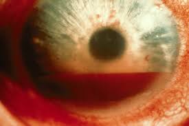
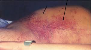
Thrombocytopenia is associated with skin/mucosal bleeding  (petechiae, gingival bleeding, epistaxis, heavy menses), also hematuria, and GI bleeding. 

Coagulopathies more commonly present with intracranial bleeding, retroperitoneal bleeding, and hemarthrosis. Girzadas  comment: I have also seen GI bleeding and deep tissue hematomas with coagulopathies.


The clinical finding most commonly associated with ITP is petechiae.   It makes sense because petechiae is in the name of the the disease “ Idiopahtic thrombocytopenic Purpura”.  Acute ITP is more common in kids.  Chronic ITP is more common in adults.


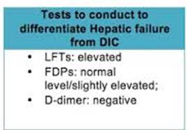
*Liver dysfunction vs DIC   Liver dysfunction will have not much elevation of fibrin degredation products and a negative D-Dimer.


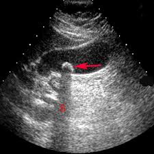
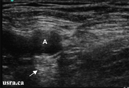
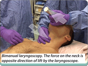
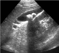
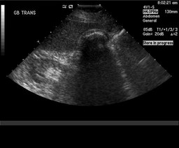
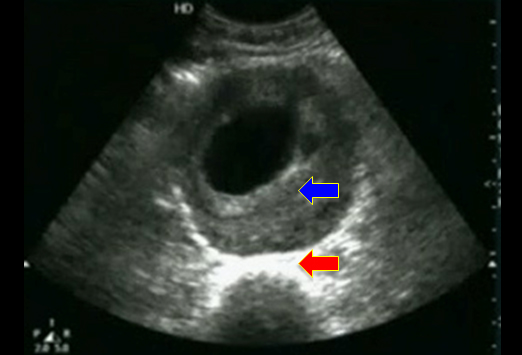
There was a discussion of performing paracentesis in patients with low platelet counts and/or coagulopathy.  Harwood comment: unless the patient is bleeding you don’t need to correct the platelet count or the INR.  Harwood added that every doctor will have a minimal platelet count threshold below which they would be unwilling to do a paracentesis.  His cut off was 50,000.  Elise added that because we use ultrasound to guide paracentesis, there is less risk of puncturing a vessel.  We then discussed doing LP in the setting of low platelets and coagulopathy.  There was consensus that LP could be done if platelets were above 50,000.  No one would do an LP if the INR was prolonged.


Reference: Severe haemorrhage following abdominal paracentesis for ascites in patients with liver disease

  1. I. Pache and
  2. M. Bilodeau


In conclusion, severe haemorrhage after abdominal paracentesis is a rare (0.19%) but potentially lethal complication. We report death following this complication in 0.02% of all paracenteses performed. Most haemorrhages occurred in severely ill patients and were rarely seen in patients with severe thrombocytopenia and/ore elevated INR. All but one occurred in the context of some degree of renal failure. The management of this complication can be conservative. Short-term prognosis is nonetheless poor and seems related to the underlying liver disease. Utility of the systematic prophylactic administration of agents that improve platelet function in patients with renal failure requiring abdominal paracentesis could be assessed prospectively. Unfortunately, this would be difficult to realize because of the very large patient sample required.


Reference:

Brief Report |

Safety of Lumbar Puncture for Children With Acute Lymphoblastic Leukemia and Thrombocytopenia FREE

Scott C. Howard, MD; Amar Gajjar, MD; Raul C. Ribeiro, MD; Gaston K. Rivera, MD; Jeffrey E. Rubnitz, MD, PhD; John T. Sandlund, MD; Patricia L. Harrison, MPH; Alberto de Armendi, MD; Gary V. Dahl, MD; Ching-Hon Pui, MD

[+] Author Affiliations

JAMA. 2000;284(17):2222-2224. doi:10.1001/jama.284.17.222

Conclusions In our study of children undergoing remission induction or consolidation therapy for acute lymphoblastic leukemia, serious complications of LP were not observed, regardless of platelet count. Prophylactic platelet transfusion is not necessary in children with platelet counts higher than 10 × 109/L. Due to the small number of patients in our study with platelet counts of 10 × 109/L or less, conclusions cannot yet be drawn for such patients.


Elise’s Reference: AASLD PRACTICE GUIDELINE

Management of Adult Patients with Ascites

Due to Cirrhosis: Update 2012

Bruce A. Runyon, M.D.

Although some physicians give blood products

(fresh frozen plasma and/or platelets) routinely before

paracentesis in patients with cirrhosis and coagulopathy,

this policy is not data-supported.19,22 Routine

tests of coagulation also do not reflect bleeding risk in

patients with cirrhosis; these patients regularly have

normal global coagulation because of a balanced defi-

ciency of procoagulants and anticoagulants.23 In a survey

of the use of blood products in relation to paracentesis,

50% of approximately 100 hepatologists

attending a conference on coagulopathy in liver disease

indicated that they either never used plasma pre-procedure

or used it only if the INR was >2.5.24 The risks

and costs of prophylactic transfusions may exceed the

benefit. Coagulopathy should preclude paracentesis

only when there is clinically evident hyperfibrinolysis

(three-dimensional ecchymosis/hematoma) or clinically

evident disseminated intravascular coagulation. A

shortened (<120 minute


Bleeding conditions occur in less than 1

per 1,000 patients who require paracentesis. There is

no data-supported cutoff of coagulation parameters

beyond which paracentesis should be avoided.19 In a

study of 1100 large volume paracenteses there were no

hemorrhagic complications despite a) no prophylactic

transfusions, b) platelet counts as low as 19,000 cells/

mm3 (19  106

/L)(54% <50,000) and c) international

normalized ratios for prothrombin time as high

as 8.7 (75% >1.5 and 26.5% >2.0).22


Acute chest syndrome is the leading cause of death in sickle cell disease and most common cause of admission as well.   There was discussion and a search of references on this point  There is controversy whether the most common cause of death is due to acute chest syndrome or infection.  There was agreement that there was overlap between the two etiologies. 

Hydroxyurea has been shown to reduce the frequency and severity of painful crises in children but has not been FDA approved.


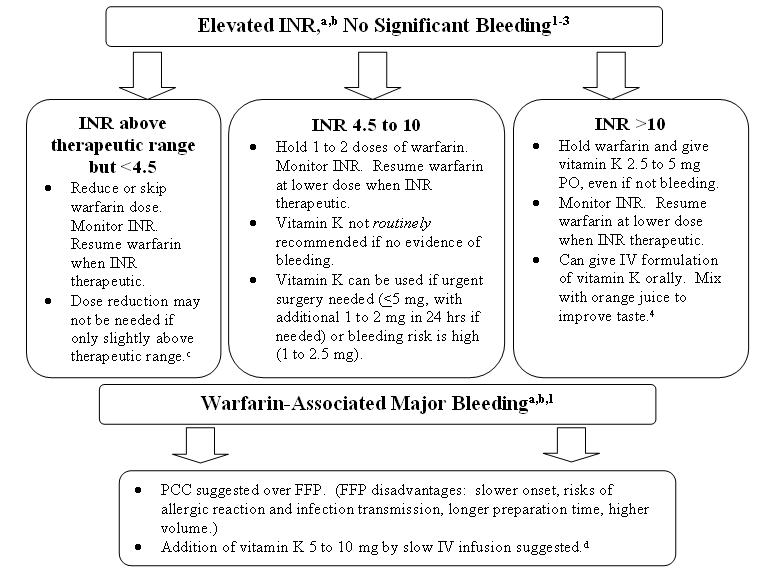
*Management of Supratherapuetic INR


Hypercalcemia treatment includes normal saline, calcitonin, zoledronic acid.


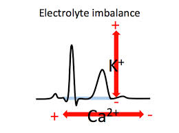
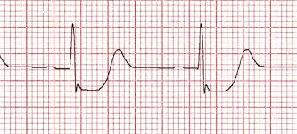
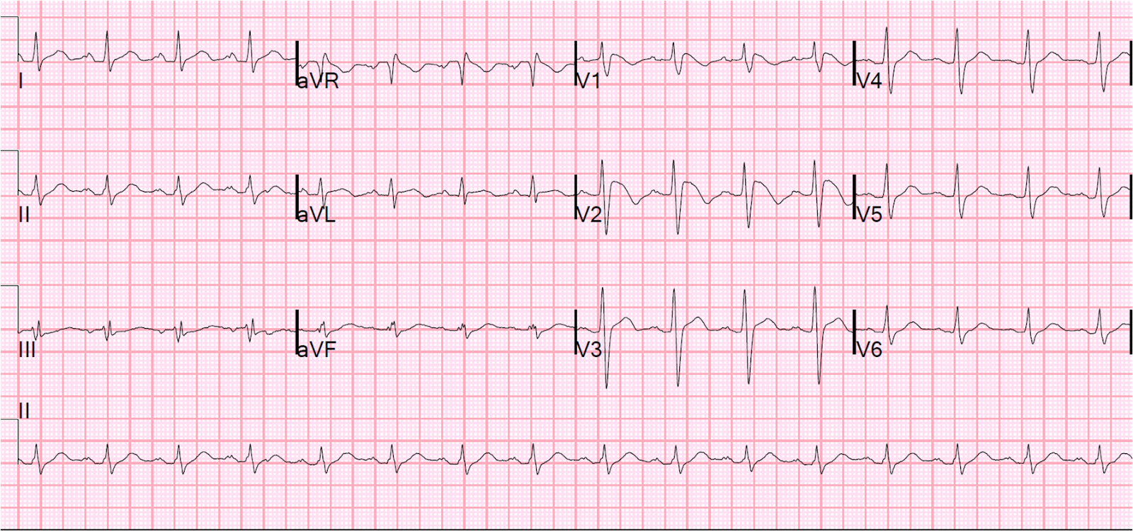
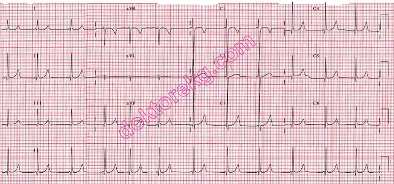
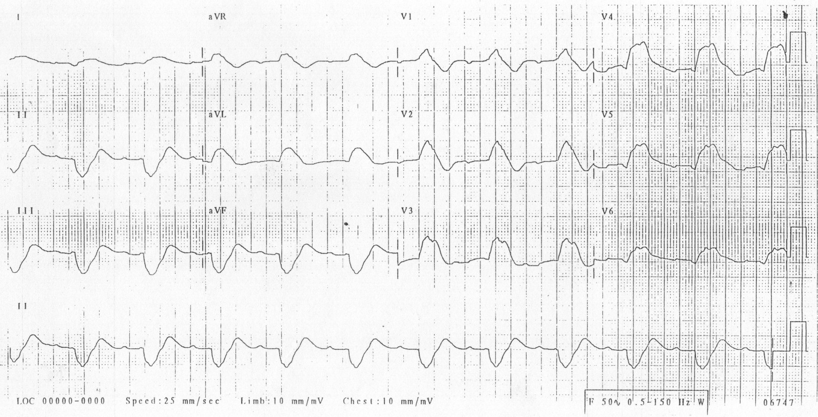
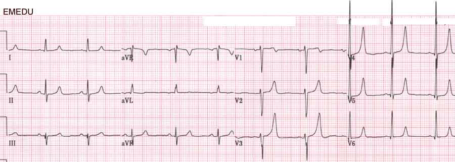
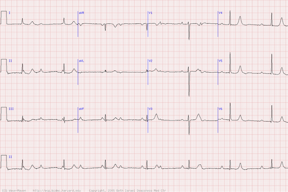
*Electrolyte abnormalities on EKG  QT lengthens with hypocalcemia and shortens with hypercalcemia.   T wave varies based on potassium level.


Bonder/Iannitelli        Workshop: ED/EICU  Care of ICU Boarders in the ED


We all worked through the new protocol for the management of ICU Boarders in the ED.  

In situ simulation of the new protocol will occur 1-13-2015.


Campanella    Comprehensive  Stroke Center


We have been successful in getting patients to CT rapidly.

We discussed various strategies to shorten the time from completed CT to administering TPA.


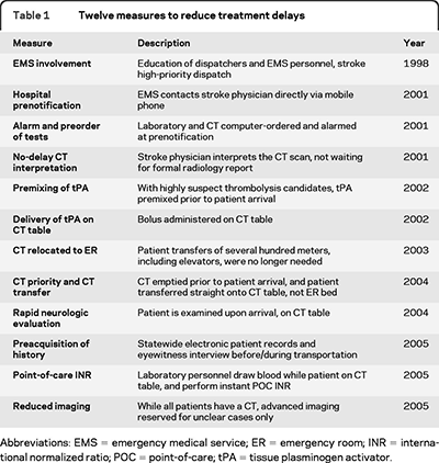
*Strategies for speeding the delivery of TPA   Dr. Campanella discussed many of these strategies.  He had some discomfort of giving TPA in the CT scanner.  He felt that it was more reasonable to use the time of transfer back to the ED to make sure the history and time of onset was as verified as possible and that the CT images were carefully reviewed. 


The eligibility criteria for TPA treatment for stroke in the time period 3-4.5 hours after onset are similar to those for persons treated between 0 and 3 hours, with any one of the

following additional exclusion criteria:

Patients older than 80

Those taking oral anticoagulants regardless of INR

Those with a baseline National Institutes of Health Stroke Scale score > 25

Those with both history of stroke and diabetes.

Findings, on computed tomography of the head, of early infarct involving more than one third of the middle cerebral artery territory


We also disussed the Mr. Clean Trial.  For patients with acute ischemic stroke caused by an  arterial anterior-circulation occlusion, intra-arterial treatment is effective and safe when given within 6 hours of stroke onset, a recent study shows. (Medscape)

This study has only been presented in abstract form.  The manuscript should be out in 2-2015.   Neurologists like it because it backs endovascular therapy for stroke.  There are however some methodological criticisms of the study. 


Beckemeyer     M&M


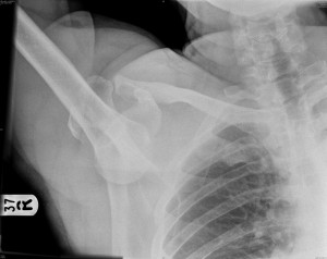
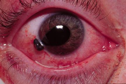
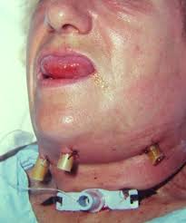
Parents bring a young child to ED for possible intranasal FB.

Patient has dried brown nasal drainage in nose but with speculum exam, no foreign body was identified.  Full visualization of the nasal passage was difficult due to swelling of the inferior turbinate.

Patient was discharged with ENT referral for the next day.


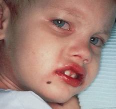
Child followed up with ENT the next day.  ENT identified a button battery in the nose and by that time the patient had suffered a severe burn to his nasal passage.  He required a nasal stent.


Aspiration of a nasal foreign body is possible but a review of case reports demonstrates that most of the time aspiration occurs with attempted removal of the foreign body.  Only rarely does foreign body aspiration occur spontaneously from a nasal lodging point.  


Button batteries are corrosive to tissue. In addition, they cause necrosis by both electrical discharge and pressure on the tissue.   Button batteries are ubiquitous.  Kids can get them from multiple toys/gadgets in the home.  Musical greeting cards are a big problem because kids can get at the button battery inside quite easily.


Case reports show that perforation and extensive burn from a button battery in the nose can occur within 90 minutes.


Based on this case, it was suggested to ask parents about the possibility of button battery insertion.   Also consider xray imaging of head to identify metallic foreign body in the nasal passage.  Consult ENT  while the patient is in the ED if there is any concern for a button battery in the nose.


Andrea, Elise, and Andrej made the comment that this was a very unusual case, “worst case scenario”.  Based on this case, they all felt that if you have a suspicion for nasal foreign body and you can’t visualize the nasal passage well, get an xray to rule out battery insertion in the nose.  Any non-battery nasal foreign body can wait until they see the ENT specialist to have it removed if you can’t get it out in the ED. 

Harwood comment: What I learned is that if the child has pain in his nose or drainage within a few hours after suspected foreign body insertion I have to strongly consider battery foreign body in the nostril and get an xray.


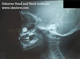
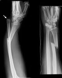
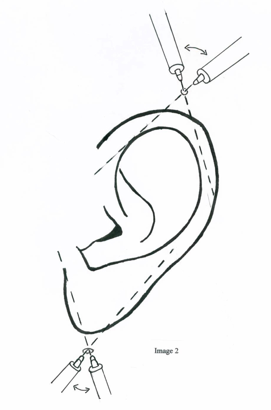
* Xray image of button battery in the nose.  You have to get this out ASAP.  Call the ENT specialist even in the middle of the night.


Balogun       ABC’s of ED management of Cirrhosis


A= Approach.  Patients present with confusion, lethargy, or bleeding.  DDX is broad including encephalopathy, cerebral edema, intracranial hemorrhage, elevated ammonia level, GI bleeding.   Patients can also present with fever.  You have to consider all potential sources of infection.   Keep spontaneous bacterial peritonitis at the top of your list,  Patients can also complain of edema and ascites.  Patients can have electrolyte abnormalities and coagulopathy.


There was a discussion of the best way to manage the airway in the cirrhotic patient.  General consensus was to do RSI with the patient in head up or RAMP position. Head up position can take some pressure of the diaphragm and improve oxygenation/ventilation.   Use rocuronium instead of succinylcholine.  Succinylcholine can cause hepatic failure in the cirrhotic.  Ketamine can also cause hepatic failure in the cirrhotic.  Most faculty however, didn’t feel there was a huge risk to using ketamine in these patients.


B= Bacterial infections    Cirrhotic patients are immunocompromised.

Spontaneous bacterial peritonitis carries a high mortality.  Diagnose SBP early by performing paracentesis in the ED.   Paracentesis is indicated for patients with fever/pain, prior history of SBP,  ascites and/or GI bleeding and/or encephalopathy.

There was faculty pushback on tapping the bellies of patients with just ascites and encephalopathy or ascites and GI bleed.  Faculty all felt patient had to have some sign pointing to peritonitis beyond ascites like fever, pain, tenderness, or leukocytosis before they would perform paracentesis.

Sole gave this follow up advice via email:  

Who should undergo paracentesis in diagnosing/treating SBP in the ED?
In my lecture, I mentioned that the following scenarios warrants a tap:
- classic sx of fever, abdominal pain/tenderness, ascites
- first time ascites*
- ascites with hx of SBP
- ascites with concomitant GI bleed and/or encephalopathy
Again, these recommendations are based on AASLD (formerly AHA, link to source below) guidelines and is even supported in Tintinalli. During my research, I came across several other sources that support variations of these recommendations. I discussed further with Dr. Raddawi after lecture and his theory regarding performing a paracentecis on first time ascites is that patients who develop ascites for the first time will often times be in decompensated liver failure, worsening their already immunocompromised status. This is also the rationale of ascites with concamitant GI bleed and/or encephalopathy. He does agree, though, that in the presence of normal vitals, no fever, no white count, absence of pain/tenderness, it is a procedure that can be performed inpatient. However, considering the significant mortality associated with SBP, it might be worth it to consider tapping these scenarios in the ED. Incidence of iatrogenic SBP when performed in sterile fashion is <3%


C= Coagulopathy.   Transfuse in platelets are less than 10. INR is the most sensitive sign of worsening liver function.    If INR is prolonged and patient is bleeding give vitamin K and FFP.


C= Circulation.   Cirrhotic patients can be hypotensive.  They are prone to bleeding, volume depletion, infection, and third spacing. Crystalloid replacement is appropriate.  Colloids are indicated only for large volume paracentesis and hepato-renal syndrome.


D= Drug considerations.   Opioids can have prolonged sedation.  Fentanyl is preferred due to its short half life.  

NSAID’s have risk of increased renal toxicity.

Benzodiazepines can have prolonged sedation.  

Zofran should be used in smaller doses.  No more than 8mg in 24 hours.  It can cause hepatotoxicity.

Treat seizures with lorazepam and keppra.  Dilantin can cause hepatic failure.


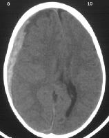
E= Encephalopathy.   Always get a head CT to evaluate for cerebral edema and intracranial hemorrhage.  Treat with lactulose.  Lactulose traps ammonia and carries it out in the stool.


E Kulstad        ATACH –II Trial


Our ED is a study site for the ATACH-2 trial,  Antihypertensive Treatment of Acute Cerebral Hemorrhage. 


The study goal is to identify if there is benefit to lowering the  BP to less than 140 or less than 180 in patients with acute cerebral hemorrhage.   BP is controlled to these levels for 24 hours.   Patients will be assessed at 30 days for neurologic outcome by blinded neurologists.


Patients are treated with IV nicardipine within 4.5 hours of symptom onset.


There is an iphone/android app for your phone to help you enroll patients in the study.   The app is called ATACH-2.    It has all the enrollment criteria and has a easy to use NIH Stroke Scale.