Radiologists are human too

An 18 y/o male presented to the ED with chest and back pain that started in the center of his chest, then moved to left scapula. Pain was sharp and worse with deep inspiration. His only associated symptoms was anxiety. No risk factors for heart disease, PE. 

VS: 101 20 136/88 20 99%

PE: normal

EKG: normal

CXR Findings: PA and lateral views of the chest are presented for interpretation. No prior studies

are available for comparison. The heart size is normal. The trachea is midline. The lung fields

are clear.

Impression: Radiographically negative chest.

Pt was discharged, symptoms persisted, so he went to another institution where he was admitted. Why? Look carefully at his cxr below

CXR1.png

As you've probably noticed, the radiologist misread the film- or dictated onto the wrong chart, or made some other mistake that we're all capable of making.  So this case serves as both a reminder to get into the habit of looking at all your own films, and a review of a neat CXR finding.  

pms.png
ptx.png

The patient has a pneumomediastinum   and pneumothorax

deep sulcus.png

with deep sulcus.

Spontaneous pneumomediastium is fairly rare. It occurs when air leaks out of a ruptured alveola and tracks along the bronchovascular sheath until it reaches the mediastinum. Air can also track to the pleural space, as in this case, or into the subQ tissues, submandibular and retropharyngeal space, or even into the spinal canal (see Dr. Permar for more information on this complication). Triggering factors can usually be identified but  are so common (crying, Valsalva, coughing, vomiting) that they don't rule in the disease.

The most common presenting symptoms will be pain wherever the air has tracked. Physical examination will likely be normal with isolated pneumomediastinum, although you may be lucky enough to hear Hamman's  crunch (crepitus heard over the heart). You may feel subcutaneous emphysema if the air has tracked there. Patient's with large pneumothoraces may have decreased breath sounds or hypoxia. 

Spontaneous pneumomediastinum is generally a self-limited disease without serious sequelae- much different than if caused by Boerhaave's or trauma. It requires no treatment except analgesia as needed. This patient was admitted for treatment of his pneumothorax.

chest pain

36 yo M, no pmhx, appears morbidly obese, no hx of HTN,but  has been told once he has borderline HTN, takes no meds. Presents by car with his father.  Patient states he had sudden onset left upper chest pain that radiated to his neck with mild sob and mild lightheadedness that started when he was walking up stairs.  He states the sob and lightheadedness have resolved and now the chest pain is mild.  It did not radiate to his back.  Nonexertional, nonpleuritic.  No hx of DVT or PE, no leg swelling or pain.   Denies abd pain.  Denies diaphoresis or nausea.  Pt is a smoker.  Denies drug use, admits social alcohol use.  Denies Fhx of early heart disease or suddent death.

PE is unremarkable

Vitals:  Hr 110, BP 122/84, RR 20, RA P02 97%, Temp 36.3

EKG: NSR no acute st or t wave changes HR 110

Inital A/P:

Well appearing morbidly obese patient with exertional onset left chest pain radiating to left neck, no radiation to back.  Denies medical hx and follows w a primary doctor.  Normal PE, no risk factors for PE.  No fmhx of CAD.  Non ischemic ekg and pain is currently mild.  He did state his pain was moderate to severe at onset.

Planned DDimer, trop x2, CXR, labs, and entering the patient into CPEP protocol with planned AM stress cardiac echo

Patient initially refused admission but was talked into staying for the protocol. wow

Results:

Tropx2 negative

DDimer negative

2 view CXR negative

CBC CMP normal

repeat ekg - non ischemic tachycardia resolved

At 7am near the end of signout I was called by the nurse as the patient just vomitted and BP was 100/55, HR was still 70s.

I reevaluated the patient and he appeared well and stated his pain returned and was still mild left CP radiating to his neck.   No other complaints besides nausea.

At this point I figured he couldnt do the stress test and changed him to an admission.  I decided to reevaluate if there was anything I was missing.  

Due to my past mental trauma from seeing 7 total patients younger than 45yo with aortic dissection in 7 years of residency/practice and 1 of those patients dying from their dissection.....I decided if I was to get any sleep I would need to get a CTA chest abd pelvis w and wo contrast prior to admission to tele.  The tipping point in my mind was why was this guy borderline hypotensive if he is in pain and morbidly obese.  Even with only moderate pain without alot of the classic symptoms of dissection.  He even had bilateral BPs done by accident which were not marked abnormal.  Also normal 2V chest and normal DDimer.  All prior 7 dissections in young people that I had seen had elevated DDimers.  It is NOT a sensitive test but with very low suspicion...sometimes to me it is somewhat reaasuring....THere is much debate about it's utility.

830am CTA results:

Large aneursymal dilatation of the proximal aorta and aortic valve.  Largest aortic diameter is 6cm.  Cannot rule out dissection within the aneursym.  Moderate size pericardial effusion vs pericardial hematoma.

Cardiothoracic surgeon immediately paged.  Cards came to the bedside.

11am Bedside ECHO results:

Large aneursymal dilatation of the proximal aorta and aortic valve.  Largest aortic diameter is 6cm.  Cannot rule out dissection within the aneursym.  Moderate size pericardial effusion vs pericardial hematoma with diastolic collapse of the RV concerning for possible early tamponade.

1pm in the OR

TEE confirms a dissection just above the aortic valve within the aneurysm.  Moderate aortic regurgitation. Moderate to severe pericardial effusion.

Cocaine negative on UTox

In the OR:

Patient had cardiac bypass/open chest sugery with replacement of ascending aorta w graft and his aortic valve was repaired but not replaced.

Post op day #1 the patient was extubated and stated he felt fine except for some incisional pain.

WOW...dodged a bullet

Almost gave this guy a stress test.  He didnt even want to stay for that.  Thankfully he vomitted and had borderline hypotension and thankfully I have a history w dissection that has made me a little more cautious....or I would have missed this.   Could've been my second death from a missed dissection...probably on the tredmill in the stress lab.  The prior death of a patient I had from dissection was a 33 yo M with a similar presentation and workup as this patient.  I discharged him and he came back on my same shift and died within 30 of arrival.  

Normal BP on arrival.  Normal bilateral BPs.  Normal 2V CXR.  negative trops and ddimers.....recipe for disaster.

Thanks to the resident and the next attending for fully taking over this guy at 1030 am and making sure he got the ECHO and the OR.  

Take homes:

- Morbid Obesity is a risk for other comorbidities (undiagnosed HTN) and dissection.  None of my young dissections appeared marfanoid but almost all were obese with probable undiagnosed or poorly treated HTN

Kyobu Geka.

 2013 Jun;66(6):437-44.

[Obesity is a risk factor of young onset of acute aortic dissection and postoperative hypoxemia].

[Article in Japanese]

Aizawa K

1, 

Sakano Y

Ohki S

Saito T

Konishi H

Misawa Y

.

- He was never truly hypotensive but borderline hypotension in a patient you would think would be hypertensive with chest pain should raise some eyebrows

- DDimer has been argued by some as a screening test for dissection but it's not sensitive enough.  This case illustrates that.  I feel that you could miss fewer dissections by using it w risk stratification, normal pulses in all four, normal 2V cxr, normal bilateral BPs etc, but if you have clinical suspicion...you have to scan

- Consider doing some documentation with chest pain patients that would make your chart more defensible in case you miss AD.  They are easy to miss and I have been there - things you could add would be equal pulses in all 4 ext, equal bilat BPs, normal CXR, and reasoning that you think the probability of dissection is so low that CTA is not indicated.

- Some experts on aortic dissection argue that its presentation can be so nonspecific that the standard of care is to not make the diagnosis.  

 Below is a CTA image showing the proximal aortic dilation and some evidence of the pericardial effusion

dissection2.jpg

Boxer's Fracture- Back to basics

A review of a bread & butter presentation based on a young man I saw last week who was vague about how his injury occured.

Boxers 1.jpg

Boxer's fractures are fractures of the 5th metacarpal neck, and typically occur from hitting something with a clenched fist.

normal.jpg

Along with the usual neurovascular exam, check for rotational deformity.

Fingers are normally parallel went hand closed

 VS,

rotational deformity.jpg

Small finger rotated or overlapping ring finger. Compare to other hand if needed.

No rotational deformity is acceptable

Reduce the fracture if there is a lot of angulation- numbers vary but generally > 40% = reduction. Function will be likely OK with more angulation but will have odd looking knuckles and a lump in palm.

boxers 2.jpg

    About 45 degrees

Reduction- pain control with hematoma or ulnar block, or procedural sedation.

For successful reduction, flex the MCP to 90. Then push on the bent finger to reduce the metacarpal head while keeping pressure on the metacarpal shaft.

boxers reduction.jpg

Apply an ulnar gutter splint with 4th and 5th fingers bent to 70-90 degrees. Buddy taping the 4th and 5th fingers together provides rotational stabilization.

82 y/o woman with hypoxia

HPI

An 82 y/o woman with multiple sclerosis, history of DVT on Coumadin and chronic indwelling Foley is sent from nursing home for hypoxia. Patient is not on oxygen at nursing home. Her pulse Ox was low (83) at the nursing home, so they sent her away to Christ hospital. EMS said they put her on 15L oxygen on non-rebreather on the transfer.

Code 44 was called by nurse. On arrival, patient said she was sleeping. She has no clue why she is sent over to the hospital. She denies chest pain or shortness of breath or any symptoms. She has not been sick. 

Physical Exam 

Vitals: SpO2-92 on 15L oxygen via non-rebreather. RR: 18, BP 146/62, P 77.


Exam:

Gen: NAD. Very comfortable. Bluish skin

CVS: RRR

Pulm: CTAB

Abd: Non-tender, soft. Foley present. Orange urine in Foley bag.

Neuro: Patient can shrug shoulders but strength 1/5. Sensation diminished below shoulders (all are baseline)


Intervention 

*Patient has hypoxia on SpO2 but she looks very good. Something does not fit quite right. ABG with panel was ordered on top of Code 44 labs.

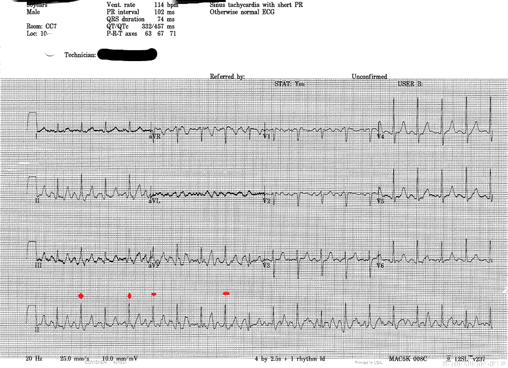
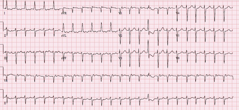
EKG: normal rate, normal sinus rhythm, no ischemia

CXR: normal without consolidation 

ABG: pH 7.44, pCO2 44, pO2 461,  HCO3 28, O2 sat 98, Carbonmonoxide 3.1, O2 content arterial 10 (Low), OxyHemoglobin 80.8, Methemoglobin 14.9 % (normal <1.2%)

Labs: normal BMP, normal CBC, and therapeutic INR: 2.3. Troponin: <0.02, BNP 30 


Patient has methemoglobinemia. Everything now fits: the low pulse Ox, maybe questionable bluish skin and patient’s lack of symptoms.

What’s the culprit? We tried to remember the meds that can induce methemoglobinemia from board exams. Nitrous or Sulfa containing meds!

Well, let’s ask our pharmacist to look at her meds list. Luckily, our patient was a healthy MS patient. Besides warfarin and Baclofen, she was given 1 other medication by nursing home. Phenazopyridine (pyridium)!

Phenazopyridine (pyridium)

Phenazopyridine is a urinary tract analgesic and available over the counter. It is supposed to be used for 3 days in an acute treatment for patients with dysuria. This poor old lady was moved to nursing home 3 months ago. NH put her on a Foley since she has urinary incontinence due to MS, and when she complained of Foley irritating her urethra, they put her on Pyridium for 3 months.

Treatment: We started pushing IV methylene blue 1mg/kg and within minutes, her pulse Ox improved to 98 on room air.

Patient update: Daughters came to thank the ER doctor months later. They moved their mom to a different nursing home. She is doing well. 

Methemoglobinemia (UpToDate)

Diagnosis: Methemoglobin level on ABG, normally methemoglobin levels <1%

Pathophys: Ferrous (Fe 2+) in heme group of hemoglobin is oxidized to the ferric state (Fe 3+). This converts hemoglobin into methemoglobin, which CAN’T bind oxygen. In addition, oxygen affinity of any remaining ferrous hemes is increased, shifting the oxygen dissociation curve to the left, so oxygen can’t be delivered to tissues. => The patient has greater functional anemia.

Acquired Causes: oxidizing agents (medications): nitrites, nitroglycerin, nitroprusside, Bactrim, benzocaine, inhaled nitric oxide, aniline and its derivatives etc. Dapsone and topical anesthetics appear to be most common precipitating agents.  

Congenital Cause: Congenital methemoglobinemia is characterized by diminished enzymatic reduction of methemoglobin back to functional hemoglobin. Affected patients appear cyanotic but are generally asymptomatic.

There is a family that lived in the hills of Kentucky known as “Blue Fugates.” They have a congenital deficiency in the enzyme causing them to have methemoglobinemia. It is autosomal recessive but this family intermarries, so viola..

http://abcnews.go.com/Health/blue-skinned-people-kentucky-reveal-todays-genetic-lesson/story?id=15759819#.T5sgQnr-mVo

Treatment:

-Asymptomatic: discontinue the offending agent

- Symptomatic or methemoglobin level>20%:  IV methylene blue 1-2 mg/kg can be given over 5 minutes. Response is rapid. A second dose may be repeated in one hour but retreatment is often not necessary. 

-Blood transfusion or exchange transfusion may be helpful in people who are in shock. Hyperbaric oxygen has been used with anecdotal success in severe cases.

Although ascorbic acid is an answer on board questions, it is a less effective treatment than methylene blue.

Symptoms:

  1. Methemoglobin>15%: cyanosis, asymptomatic
  2. >30%, fatigue, headache, dizziness, tachycardia, weakness
  3. >55%: dyspnea, bradycardia, seizures, coma
  4. >70%: death


What about pyridium?

- Pyridium has been reported sparingly in medical literature as a cause of methemoglobinemia, either as an acute overdose or in patients with prolonged therapeutic use.

Take Home Points:

- Methemoglobinemia on board questions: pulse Ox 87 no matter how much oxygen is given. Blood itself is chocolate-colored. Get ABG with panel.

- It is often drug induced. Topical anesthetic (patients coming from GI procedure) or TMP-SMX are common meds.

- Treatment: IV methylene blue 1-2mg/kg x 1


56 YO MALE WITH SOB

HPI: 56 yo male with small cell lung cancer on chemotherapy (3 days ago, first tx) presents to the ED with SOB over the past day. No chest pain, LE edema, orthopnea. No fevers, chills or hemoptysis. +Chronic dry cough.  He also complains of generalized weakness with ntermittent nausea, vomiting and diarrhea since chemotx. No abdominal pain. No urinary changes.

Physical Exam:

VS: HR 124, BP 102/69, Sat 89% on RA, RR 30, T 36.5 R

GEN: Alert, mild-mod distress, thin

HEENT: MM dry

CV: tachy, regular

PULM: Course bilaterally with wheezing. Tachypneic. No rales

ABD: Soft, NT

EXT: No LE edema

INTERVENTION #1: O2, BIPAP, nebs, 2 L NS bolus

EKG: Sinus Tach. No STE or right heart strain

CXR:

CXR1 1.jpg

“There are extensive new airspace opacities at the lung bases and left upper lobe . . .”

INTERVENTION #2: Vanc, zoysn and azithromycin for HAP. Unsure of WBC at this point. HR and RR improving with IV fluids, Bipap and O2. BP is stable. Still somewhat tachypneic.

LABS:

Na

132

WBC

57.2

UA =

unremarkable

K

2.8

Hb

11.2

BNP

314

Cl

90

Hct

33.7

Trop

0.37

Bicarb

19

Plt

498

Flu

Neg

BUN

52

PMN

95% = 56

C diff

Neg

Creat

2.31

Bands

3

L.A.

3.0

Ca

5.6

Glc

184

***So why is the calcium so low? Kessen, who is sitting next to me and fresh off of his oral boards beat down by Kerwin (ONC emergencies) says, “tumor lysis syndrome”.

-But what about the potassium? Shouldn’t it be higher?

-So we add-on a few labs:

Uric Acid

9.2 (3.5-7.2)

Phos

7.8 (2.4-4.7)

Mg

0.8 (1.6-2.4)

-Well shit, it is tumor lysis syndrome (TLS), right?

INTERVENTION #3:|

-Consult oncology

-Replace K, Mg, Ca.

-IVF hydration

-After a discussion with Neil (pharmD), we order Rasburicase for the elevated uric acid (Apparently it’s very expensive ~$20,000)

-Oncology agrees with assessment and plan. States that TLS is associated with small cell lung tumor

-Admitted to MICU

HOSPITAL COURSE:

-Repeat Uric Acid is 3.6 after 6 hours

-other electrolyte abnormalities are more difficult to correct, but do improve by day 3

-Pts respiratory distress worsens and he is intubated the 1st day in the ICU

-he become neutropenic and pancyotpenic on day 3-4 (~7 days after chemotx). Possible DIC

-Pt dies on day 7

 ---------------------------------------------------------------------------------------------------------------------

TUMOR LYSIS SYNDROME (TLS): from uptodate.

Massive tumor cell lysis with the release of large amounts of potassium, phosphate, and nucleic acids into the systemic circulation. Catabolism of the nucleic acids to uric acid leads to hyperuricemia.

TLS most often occurs after the initiation of cytotoxic therapy in patients with high-grade lymphomas and acute lymphoblastic leukemia. However, TLS can occur spontaneously and with other tumor types that have a high proliferative rate, large tumor burden, or high sensitivity to cytotoxic therapy.

-Hyperuricemia: a consequence of the catabolism of purine nucleic acids to hypoxanthine and xanthine and then to uric acid via the enzyme xanthine oxidase. Results in the precipitation of uric acid in the renal tubules leading to acute kidney injury

-Hyperphosphotemia: rapid tumor breakdown often leads to hyperphosphatemia which can cause secondary hypocalcemia. This can lead to AKI by calcium phosphate deposition in the renal tubules and also to cardiac arrhythmias.

DEFINITION:

Cairo-Bishop definition of laboratory tumor lysis syndrome

Element

Value

Uric acid

≥ 8 mg/dL

Potassium

≥6.0 mmol/L (or 6 mEq/L)

Phosphorus

≥ 6.5 mg/dL for children or 4.5 mg/dL for adults

Calcium

≤  7 mg/dL

- Two or more laboratory changes within 3 days before or 7 days after cytotoxic therapy

TREATMENT:

-Correct electrolytes and monitor q 4-6hrs

-Hyperkalemia: typical treatment, including dialysis if severe

-Hyperuricemia: IVF and rasburicase (0.2 mg/kg, repeat as necessary). Rasburicase is urate oxidase (uricase), which catalyzes oxidation of uric acid to the much more water-soluble compound allantoin. Can also do Allopurinol if rasburicase is not available.

-Hypocalemia: Treat only if symptomatic. Correct elevated phoshate first if possible

-Hyperphoshatemia: Oral binding agents (not very effective)

-Kidney injury: IVF rehydration, treat elevated uric acid. May need dialysis.

GCBH2...57 yo F...psych complaint

57 yo F brought in by brother-in-law for purported suicide attempt. He tells me she's done things in the past, but nothing serious, and that he believes she is just trying to get attention. Pt is somnolent but arousable, I do my quick standard psych H&P, patient reports that she drank 10 ounces of windshield wiper fluid approximately 12 hours ago with the intent of killing herself. She is mildly tachypneic and diaphoretic, so the first the thing we do is order an ABG:

pH 7.06, HCO3 6, and anion gap of 32

ELEVATED ANION GAP ACIDOSIS!!

Remember MUDPILES?

M-methanol

U-uremia

D-DKA

P-propylene glycol

I-infection/isoniazid/iron tox/isopropyl alcohol

L-lactic acidosis

E-ethylene glycol

S-salycylism

Re-evaluation of patient showed decreasing GCS and increased WOB, so we moved her to CC room and intubated her. Patient was started on fomepizole drip (I wanted to started pouring whiskey down her OG tube but Erik wouldn't let me) for presumed toxic alcohol toxicity, vascular surgery was called to place catheter for emergency HD,  and nephrology was emergently consulted.

Toxic alcohol levels showed an initial methanol level of 145 mg/dL; no ethylene glycol or isopropyl alcohol detected.

Nephrology recommended adding bicarb drip, and within a couple hours hemodialysis had begun. After 3 hrs of HD, methanol levels were down to 68, pH increased to 7.50, and the anion gap was closing. Pt then went to ICU.

She underwent a total of 12 hours of HD. On day 2 of hospitalization, methanol level was <25, so fomepizole and HD were dc'ed. Pt was shortly therafter extubated, and transferred the next day to psych floor. Interestingly, she never developed any opthalmologic complications.

One final footnote: Before she crunked and got tubed, she was trying to remember the brand of wiper fluid she'd guzzled. All she could remember that it had the word "Blast" in it. After some internet research, I read that there was indeed a company based out of Minnesota that made a wiper fluid called "PowerBlast." Interestingly, they were currently being sued by another windshield wiper producer for "watering down" their product, ie, using less methanol, to save on manufacturing cost. I found it interesting that perhaps by working to improve their bottom line, the company had inadvertently made their product less toxic, and as such perhaps prevented this particular patient's demise (although who knows the M&M that resulted from spinouts out on icy roads by drivers unable to see out of frozen, wiper-fluid-encrusted windshields).

17 y/o with hip pain playing basketball

17 y/o male brought to PED2 by EMS after hurting his hip playing basketball. He was running down the court when his friend collided with hip, hitting friend's elbow on patient's bony pelvis. Patient said he couldn't walk afterwards, his dad seems to think he's being overly dramatic. No PMH. VSS. Exam shows tenderness on right side of bony pelvis without brusing or deformity. No other abnormalities on exam.

AP pelvis XRAY

ASIS.jpg

What's the diagnosis? Treatment?

OK, now that you've thought about it a while, I'm sure you've diagnoses an avulsion fracture of the anterior superior iliac spine or ASIS. These tend to occur in athletic adolescents who have a forceful contraction of the muscles that attach there (sartorious and tensor fascia lata). They fracture because the apophyses are forming in puberty. Other areas prone to avulsion fractures are ischial tuberosity, and AIIS (anterior inferior iliac spine). 

Xrays are often not obtained as the mechanism (sprinting, jumping) sounds like it wouldn't cause bony injury, and the injuries are misdiagnosed as a muscle strain. The good news is that the treatment is generally similar- rest with partial weight bearing on cructhes, pain control, followed by PT. If the bony fragments are more than 3 cm appart or healing is delayed, the patient is a candidate for surgical repair.

50 year old male with syncope, HR 220

This 50 year old male presented to the ED folowing syncope. He had sudden onset of severe palpitations associated w/ throat fullness, diaphoresis and nausea. He had a syncopal episode at home and then presented to the ED.

Inital Triage VS:

HR 227

BP 124/86

RR 28

SpO2 100% RA

He's roomed immediately, here's the EKG

EKG1.png

Very fast, irregular narrow complex tachycardia w/ varying QRS morphologies. Rhythm is atrial fibrillation, with some consecutive beats conducted at a rate of nearly 300 bpm (R-R interval 200 msec), which is nearly diagnositic for the prescence of an accessory pathway--the AV node simply cannot conduct that fast. Atrial Fibrillation w/ WPW, this is the one we train for.

The patient notes a history of Rheumatic Fever as a child, but no other medical problems and no recent stimulant or other drug use. On exam, he's awake and alert and appears uncomfortable and clutching his chest. He denies chest pain or shortness of breath, just very uncomfortable palpitations. On the monitor, there were frequent runs of nonsustained Wide complex beats concerning for immenient degeneration into Vfib.

Management options?

Adenosine?

No! Ineffective at best and could possibly cause increased conduction through the Accessory pathway

Rate Control?

No!

Medical Cardioversion?

Potentially an option for the stable patient. But this patient, while not meeting any of the strict criteria typically used to define 'unstable' in ACLS, was clearly electrically unstable. N.B. The two options typically listed for cardioversion are procainamide and amiodarone, however numerous case reports and been published showing increased ventricular response and even Vfib during infusion of amiodarone have been published, so best to stick w/ Procainamide.

Electrical Cardioversion?

Yes! Undoubtedly the fastest and most efficacious method, it's also probably the safest as well. We sedated the patient using etomidate and successfuly performed syncronized cardioversion w/ 200 J. Upon waking the patient reported complete relief of symptoms.

Post-Cardioversion EKG

EKG2.png

A lot of motion artifact, but Sinus Tachycardia with a short PR interval w/ delta waves evident in some of the complexes. Interestingly, these were not seen on subsequent EKGs.

51yo Male w/ Nasal Congestion

51 yo male w/ no PMH presents w/ nasal congestion. Went to local outpatient care station for cough & congestion x 4-5 days. afebrile. Care station VS showed HR in 180's, sent to ED. No CP/SOB/Syncope/Dizziness/Palpitations. Patient had no history of arrhythmia. BP 130/78. Unremarkable physical exam.

ekg1.JPG

EKG showed a Wide, Regular Tachycardia, and the patient had a normal SBP and was essentially asymptomatic so this patient was considered "stable".  Differential includes Vtach vs SVT w/ abberancy.  Treatment options include Adenosine, Procainamide, Amiodarone, electricity (Adenosine should/can only be used in wide complex tachycardias that are regular).  If the patient was unstable, electricity would have been the first choice.  No one will fault you for treating all wide/regular tachycardias as VT, but the treatment of choice for stable VT is up for debate.  It is not recommended to use both Amiodarone AND Procainamide because their side effects can be additive, so pick one and stick with it.  Our patient got Amiodarone, which did had no significant effect. The patient then got 6mg Adenosine, which had no effect.  12mg of Adenosine then converted our patient into the EKG below.

ekg2.JPG

WPW. 51 yo and had never been diagnosed. Son of a bee sting. His accessory pathyway was ablated 2 days later and was discharged without further complication.

If I could do it again, I would have started with Adenosine, then Procainamide would have been my 2nd choice, and I would have used electricity if those had both failed.

WTF...Diagnosis Wenkebach??

This is a case from the other day I'm still not sure I completely understand and need some help from all of the briliant minds out there.

atrial tach retrograde p.jpg

78F, under treatment for multiple myeloma, recently started on hemodialysis, COPD, DM, came to the hospital for an outpatient IR fistula procedure. She was sent to the ER for tachycardia, thought to be a-fib. Pt has had some epigastric tightness for a couple of days and some mild shortness of breath, but no palpitations or real chest pain. Initial EKG below:

Definately not a-fib (too regular, weird p-waves present, look at V1). A few thoughts went through our heads: AVNRT (retrograde P's), a-flutter (ventricular rate is almost exactly 150), SVT....

We went with a-flutter, started on diltiazem 10mg bolus, 5mg/hr drip.

Check on pt a bit later, rate in 80s. Yay, we fixed her! Let's get a repeat EKG!

a tach with wenkebach av block.jpg

Uh...WTF is this? Still not a-fib, P's march out regular with single morphology. Does not seem to be an association between Ps and QRS. 3rd degree heart block? Can't be, QRS is narrow so ventricular rhythm orignates above the AV node, also the rate is norma, no bradycardic. Is there a 2nd degree AV node block of some sort? The groupings of QRS complexes are regular.

Cardiologist in ED to see pt. Answer: Uh...WTF is this? Wenkebach? (sigh of relief I'm not the only one that can't figure it out). Let's get an EP consult

EP consult: Atrial tachycardia with AV block suggestive of Wenkebach periodicity. Neg P waves in lateral leads suggest atrial tachycardia, periodicity of QRS suggests Wenkbach

Some more info on Wenckebach:

2nd degree heart block, Mobitz type 1. PR interval gradually increases until there is a non-conducted P wave. P waves tend to be regular. QRS complexes are clustered, usuall in a consistent pattern (P:QRS of 3:2, 4:3, 5:4).

I still don't completely understand this EKG. If you have any thoughts, please comment on this post (click below, don't reply to email) for everyone to see and discuss.

View this video for some inspiration: http://www.youtube.com/watch?v=GVxJJ2DBPiQ

Louis

Update

on 2013-11-29 16:23 by Robert Harwod

Louie-->

Great case, but I'm not sure why you are feeling so out-of-sorts.

EKG#1: A.Flutter @ 143 (Atrial rate=286 best seen/not well in lead III) with a 2:1 block. If you are looking for A. flutter 2:1 block (i.e. regular tachycardia 140-160) look for flutter waves in inferior leads or V1/V2. 

EKG#1: You got it! Go head & treat. You did with CCB! 

CCB's slow can slow atrial rate & WILL increase AV block. This is why it works so we'll in slowing P.A.Fib & why we use it everyday. It works.

EKG#2: show group beating. Group beating is Wenkebach/type 2 block. 99.9% of the time this is 2nd AV block (0.01% it is SA nodal block, sometimes called Sinus exit block, which is grouped within sick sinus syndromes).

EKG#2: What's the p rate? Before meds it was flutter @ 286. Now it is 123. Could be sinus @ 123, but P wave configuration makes non-sinus atrial tachycardia @ 123.

EKG#2: Examining the lead V1 rhythm strip (2nd from bottom), you see QRS #1, then p#1 with drop (missing QRS). P#2/P#3 conducted with increasingly PR interval until another dropped beat. This 3:2 group beating 2nd HB/Wenkebach continues across the EKG. The is an isolated 4:3 group in the middle of the strip.

EKG#2: PAT with block (classic for dig toxicity, by the way). You got it!

If this was 2 pts with 2 EKG's, you'd be "good." Maybe your discomfort is these 2 EKG's in the same pt?

OK. So go back to the pt. ESRD on HD, MM, COPD. This pt is a train wreck. Even if her K+, Mg++, Ca++ & phos were normal in her serum, do you believe they are are perfect in her heart tissue? Her SA node? Her AV node? Then you give her an IV CCB. It does what it is supposed to do. It slows & blocks. Sherlock Holmes consult? Not really needed.

A final point about P.A.Flutter. This only rarely is a chronic condition; it seems like it is an inherently unstable rhythm. It usually ends up as chronic A. fib or sinus. What rhythm did she end up in with long-term?

-->Harwood

49 yo F

The pt is a 49 yo F with a hx of uncontrolled hypertension and heavy smoking, presnting for a week of nausea, vomiting, diarrhea, and now abdominal pain. Pt BIB EMS, found by the to be hypotensive, on arrival to ED, pt is ashen, exrtremely pale, in distress, saying "I cant breath", hyptensive to 80s systolic, tachycardic to 130s. Her abdomen seems distended and is pulsatile on palpation. Ultrasound machine is brough into the room, shows a 9cm structure at and below the umbilicus (AAA? but too low?). Pts vitals improve enough after 2L IVF on pressure bags for her to be taken to CT, she is in the scanner 30min after arrival in ED, vascular surgery contacted before she goes to CT and actually meets her there. Findings are below.

aneurysm1.jpg
aneurysm2.jpg

Pt had a ruptured ILIAC artery aneurysm. Her initial pH was 7.07 and lactate 19.5. Hgb 6.2. Upon return from CT, blood was waiting for her, her vitals improved to SBP in 120s and HR  around 100. She was less pale. She was taken to the OR just over an hour after arrival in the ED. Currently in SVTU, doing well as far as I can tell.

68 yo F with left lower face pain and swelling x 5 days

HPI: The pain/edema was initially noted in the left lower lateral face and progressed to include anterior tongue. The patient was tolerating her secretions though it was painful for her to open her mouth. There was no history of oral trauma. No fevers, chills, headache, dizziness, vision changes, skin changes, and shortness of breath.

PMH: Rheumatoid arthritis on Methotexate, Adalimumab, and Leflunomide

PE:

-VS: T 37.1.C; BP: 156/98 mm Hg; HR: 115 beats/min; RR: 18 breaths/min, pulse ox: 96% on RA

-Gen: Well appearing

-HEENT/Neck: left submandibular edema with TTP. No overlying erythema. Tongue with mild distal edema, but no fluctuance; there was no tongue protrusion, trismus or drooling. Full ROM of the neck, with mild pain on neck rotation. No other oropharyngeal edema. Normal dentation without evidence of oral trauma

CT Max/Face W/Contrast (click on image to make bigger): 

Diagnosis????

Answer:

Tongue Abscess. Likley secondary to immunosuppression. Radiologist called to say he had never seen this before and according to the limited literature, it is rare. Less than 30 cases reported in the US.

Backround info: Spontaneous lingual (tongue) abscess is a rare life-threatening medical condition that may result in acute airway obstruction. The rich vascular supply, thick mucosa, and the anti-infectious characteristics of saliva make lingual abscess uncommon. Symptoms of a lingual abscess include tongue pain and swelling, odynophagia, dysphagia, difficulty speaking, changes in voice, tongue protrusion, and fever. 

The responsible organisms are usually normal flora from the oral cavity. The most common cause of a lingual abscess is direct trauma, whether this be from the teeth or a foreign body, and can also occur in association with dental infections. Immunocompromised state is considered a predisposing risk factor.

The differential diagnosis for lingual abscess includes aneurysm of the lingual artery, neoplasm, hemorrhage, infarction, cyst and angioedema. CT scan with IV contrast is generally recommended for improved characterization of the lesion, though it may be difficult to determine the etiology without surgical exploration and biopsy.

The treatment of a TA begins with airway assessment. Once airway stability is confirmed, further management includes broad-spectrum antibiotics to cover oral flora and consultation with either oral surgery or ENT to discuss drainage of the infection. 

7 y/o

7/ o m presents to the ED after coughing while playing with legos. He is satting 100% on room air, comfortbale playing in the room. His CXR shows:

"There is no acute cardiopulmonary process. there is no evidence for

radiopaque foreign body. There is no significant evidence for air-trapping."

Decubitus films:

No evidence for air-trapping

We take a look at the CXR ourselves, do you see anything pecuilar? (CLICK ON XRAY its a thumbnail)

Look at the right mainstem area, can you make out the pinholes of a lego piece? Apparently there is no air trapping because the lego was not solid but had a hole in the middle of it!

It helps that he had unilateal wheezing on that side and stridor, but either way a piece of lego was removed form his right mainstem bronchus.

-dave

62y/o AMS weakness, sob

62y/o M presents to the ED with AMS, bradycardia. We cannot get an IV but he looks liek S***. He is short of breath, DEXI is 42, no IV access, Im putting an IO since he is awake but clearly confused. We are trying to still get a full set of vitals and an EKG (since he was a code 44 at 540am). He is defintiely intubatable, but it looks like pulse ox is 100% on NRB, so we have a minute here. 

I am shooting a quick EKG while pushing an amp of D50 into the IO (which by the way, he didn't complain at all while putting in the IO, but he DID NOT like the D50 getting pushed in). 

Just then the EKG  tech says hey his EKG is looking really funny, just as the patient goes unresponsive:

Barounis-EKG1.png

EKG 1.

Not so good, 2 amps of calcium chloride, 2 amps of bicarb, 30 seconds of CPR followed by intubation with rocuronium (he was still breathing.)

EKG 2:

Barounis-EKG 2.jpg

Potassium came back 8.0, acute renal failure, got dialysis a few hours later and despite our best efforts died in the MICCU. Hyperkalemia is scary, but treatable, first and foremost diagnosis in ANYONE with bradycarida without P waves should be hyperK. 

17 y/o with chest pain

This guy developed chest pain about 1 week ago after playing basketball, felt SOB with exercise only. He saw his PMD on day of presentation who did outpt xrays. Below is his repeat ACMC CXR.

ptx.png

The reading is a complete left sided PTX, if the image isn't clear. So, how do you treat it? It's a simple PTX, not trauma. Does it require a chest tube, catheter aspiration, or even needle aspiration?

At the time, I thought it was too big for anything other than a chest tube. It had also enlarged from a few hours earlier (from about 70% to complete collapse), although the patient was stable (comfortable at rest, normal VS, and normal O2 sats). Looking it up afterwards, some teaching points.

1) Aspiration is successful in about 65-80% of simple PTX (no underlying lung disease, no trauma). The rate of PTX recurrence is the same as if a chest tube was placed.

2) If you put a catheter in the chest, it should be small (14 F or smaller). A chest tube should also be small (16 to 22 F).  You can put a Heimlich valve on the end if the lung seems to expand well, instead of suction.

3) For a small (<2 cm) PTX that isn't causing "breathlessness", don't do anything but monitor per British Thoracic Society.

4) BTS advocates needle aspiration for all simple PTX regardless of size. Most studies (few patients) had patients with small to moderate PTX. Either way, the patient will need 6 hrs obs in the ED and repeat CXR. Leaving a catheter in place so more air could be aspirated is a nice option.

5) Patients with underlying lung disease (eg COPD) probably won't do as well, but BTS advocates giving aspiration a shot.

What did I do? Put in a 20 F chest tube (with ketofol) to wall suction. His lung went to 95% inflated immediately. Maybe next time I'll try 14F catheter aspiration. see http://emedicine.medscape.com/article/1959416-overview#aw2aab6b4 for technique

New onset-seizure in a 9y/oF

Patient is a 9y/o F with a PMH of sickle cell disease who presented to the emergency department with new onset seizure. Parents thought patient had a tactile fever and cough at home over the past two days. At the OSH CBC demonstrated hgb of 6.3 (baseline 7.0), and with normal reticulocyte count base don disease.

On our exam here patient  had right sided weakness RUE > RLE, and "knew the words she wanted to say, but was unable to effectively communicate them" (what type of aphasia and where is the lesion?)

CTH was unrevealing and patient was transferred to our PICU.

MRI/MRA of the brain was completed that demonstrated:

MRI  brain  without  and  with  contrast:  Patchy  areas  of  restricted  diffusion  and

hyperintense  FLAIR  signal  throughout  several  portions  of  left  cerebral

hemisphere  primarily  at  the  left  middle  cerebral  artery  territory  (including

left  parietal  lobe  cortex  at  superior,  mid,  and  inferior  levels,  left  lentiform

nucleus,  left  internal  capsule  posterior  limb,  left  caudate  body/periventricular

region,  and  superior  left  frontal  lobe  cortex).  Increased  vascularity  of  left

cerebral  hemisphere  on  postcontrast  series  in  the  affected  regions  may  represent

luxury  reperfusion.

Treatement?

Exchange transfusion was begun with placement of a quinton catheter and a goal HgS < 40%, and a Hct > 21%.

Heparin-Induced Thrombocytopenia

62y/o F 12 days s/p CABG presents to the er with a swollen hand to GCB hall 2. Patinet says she went to bed last night and awoke this morning with severe hand numbness, a bluish hue, and edema. Per the patient it was "completley normal last night" She has a small back escar over the area where a radial A-Line cutdown was completed. patient has only the complaints of numbness in the hand, but has normal motor functions. 

PE: 36.6, 102, 12, 140/64, 99% on RA

HAND: patient has NO palpabale or dopplerable radial PULSE, she does have a triphasic ulnar pulse. her fingers are blue, and insensate at this point. her motor exam is unremarble. her entire left upper ext appears edematous as does her lower ext. 

Labs start rolling back in:

CBC: 8<11.1>18K   CBC (5days prior): 9<10.1>220K

Chart biposy shows that patient was on a heparin drip as an inpt which caused MASSIVE vaginal bleeding and required holding the heparin transfusion. She no longer was placed on heparin post-CABG, however HEPARIN is known to be in the flushes that nursing uses on a Central line.

So by now most people have the diagnsois of heparin-induced thrombocytopenia, but there are several important points about this case, because like STEMI's time is tissue and if you miss the diagnosis or just think that they can figure it out onthe floor the patient may loose their arm. 

1. There is an EXTREMELY high mortality rate in patients that develop HIT with thrombosis. This lady had a radial aa thrombus, b/l UE DVT's and 2 LE superficial vv thrombuses, and it's only time before she develops clots in places that cells cannot be regenerated (myocardium, brain parenchyma, lung). 

How do you diagnose this?

1. High index of suspicion in patients with thrombocytopenia even if they are NOT RECIEVING HEPARIN currently. This is an IgG mediated phenomenon, and it can be dalyed up to 5-14 days after last dose. 

4 T's of HIT

1. Thrombocytopenia: 2 pts if plt count fell >50% of previous value, 1 pt if 30-50%

2. Timing: 2pts if fall is betwn 5-10days after exposure, 1pt after day 10

3. Thrombosis: 2pts new thrombosis, skin necrosis, or systemic rxn. 1 pt if progressive or recurrent thrombosis

4. alTernative Cause: 2pts if no other cause, 1 if there is another possible, 0 if no definite other cause

0-3 Unlikely, 4-5 intermedia, 6-8 HIGHLY probable (our ladies score was 8). 

Why do they clot when they are thrombocytopenic?

The immune complex of the patients IgG ab's to the heparinoid complex(heparin and plt factor 4 DELETE FROM MEMORY PLEASE), bind to receptors on the plts, activating them and causing thrombosis which from blood clots and cause the platelet count to drop. Think of this like the ADAMS factor in TTP causing plts to aggregate. 

How do I treat this???

1. Agatroban drip is LIFE/LIMB-SAVING. Approximate RR 0.2, with relatively narrow CI. It sounds strange that an anti-coagulant would be life saving in a patient with thrombocytopenia, but think of this entitiy similar to TTP.

AVOID plt transfusion in these patients as well.

So how did our lady do?

-Saved her limb, she dveeloped color and flow to her hand, and regained some of her sensation in LUE.