Conference Notes 12-31-2013

Happy New Year Everyone!   Conference switches to Wednesdays next week.

Williamson              Study Guide

I am sorry I missed this excellent lecture.

Fort    M&M

Pt is on warfarin.  He has a RLQ abdominal mass and is hypotensive.   Pt had gingival bleeding over the last few days.   Busy ED shift and there are patients in the queue for CT scan.

Ct abdomen shows markedly enlarged right kidney with large perinephric hematoma and ascites.

Dialysis arranged, 2u of PRBC’s started.  FEIBA and Vitamin K given.  Surgery consulted. 

Pt goes to OR after receiving 3 units of PRBC’s.  Dialysis was deferred until after surgery. 

Pt arrests post op.

Opportunities for improvement: Double check orders and be sure you have ordered all the labs you need.   Be an advocate for your patient and push the workup through a crowded ED system.  Get consults involved early.  

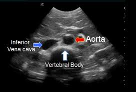
Non-traumatic retroperitoneal hemorrhage:  Most patients with non-traumatic retroperitoneal hemorrhage are on anticoagulation and their INR’s are usually in the therapeutic range.   Dialysis increases the risk in anticoagulated patients because these patients receive heparin and their platelets are dysfunctional.    Cardiac caths can iatrogenically cause retroperitoneal hemorrhage.   The posterior wall of vessel can be punctured.  Retroperitoneal surgery can also cause retroperitoneal bleeds.    Retroperitoneal hemorrhage can also be due to rupture of vascular aneurysms.   Retroperitoneal bleeding can cause abdominal compartment syndrome.  This adversely affects renal function, limites respiratory function ,  impairs vascular flow to bowel and compresses  the IVC reducing preload.  If abdominal compartment syndrome is present, the patient needs to go to the OR.  CT is best test to identify retroperitoneal bleeding.   Management of non-traumatic retroperitoneal hemorrhage is fluids, prbc transfusion, reversal of anticoagulation.  Surgery has the risk of removing tamponade effect and increasing bleeding.  IR embolization can be used for hemodynamically unstable patients.   Unstable patients in which IR is unsuccessful or in patients with abdominal compartment syndrome should go to OR.

Harwood comment: SICU nurses know how to measure abdominal compartment pressure with specialized Foleys and arterial pressure monitor.  Call up to the ICU for the special foley and possibly the expertise of the ICU nurse.    Things to do in the ER are drain bladder, place NG in stomach, and drain ascites to decrease intra-abdominal pressure.      Elise comment: There is a specialized foley made by Bard that has a pressure manometer for measuring abdominal compartment syndrome.   You have to identify your sickest patients and mentally visualize that you are moving those very sick patients through the system while continually following up to make sure tests, procedures, and consults are getting done.

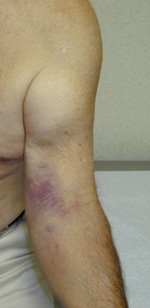
Right side retroperitoneal hematoma


There are five signs suggesting retroperitoneal bleeding. They generally appear 24 hours after bleeding starts, occurring when blood extravasates along ligamentous connections between the retroperitoneal space and skin surface.

1. Grey-Turner Sign: Named for the surgeon who identified it in 1920; a bluish hematoma across the lateral abdominal wall when blood from the pararenal space leaks along the quadratus lumborum.

2. Fox’s Sign: Described by Dr. Fox in 1966 in two patients, (ruptured AAA and pancreatitis); ecchymosis over anteromedial thigh secondary to blood seeping along the fascia of psoas and iliacus.

3. Cullen’s Sign: Dr. Cullen (gynecologist), described this in 1918 as a sign of a ruptured ectopic pregnancy; bruise around the umbilicus from retroperitoneal blood tracking along falciform ligament.

4. Bryant’s Sign: Dr. Bryant initially described this sign; ecchymosis of the scrotum from blood tracking down the spermatic cord.

5. Stabler’s sign: Ecchymosis over the inguinal ligament

Z. Dezman  University of Maryland


Advanced Wound Management Workshop


Conference Notes 12-17-2013

Happy Holidays Everyone!  No Conference next tuesday. 

Cash                Trauma Lecture

47 yo male struck by an auto.  Vitals were stable and no injuries to head/chest/abdomen.   Pt did have severe mangling injury to right lower extremity.

Control hemorrhage with direct pressure or tourniquet.   Tourniquets have been shown to improve survival.  Transient nerve palsy can be a complication from a tourniquet.

Don’t miss a second injury.  The patient was found in surgery to also have an open fracture and tendon injuries in the left lower leg.    Harwood comment: To avoid being distracted by a horrific injury, cover up the injured limb with a sheet or blanket and focus on the ABC’s.  After ABC’s addressed, uncover severe injury and address it.

Testing the peripheral nerves of the upper extremity: Rocks-Paper-Scissors.    Rocks checks the median nerve,   Paper checks the radial nerve, and scissors checks the ulnar nerve.    Girzadas comment: You also need to have them do the  OK sign as well to check the anterior interosseus nerve.

ABI’s check for arterial injury:  if <0.9 you need to do some imaging to assess for vascular injury. 

Open fracture increases the risk of osteomyelitis and limb loss.   Start antibiotics at time of initial evaluation.  Irrigate with NS, apply moist sterile dressing, spint and update tetanus.  These have to go to the OR within a few hours.

Open Fracture Grading System

Harwood comment: This system is not intuitive for the average ER doc.  This injury was severe and patient had to go to the OR no matter what the grade.  If the consultant is giving you a hard time about your  description of the injury, text them a picture.

Factors that increase risk of limb loss: delay in revascularization, blunt trauma, high velocity bullet, older patient, shock, limb ischemia, resource limited environment, and multi-casualty event.

By definition, the mangled extremity has 3 of 4 anatomical components injured.  (bones, vessels, nerves, soft tissue)

Mangled Extremity Score of 7 or higher suggests a low likelihood of limb salvage. With severely mangled extremities, functional recovery of the patient is more likely with amputation.

Protecting amputated digit or extremity in the ED: Wrap part in saline soaked gauze.  Place wrapped part in plastic bag.  Place plastic bag on ice in a cooler or other container.  Keep the container holding the amputated body part with patient on their cart so body part  is not lost.  

The patient in this case elected to attempt slavage of his limb despite orthopedic recommendation to amputate.  51 days later pt underwent BKA following multiple surgeries and complications.

Case 2: 60 you male with leg trapped under car when the car slipped off jack.  Pt looked well and limb seemed to be ok as well,  but pt was admitted to observe for rhabdomyolysis.    Pt did develop rhabdomyolysis.   CK is usually 5X normal with rhabdo.   Pt’s can be hypovolemic and develop hyperkalemia.  Patients can develop renal failure.   Follow K+ to avoid severe hyperkalemia and possible cardiac arrest.  Treat with IV fluids as soon as possible.  Even start IV fluids in the field prior to limb rescue from crush mechanism if possible.   This optimizes hydration of kidneys prior to  CK and potassium release.

Compartment syndrome: perfusion is compromised when compartment pressure gets within  30mm/HG of diastolic BP (Delta pressure).    Normal compartment pressure is 0-8mm/HG.  Pt will need fasciotomy if delta pressure is less than 30.


Febbo          Management of the Agitated Patient

Violence is usually preceded by anger, resistance, and verbal confrontation but still can be difficult to predict.  Risk is increased with males, substance abuse and previous history of violence.

Attempt de-escalation with clear, calm speech, offer food, agree with patient as much as possible, and remove aggravating factors from situation.

Physical restraint is a bridge to chemical restraint.

Search for underlying causes of agitated  behavior.  FIND ME mnemonic:   functional, infectious, neuro, drugs, metabolic, endocrine.

Quick Screening for organic disease: Disorientation, abnormal vitals, clouded consciousness, age over 40 with no prior psych history.

You ideally should have 5-6 people to physically restrain a person.   Leg restraint should be tethered to opposite side of cart to prevent  pt from kicking  leg laterally.

Chemical restraint:  Much  discussion about different approaches to chemical restraint.   All faculty felt Geodon (ziprasidone) and ativan were the most common combination for sedation but Brian made the point that this combo is not recommended by the most recent ACEP guideline.  This was based on expert’s  concern about combining atypical antipsychotics and benzos.  (A quick/very imperfect google search  could not find a direct recommendation against this combo, so I don’t know what the right answer is about this combo.  Be careful to monitor for over sedation and arrhythmias if you use it, I guess. Attendings' anecdotal experience has been that it is safe. )    Geodon is well known to cause prolonged QT (don’t give it IV/more likely to cause prolonged QT).   Benzos are a class D in pregnancy.   Everyone felt succinylcholine was virtually never needed as a last resort unless you don’t have enough security support in a very violent patient.  Elise felt ketamine  4mg/kg im is now the last resort drug instead of succinylcholine.     Ketamine is a bridge to getting IV access and dialing up other antipsychotics.

Excited delirium syndrome:  Usually male, underlying psych disorder and using sympathomimetic drugs.  Pt’s are severely agitated and have super-human strength.  Hyperthermia, hypovolemia, acidosis and rhabdomyolysis are frequently present.    High risk for death.   Terminal rhythm is most commonly brady-asystolic.

Attending discussion about the medication choices for chemical restraint.  They all have downsides.

Drug     

Dose (mg)     

Onset (min.)     

Half-life     

Disadvantage     

Haloperidol  

 2-10 IM

 30-45

 20 Hours

 EPS

Droperidol 

 5mg IM      

 15-30

 20 Hours      

 FDA Warning

Risperidone 

 2mg ODT 

 30-60 

 20 Hours

 Oral

Olanzapine 

 5-10 mg IM

 15-45 

 30 Hours 

 CNS Depression 

Ziprasidone 

 10-20 IM 

 15-30 

 2.5 Hours 

 QT prolongation 

Aripiprazole 

 5.25-15 IM

 30-45

 75 Hours 

 Cost

Lorazepam 

 2-4 IM 

 30-60 

 14 Hours

 Sedation 

     * Source: “Physician’s Desk Reference” www.pdr.net

ACEP Article resource

DenOuden                   Progressing from Resident to Attending

Identify what clinical situations you fear , write those down , and then specifically work on those areas.

Identify the weaknesses of the system of your new ER.    Prepare for those weaknesses.

Issues identified by recent grads: No such thing as bypass at other hospitals.  You will have to respond to codes in the hospital.  Some times there will be no videolaryngoscopy.  You likely will have to learn to work with midlevels.   Ultrasound is not available in every ED.  You will have to transfer more patients.  

Crash Cart medicine: 

Use IO when you need it.

Case 1: 22yo with severe asthma.  Diaphoretic, hypoxic, altered mental status slightly.     Altered mental status is due to hypoxia, hypercarbia, and exhaustion.  Bipap was started. Usual asthma meds given (albuterol, solumedrol, magnesium)  Consider IM epi as well prior to IV access (2 epipens gives optimal dose).  Use passive oxygenation if you attempt intubation.  Put a high flow nasal cannula in place for all phases of intubation.  It significantly prolongs the time to desaturation.    Another strategy is Delayed sequence intubation using Bipap and ketamine (low dose 0.5mg/kg). Goal is to improve conditions (full oxygenation/lowest possible pCO2) prior to intubation.   You can combine both these strategies and use a high flow nasal cannula under the bipap mask.

Case2: 66 yo female with fever, pneumonia on CXR and elevated lactate.  Sepsis management begun.   Pt coded 6 hours later.  Ct showed diffuse colonic thickening.  Stool showed C-Diff.   Should we consider C-Diff in all older patients with abdominal pain, fever, diarrhea?  Should we treat empirically in high risk patients?   Send stool for testing!

Tricks of the Trade:  When getting the rectal temp also do a hemoccult and put some stool in a cup to send to lab.   You can add sterile saline to hard stool to soften it up for the lab so they can run the test.  Christine comment: there are patients who are C-diff positive who are not clinically ill with diarrhea so you have to use some clinical judgement if you are using this saline trick.

No sedation shoulder reduction:  Cunningham technique with shoulder massage.  You can also do intraarticular injections of lidocaine.

Ring removal: Wrap elastic tourniquet around finger for a few minutes, have pt hold their hand above their head.  It frequently removes enough edema to get ring off finger.

Painless abscess I&D: Aspirate fluid from abscess first to relieve pressure. Then inject lidocaine.  Let is take effect for a few minutes and then I&D.

Ntg spray has 400mcg per spray.  Good way to get quick ntg in a CHF patient prior to having IVNTG drip started.

Case 3: 70 yo male, hypotensive in extremis.  Diagnosis is sepsis.   Crash cart pressor initial dosing:  EPI 1mcg/min, Norepi is 10mcg/min, Phenylepherine is 100mcg/min.  A milligram of any of these drugs diluted in one liter of saline give you 1mcg/ml.

Case 4: 34 yo female bit by a bee and develops anaphylaxis.  Epi is the key.   Epipen is the place to start and most of the time will be all you need. If you need an epi drip; 1mg of epi in a liter of saline makes 1mcg/ml.   Give 2-4 mcg/min.    I drop per second is 3mcg/min.     

Carlson                       Toxicology Case Conference

Suicide rates decrease during the holiday season.

Unintentional toxic exposures to young children increase during the holidays.

Case 1: Unresponsive 3yo found on living room floor.  Dexi=20.   Patient had ingested eggnog spiked with etoh.   Pediatric etoh exposures increase during the holidays.  Kids are very sensitive to etoh.   Hand sanitizers have been reported to cause severely high etoh levels in kids.   One adult ingested hand sanitizer and had an etoh level of 800!   Hypoglycemia is common with etoh ingestion in kids due to their limited glycogen stores.

Case 2:  2 people at a party with nausea and vomiting after eating creamy appetizer.  It turns out it wasn’t due to food.   They were CO toxic  because the flue of the fireplace was not opened properly.  Elise comment: Use the finger probe device or get a venous blood gas to check CO level.  No need to do an arterial blood gas.   Andrea comment: Check the most symptomatic person first. If they are ok everyone else is likely to be as well.

Case 3: Poinsettias ingestions are very unlikely to be life threatening.   They can cause GI upset but are not lethal.   You would have to eat hundreds of  leaves to get sick. Andrea said there is no risk of lethality from ingestion.    There is a latex analogue in poinsettias that can cause allergic reaction in latex sensitive.  

Case 4:  European Mistletoe berries can be toxic.  American berries are not toxic.    No reported cases of death from ingestion of European mistletoe.    Mistle toe berries (European and American) can cause gi upset and mild neuro findings.  Not considered lethal.

Case 5:  Holly leaves are non-toxic.   Berries can cause GI upset and drowsiness. 

Pretty much all Christmas plants are not going to cause any serious toxicity.

Case 6: Kid ate a Styrofoam gingerbread ornament and some lights.   Christmas tree bubble lights contain methylene chloride.  Methylene chloride gets metabolized to CO.  Kids can get pretty high CO levels from methylene chloride.  Methylene chloride is also found in decorative spray snow.  

Case 7: Kid was drooling after playing by Christmas tree.  The child was found to have an esophageal ornament impaction.  Kids will also swallow button batteries.  These need to be removed in a few hours.  Recently battery companies have made improvements of batteries.   They are smaller and pass the pyloris more reliably.   The battery seals are much better and won’t leak for weeks.   Esophogeal batteries need to come out in a few hours.  In the stomach and bowel, kids can be observed.    Bill Schroeder comment: If a child ingests rare earth magnets, they can cause perforation of the bowel.  They frequently  require surgical removal.    In general  foreign bodies  get stuck at C6 chricopharyngeus,   T4 aortic arch,  GE junction T10-11.

Fort/Watts                         Oral Boards

Case1  Epistaxis due to thrombocytopenia secondary to leukemia.  WBC=47   Plt=16.  Management: Platelet transfusion given. Nasal packing placed.  T&C in case prbc transfusion is needed.  

Case2   Subdural hemorrhage on Pradaxa.   Management:  FEIBA, vitamin K and neurosurgical consultation.   Surgery is indicated for big subdurals (5mm), mass effect, severe symptoms.  There was a discussion about management of Pradaxa bleeding that is life threatening.  There is a lot of controversy on this topic.  However it is recommended to give high dose FEIBA 25u/kg.   Can give factor 7 as well.   Vitamin K is not indicated with Pradaxa bleeding.  This is different than the warfarin coagulopathy protocol which recommends vitamin K in addition to FEIBA for warfarin coagulopathy.

Case3  Cyanotic infant.  Pt with recent URI symptoms and cough.   DX=Pertussis.  CXR is usually normal.  Treat with macrolides or bactrim.  Kids less than 6 months can have apnea and bradycardia.   Labs can show a high lymphocyte count.

Conference Notes 12-10-2013

Please consider giving to the ED “Adopt a Family Fund”  by 12-17

Thanks to Christine Kulstad for her help with this week’s Conference Notes.  She better watch out or she’s going to become a regular contributor!

 Fakhouri      Elbow Injuries

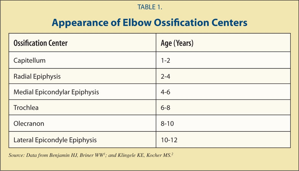
Pediatric ossification centers of the elbow- use the mnemonic CRITOE (capitellum, radial heal, internal [medial] epicondyle, trochlea, olecranon, lateral epicondyle) ossify at ages 1-3-5-7-9-11 in girls, add a year in boys.


CRITOE

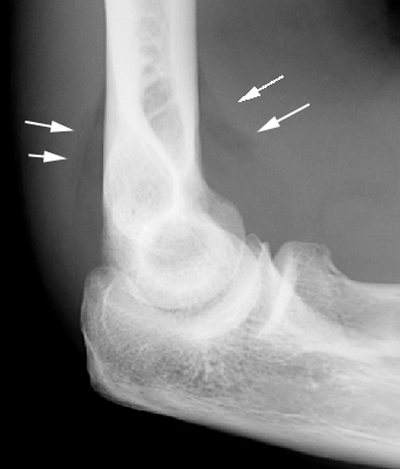
Posterior fat pad is always abnormal. The posterior fat pad is part of the elbow joint, it exists within the deep olecranon fossa normally. When fluid accumulates in the joint, it pushes it out and shows up on xray. Anteriorly, the fossa is much more shallow so the fat sometimes shows up on xray in a normal joint.

Posterior fat pad=fracture

Distal humerus fractures- supracondylar, medial epicondyle, lateral condyle.

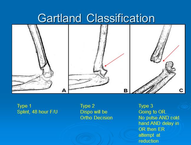
Supracondylar fractures- if non-displaced can be splinted at 90 degrees of flexion and discharged IF you arrange ortho f/u in 2 days and have a reliable patient/family. Any displaced fracture needs surgical intervention. Generally not a good idea to reduce in the ED- should be done by ortho in the OR or with fluoroscopy (c-arm).


Gartland Types of Supracondylar Fractures

Splint the displaced supracondylar fracture in the position of comfort for the patient.  Do not force the patient’s elbow into 90 degrees of flexion.

In subtle supracondylar fractures, the anterior humeral line will bisect the anterior 1/3 o the capitellum instead of where it normally bisects the junction of the anterior and middle 1/3’s of the capitellum.

Normal alignment of the anerior humeral line


Supracondylar fracture with posterior fat pad and abnormal anterior humeral line

Heterotopic bone formation can develop around the elbow following supracondylar fractures.

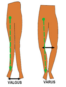
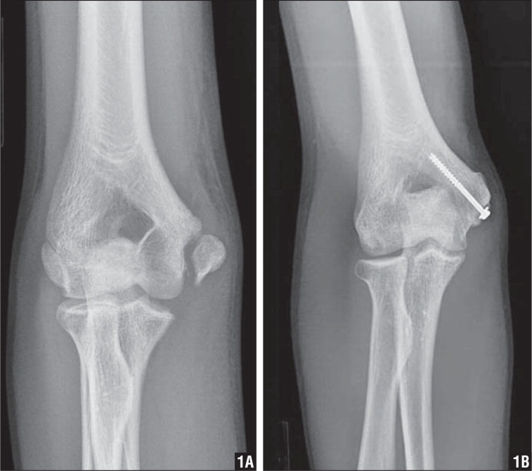
Lateral condyle fractures usually occur when a patient falls on the arm and the arm is in varus position.  The tendon can avulse the lateral condyle off the distal humerus.   If these injuries are not repaired you develop a valgus deformity and the patient can develop an ulnar neuropathy

There was a discussion of the terms varus and valgus.   Varus deformity describes the distal forearm pointing medially toward  the midline.   Valgus deformity describes the distal forearm pointing lateral from the midline.   Mnemonic is to remember the L in valgus reminds you that the distal forearm is pointing laterally from the midline of the patient’s body.


Varus and Valgus


Varus and Valgus

Medial condyle fractures  

Normally the medial condyle should be just below the Shenton’s line.  If it is not in that location  you have to be suspicious for medial condylar fx.

Medial Condyle fracture

Radial head fractures

Splint these injuries initially.  Ortho will mobilize them early if non-displaced.    2mm or more of step off or separation of radial head fractures requires surgery.

Olecranon fractures

Any step off or separation of more than 2mm requires surgery.

Elise comment: Of all the above fractures, supracondylar fracture is the most high risk injury that can result in deformity, compartment syndrome, or neurovascular   injury.  Dr. Fakhouri agreed.

Elbow dislocation

Multiple attempts at joint reduction injures the cartilage.   Dr. Fakhouri prefers the prone position technique.  If you see an elbow dislocation in kids look for an associated fx.

Distal biceps tendon ruptures always require surgery.  Pt will feel a pop or a snap of a rubber band.  These occur mostly in patients around 30-40 yrs of age.   Older patients will have a proximal tendon ruptures.  Proximal tendon injuries don’t need surgery.     Both types cause a popeye deformity of the biceps muscle.   In proximal ruptures, the muscle will bunch up distally.  In distal ruptures, the muscle will bunch up proximally.   Treat with a sling in the ED and refer to ortho for surgery.   Harwood comment: why not surgically repair proximal ruptures.   Fakhouri response: The outcome is the same with non-operative management .  Patients actually get back to work more quickly without surgery for proximal injuries.   To be clear, distal ruptures all require surgery.

Distal biceps tendon rupture

Erickson    M&M

Elderly  female with history of DM, HTN, COPD presented with fever, cough, and SOB.   Lung exam demonstrated bilat wheezing.  Heart was irregular and tachycardic.  

At outside clinic patient received med neb and steroids.   CXR showed possible pneumonia.

In ED patient received med nebs and IV antibiotics.  Labs showed mildly elevated Troponin and minimal bump in creatinine.    EKG showed new onset afib.  Heart rate was controlled with diltiazem.   Heparin was started.  Patient was admitted to floor.

While in hospital, pt developed a left rectus sheath hematoma.  With that finding, heparin and warfarin were discontinued.   Pt coded on day 2 of hospitalization.  She was resuscitated.  By  Day 3 her abdomen was severely distended and had intra-abdominal fluid.  It was hypothesized that pt may have had further bleeding in her abdomen.  

Anti-thrombotic therapy for AFIb:  Use CHADS2 score for risk stratification

CHADS2

Linsey identified a potential system based error with the heparin order set.

Harwood comment: The higher bolus given in the ED is very unlikely to have caused this patient’s outcome.   There was generally faculty agreement that the initial heparin bolus was not the cause of the patients death.   The decision to anticoagulate is appropriate based on the CHADS2 score.   Maggie comment: Unfortunately a high risk CHADS2 score also carries a high risk for bleeding from warfarin.

Take home point:  Double check your orders.   Verify the patient, the dose.  Discussion strayed into other examples of medication errors in kids and adults.  One further point,  be careful to not be duplicating someone else’s order.

Elise comment: Strong plug for PediStat App for your smartphone.   It is a great App for getting ped’s resuscitation drugs right.    Harwood comment: We should probably change the Order Set wording for heparin to “Cardiac”  and “Non-Cardiac” dosing.  Elise comment: Be sure to know dosing of common drugs so you have some reference point to identify incorrect dosing.   Harwood comment:  This is an elderly woman with influenza who had an nstemi and afib.  She then develops a rectus sheath hematoma from coughing and/or heparin.   If is totally unclear what was the cause of death.

Lambert     Study Guide  Resuscitation

Best predictor of survival in setting of cardiac arrest is time prior to resuscitation.

In setting of hyperkalemia, get EKG first to identify cardiac effects of hyperkalemia.

Why use cuffed ET tubes in kids: higher probability to correctly size the ET tube at first intubation attempt.   New technology  cuffs are also less likely to produce pressure injury.

Mnemonic for sizing peds tubes 1-2-3-4

1x= Et tube size is (age/4) + 4   ,   2X =NG size,   3X = Depth of ET tube,   4X =chest tube size.

Atropine is no longer indicated for PEA

Antiarrythmic drugs are not supported in the literature for ventricular arrhythmias in the setting of hypothermia.

Child pulled out of a lake with weak, slow pulse.  What is initial management?   CPR and ventilation.

Avoid adenosine in pediatric patients with irregular wide QRS tachycardia.   Gotta suspect WPW in this patient and procainamide is a better choice.

Bradycardia- maximum dose of atropine is 3 mg

In witnessed VF, the first treatment is CPR. Defibrillation should be done as soon as is available.

Monophasic defibrillators provide more energy than biphasic. Biphasic ones deliver the energy more effectively, so can use less.

After treating paroxysmal SVT with adenosine, the recurrence rate is 50-60%. This is based on one small study and attending experience is that the rate is not this high.

Cuffed pediatric ETT are recommended because the seal is better so airway protection is better. It can be harder to pass the tube through the cords.

Disposable end-tidal CO2 monitors are very (reported at 100%) sensitive in non-arrest patients, and can detect CO2 >2%.  

A patient in persistent VF, consider doubling the energy for defibrillation (Lambert used 2 defibrillators at the same time [or 0.5 s after]  to successfully get someone out of Vfib after 30 shocks. There was also an EP case series where it worked >50% of the time , in about 18 pts).

Adenosine is dosed 0.1 mg/kg initially, the second dose is 0.2 mg/kg. Remember, it should be pushed very quickly and followed with a saline bolus. The half-life is seconds.

Ventilator settings for a post-arrest patient should be tidal volume of 6-8 ml/kg and rate of 10-12 breaths per min. Use lower lung volumes for patients at risk of ARDS.

Atropine should be used with caution in a patient with Mobitz type II AV bloc with associated BBB due to a potential increased risk of converting to 3rd degree AV block.

The rhythm with the highest chance of survival for an out of hospital arrest is VF (other choices were PEA variants)

Epinephrine use in cardiac arrest is based on animal studies and anecdotal clinical evidence.

The most common adverse effect of IV amiodarone is hypotension- likely due to the diluent. Pulmonary fibrosis is a common long-term side effect.

Procainamide is dosed at 20-50 mg/min to a max of 17 mg/kg, QRS widening, conversion of rhythm, or hypotension.

Pediatric CPR should avoid excessive ventilation, compress the sternum 1.5 inches in infants and 2 inches in children, have a compression rate of 100/min

The most common cause of mortality in AMI patients is cardiogenic shock.

The objective of CPR in PEA/asystole is to delay tissue death until a reversible cause can be found.

In a choking victim, if they can speak one word and cough- you should only encourage spontaneous efforts. When they can’t breathe, you intervene.

When using the ETT for medication administration, increase the dose 2 to 2.5 times.

When you put your defibrillator to the SYNC mode, it delivers energy with the sensing of the QRS complex.

Respiratory distress in peds is best described as increased work of breathing. Signs of respiratory failure includes apnea, bradycardia, cyanosis.


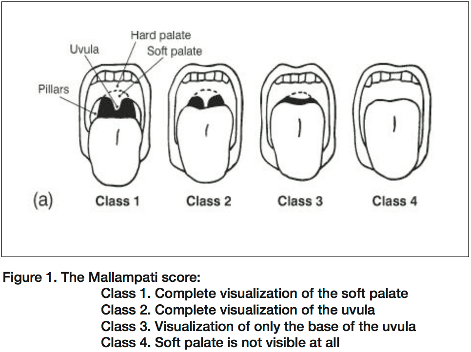
Mallampati score

Balogun      Congenital Heart Disease

Cyanosis + Tachypnea + Shock + <1 month = left obstructive CHD until proven otherwise

50-60% of CHD diagnoses after hospital discharge, 30% present to ED in first month of life for 1) poor feeding, 2) cyanosis 3) tachypnea/tachycardia or 4) shock

Ductal dependent lesions present in the 1st month.

               -Right heart lesions- pulmonary flow depends on DA- include Tetrology of Fallot, tricuspid atresia

               -Left heart lesions- systemic flow depends on DA-  include aortic stenosis, hypoplastic left heart,  

 The ductus arteriosos usually closes 12-15 hrs after birth due to increase O2 and decreased prostaglandin. It is permanently closed by about 3 wks. Delayed closure occurs due to decreased PaO2 in right sided lesions. Baseline O2 sats 75-85%, which drop as ductus closes. The ductus stays open in left sided lesions due to increased pressure. When it closes, kids present in shock, mild cyanosis

Left-right shunting lesions present between 2 and 6 months- VSD, truncus arteriosis. For the first 2-3 months, pulmonary and systemic vascular resistance are equal. At 2-3 months, the pulmonary resistance falls which triggers the shunting and symptoms.

Kids with CHD will also have problems with concurrent illness or metabolic derangements or during physiologic anemia of newborn period.

ED management for CHD kids- Intubate early, keep O2 sats at 80-90%, optimize systemic perfusion.

Airway- anticipate adverse events, Pretreat with atropine 0.01 mg/kg (min 0.1 mg).

               -Ketamine- avoid as it can cause pulmonary HTN

               -Benzodiazepines- avoid as it can cause hypotension

               - Propofol- avoid as it can cause hypotension

               -Etomidate- yes!

               - Succinylcholine- avoid if possible

               -Rocuronium is preferred 1 mg/kg, fast onset


Breathing- keep sats low (80-90%). There is a technical explanation involving Fink’s principle that has to do with balancing flow to pulmonary and systemic beds, but just remember the number

 Ventilator settings- Assist control, tidal volume 6-8 ml/kg, rr 20-50, peep 5, FiO2 start low and titrate up

 Give PGE1- life saving! Re-opens and maintains patency of ductus arteriosis. Start at 0.05 mc/kg/min and titrate up every 15-20 min until desired effect.

Circulation- Target MAP of at least 40.  Bolus with 10 cc/kg. If fluid seems to make them worse, then add dopamine infusion  at 5 mcg/kg/min and titrate up to a max of 10 mcg/kg/min.  Second line is epinephrine infusion (vs all other situations we use pressors).

 Work-up- ABG, CXR, EKG, blood cx, trop, bnp, tsh, cortisol, coags, T&S, CMP, CBC.

Start empiric antibiotic therapy. Give bicarb while trying to restore perfusion (1-2 mEq/kg).

 Watch for hypoglycemia and hypocalcemia and treat if they occur.

 Regulate the temperature to normal. NG, Foley to monitor UOP, arterial line if able

 Resuscitation is critical for post-operative course. Patient must be transferred to center capable of correcting lesion with PICU/PSHU.

 Tet spells- in kids with mild TOF. They present with Tet spells, cyanosis treat with COINSPIRES

C- comfort- parents hold child, knees to chest position

O-oxygen

IV fluids- 10 cc/kg

Narcotics- morphine 0.05 -0.1 mg/kg

Sedation- more morphine

Phenylephrine- 10-20 mcg/kg/dose

Intubate (last Resort)

Esmolol- 500 mcg/kg load over 1 min, then infuse 50-300 mcg/kg/min

Harwood comment: the pathophysiology/pharmacology of tetrology is hard to understand.  I know I need knee-chest position, oxygen, morphine and a cardiologist.  The rest, I don’t know.  

Elise comment: If you are giving prostaglandin,  be prepared to intubate.  Prostaglandin can cause apnea.   Pulse ox differentials in upper and lower extremities suggests coarctation.

Give prostaglandins for infant with cyanosis, murmur, weak pulses. (any two)

Conference Notes 12-3-2013

Kessen/Kerwin      Oral Boards

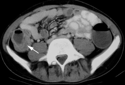
Case 1.  Adult male with neutropenia due to chemotherapy presents with abdominal pain and sepsis picture.   Diagnosis was typhlitis, otherwise known as  neutropenic  enterocolitis.   There is compromised integrity of bowel wall that leads to sepsis.   Treatment is hemodynamic support and big gun antibiotics.


Typhlitis

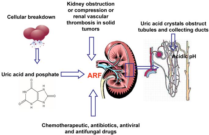
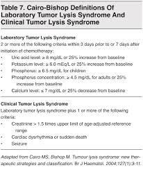
Case2.  Pt  receiving chemotherapy for lung cancer. Pt had a seizure.   Labs showed an acute increase in Creatinine, hyperkalemia, hyperphosphatemia, hyperuricemia, and severe hypocalcemia.   Dx was Tumor Lysis syndrome.    Management is hydration, manage hyperkalemia, give calcium.  Be cautious with bicarb because it can increase precipitation of calcium.  Rasburicase is a medication that can lower uric acid levels.  Kerwin states this is the money drug for Tumor Lysis Syndrome.


Tumor Lysis Syndrome

Tumor Lysis Syndrome

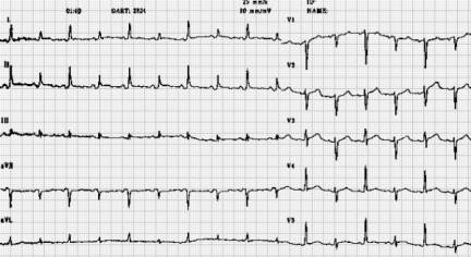
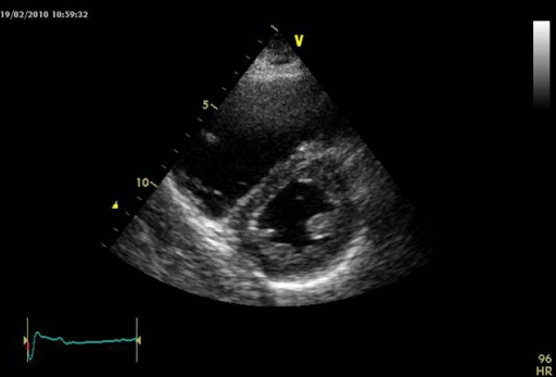
Case3.  72yo male presents with dyspnea.   90/65   T=37.2  HR=116   R=25  Glucose=90   Cachectic.  Hx of COPD and mesothelioma.  Ultrasound showed pericardial effusion.   EKG showed electrical alternans.  Pt had malignant pericardial effusion causing tamponade.   Pericardiocentesis was performed with improvement in clinical condition.


Electrical Alternans

Elise comment:  Great set of cancer related oral board cases.  There is a really nice reinforcement of the management of cancer emergencies on a recent EM Rap.

McGurk        Billing and Coding

Cindy Chan comment: You have to be careful with template charting to make sure there aren’t unintended  findings automatically listed on the chart.   McGurk response: We have seen charts in which elderly female patients were noted on ROS to deny scrotal edema.  Felder comment: We have seen stroke case charts with ROS for neuro  listed as negative.  So be careful with template charting.

The line “reviewed as documented in chart”  is not helpful for billing without a date listed as to what date of records were reviewed.   Harwood comment: We need to change the template to say “reviewed as documented in patient summary screen”.

For an EM level 5 you need PMH/Surg HX and either past social hx or family hx. You don’t need both social and family hx.  Harwood comment: Just ask/document   No asthma, lives with parents or HTN and smoker.     McGurk comment:  Lives independently,  lives in a NH,  assisted care are all usable comments for social hx.

There is typically a 5 day lag between when you see the patient and when the coders get to the chart.  Get you charts done and signed within 5 days.   Girzadas comment:  Get your charts done in less than 5 days.   There’s an old Joe Wood comment, “The longer you take to finish your charts the more it starts to look like fiction.”

Chan   Stroke Core Measures

The important core measures for stroke in the ED are:

  1. Thrombolytic therapy given when appropriate,
  2.  Patients with afib/aflutter receive anticoagulation therapy,
  3.  ASA or other anti-thrombotic therapy  by day 2.

Harwood comment: Many times there may be concerns by neurology about starting heparin in the ED. So giving heparin in the ED for afib and stroke may not be indicated in many cases.    Possibly documenting in the chart the contraindication or relative contraindication for heparin may help meet the core measure. 

Beckmeyer     Safety Lecture   Improving the Care and Safety of Patients Boarding in the ED

Strategies to improve the care/safety of boarding ED patients:

Increasing face to face communication in the ED between ED physicians and Intensivists

Ask intensivists to round in the ED in the AM

Bedside rounding

At sign out be sure antibiotics/meds/labs are ordered

Improved order sets for boarding patients

Push back on soft ICU admits and try to admit to step down or tele

Downgrade patients to floor admits whenever possible

Use the SVTU or SINI as an area to overflow patients

Tranfer to outside ICU?  

Urumov/Harwood comment:  Transfer to outside hospital ICU can be done but has risks and negative poltical/financial consequences.   It can be done on a case by case basis for ICU players that are stabilized and not requiring super high level of care like 3 pressors.    C.Kulstad comment: Be really careful about transferring out ICU players.  You are not transferring to a higher level of care.  It is really the same level of care so if patient crumps in route you are responsible for that decision.

5W step down is basically an ICU without resident coverage.   C. Kulstad comment: 5W doesn’t have the same nursing staffing as does an ICU.    Staffing ratio is 3:1 on 5W.    SVTU is SINI overflow with no resident coverage.

ACMC clinical tools on the web has a parenteral administration guide.  It will tell you what drips can be managed on what unit.  

DKA must go to an ICU.   Non-DKA on an insulin drip can go to any unit.   Maletich comment:  Endocrine consults can help with getting access to non-ICU settings for non-DKA patients.

The SSU can take 1-2 ICU level patients.   Maletich comment: May be a good unit to put a head injury patient with a minor finding on CT head who needs observation.

Remember to report errors/near misses.    Go to Advocate homepage>> culture of safety >> safety event reports forms.

For unattached ICU admits,  the admitting/attending physician should be the on-call  Intensivist.   The Adult Medicine Attending on call should not be listed at all for ICU admits.

Suter                 AAEM  Guest Speaker,    Pediatric Trauma “Why Kids are Different”

From a physiologic stand point, kids under 8 years of age are different than adults. 8 years of age seems to be the cut off that really differentiates kids from adults from a physiology perspective.    Kids over 13 are really like little adults.

Physiological differences in kids: Force is distributed to a smaller area.  Greater relative body surface area results in greater heat loss.  Smaller and more anterior airway.   Greater airway resistence in kids.  Relatively larger head to body ratio.  More flexion and extension ligamentous injuries due to head size.  Organs are more anterior and less protected making them more vulnerable.  Greater capacity to compensate for shock. Salter harris fractures.  More sensitive to radiation.  Infants are nose breathers. 

During intubation of young child, don’t put pad behind head.  It puts the neck in flexion and limits airway visibility. Miller (straight) blade is preferred in kids.  Atropine is indicated in kids less than 8 to prevent bradycardia during intubation.

Pulse is a more reliable guide to cardiovascular instability than BP due to kids’ ability to compensate for volume loss.

Don’t be hesitant to place an IO line in patients with difficult venous access.

Leading cause of death is head injury, but 90% of head injuries are minor.


Glascow Coma Score for Kids


C. Kulstad sent out the PECARN Head injury Rules to everyone. This is a synopsis image. 

Cspine injuries in kids <8yo are more likely to be high cervical injuries above C3.


Pseudosubluxation

Kids with head/neck injury that  have paresthesias in the extremities and have  negative c-spine films. They need an MRI to rule out SCIWORA.

Harwood question: At what age cutoff do you need to start worrying about aortic injury due to deceleration injury?   Response:  somewhere around 8yo.  Harwood comment: I was thinking somewhere around 12yo.  Both agreed literature was scant on this issue.

FAST exam is not as reliable in kids than in adults.   Lam comment: I still feel FAST has utility to identify serious injury in the injured child.   Response: Agree, just remember if no abnormality is identified it doesn’t mean there is no injury.

Transfer decisions to Trauma Center can be aided by Pediatric Trauma Score.  Score less than 8 indicates need for transfer to Trauma Center.

Elise comments: "Cuffed Versus Uncuffed Endotracheal Tubes

Both cuffed and uncuffed endotracheal tubes are acceptable for intubating infants and children (Class IIa, LOE C). In the operating room, cuffed endotracheal tubes are associated with a higher likelihood of correct selection of tube size, thus achieving a lower reintubation rate with no increased risk of perioperative complications.88,–,90 In intensive care settings the risk of complications in infants and in children is no greater with cuffed tubes than with noncuffed tubes.91,–,93 Cuffed endotracheal tubes may decrease the risk of aspiration.94 If cuffed endotracheal tubes are used, cuff inflating pressure should be monitored and limited according to manufacturer's instruction (usually less than 20 to 25 cm H2O).


In certain circumstances (eg, poor lung compliance, high airway resistance, or a large glottic air leak) a cuffed endotracheal tube may be preferable to an uncuffed tube, provided that attention is paid to endotracheal tube size, position, and cuff inflation pressure (Class IIa, LOE B)."


Harwood comments:  Here's the Peds blunt trauma rules. PECARN no longer recommends  that finding 20 RBC's in urine mandates CT abdomen/pelvis.

Ann Emerg Med. 2013 Aug;62(2):107-116.e2. doi: 10.1016/j.annemergmed.2012.11.009. Epub 2013 Feb 1.

Identifying children at very low risk of clinically important blunt abdominal injuries.

Holmes JFLillis KMonroe DBorgialli DKerrey BTMahajan PAdelgais KEllison AMYen KAtabaki SMenaker JBonsu BQuayle KSGarcia MRogers ABlumberg SLee LTunik MKooistra JKwok MCook LJDean JMSokolove PEWisner DHEhrlich PCooper ADayan PSWootton-Gorges S,Kuppermann NPediatric Emergency Care Applied Research Network (PECARN).

Source

Department Emergency Medicine, UC Davis School of Medicine, Sacramento, CA, USA. jfholmes@ucdavis.edu

Abstract

STUDY OBJECTIVE:

We derive a prediction rule to identify children at very low risk for intra-abdominal injuries undergoing acute intervention and for whom computed tomography (CT) could be obviated.

METHODS:

We prospectively enrolled children with blunt torso trauma in 20 emergency departments. We used binary recursive partitioning to create a prediction rule to identify children at very low risk of intra-abdominal injuries undergoing acute intervention (therapeutic laparotomy, angiographic embolization, blood transfusion for abdominal hemorrhage, or intravenous fluid for ≥2 nights for pancreatic/gastrointestinal injuries). We considered only historical and physical examination variables with acceptable interrater reliability.

RESULTS:

We enrolled 12,044 children with a median age of 11.1 years (interquartile range 5.8, 15.1 years). Of the 761 (6.3%) children with intra-abdominal injuries, 203 (26.7%) received acute interventions. The prediction rule consisted of (in descending order of importance) no evidence of abdominal wall trauma or seat belt sign, Glasgow Coma Scale score greater than 13, no abdominal tenderness, no evidence of thoracic wall trauma, no complaints of abdominal pain, no decreased breath sounds, and no vomiting. The rule had a negative predictive value of 5,028 of 5,034 (99.9%; 95% confidence interval [CI] 99.7% to 100%), sensitivity of 197 of 203 (97%; 95% CI 94% to 99%), specificity of 5,028 of 11,841 (42.5%; 95% CI 41.6% to 43.4%), and negative likelihood ratio of 0.07 (95% CI 0.03 to 0.15).

CONCLUSION:

A prediction rule consisting of 7 patient history and physical examination findings, and without laboratory or ultrasonographic information, identifies children with blunt torso trauma who are at very low risk for intra-abdominal injury undergoing acute intervention. These findings require external validation before implementation.


There was a discussion about Cspine CT vs Plain films in injured kids.  

Trale Permar comment: CT Cspine is more sensitive in kids. See graph below


Conference Notes 11-26-2013

Happy Thanksgiving to everyone!  Feast on this week's EM discussions. 

Steffenson     Status Asthmaticus

This year has seen a record high number of intubations for pediatric asthma at ACMC.  All clinicians need to treat asthma patients aggressively to avoid having to intubate them. 

Use a Pediatric Asthma Score to assess & trend patients during their ED stay.   Respiratory therapists will calculate the Woods Asthma Score on patients in the ED.


Woods Pediatric Asthma Score   If after initial treatment in the ED, the patient's score is more than 4 probably you should be considering an ICU admission. 

No benefit of xopenex over albuterol for status asthmaticus.  There incidence of adverse effects is not lessened with xopenex.    If you are getting too much tachycardia with albuterol, you can cut back the dose instead of switching to xopenex. 

D5.9NS with 20K is recommended for maintenance fluid in status asthmaticus.   Kids tend to be alittle volume depleted in status asthmaticus so maintenance fluids are usually indicated.  

Give atrovent Q2 hours for moderate and severe asthma.   It has been shown to decrease ICU admissions.  Elise comment: how long?  Response: Give atrovent Q2 hours until patient is off continuous med neb treatments.

Steroid dose should be  2mg/kg first dose either oral or IV.

Non-Invasive Ventillation should be strongly considered early for severe asthma.   It decreases muscle fatigue and it keeps alveoli and small airways open.   Harwood question: Can we give heliox through the bipap machine?  Response: yes

Magnesium IV 50mg/kg over 30 min.  (2gm max)  Can give Q6 hr doses or give 20mg/kg/hr drip.   Goal is serum mag level of 3-5.5.

Terbutaline  IV infusion is a second line therapy.

In the PICU, aminophylline is used if a kid is heading toward intubation.   The safety profile is better for kid than adults when using aminophylline.   Probably not a drug we will be using in the ED unless a status asthmaticus patient is in the ED for a prolonged time.

Elise question: How ‘bout using epi?  Response: Optimizing the other treatments mentioned above are probably preferred choices.  Especially giving more albuterol. 

If you have to intubate,  ketamine as a sedative may give you some added bronchodilation. 

In intubated patients, inhaled anesthetics can give smooth muscle relaxation resulting in bronchodilation. 

ECMO can be lifesaving as a last ditch modality.  83% of kids placed on ECMO survive.

Harwood question: Why do you guys get troponins in these patients.   Response: If the troponin is elevated we will stop terbutaline.  We would also more strongly consider intubation.

Frazer      U/S Guided Peripheral Nerve Block

Peripheral nerve blocks have the advantage of Giving  pain relief without the side effects, time, and monitoring that go along with procedural sedation.

Interscalene brachial plexus nerve block:   Can give anesthesia to upper arm, humerus, and elbow.  Position patient like you would for an IJ line.   Put U/S probe more laterally on the neck to identify the group of 3 nerve bundles (hypoechoic or honeycomb appearance on U/S).  Depth of needle puncture is similar to the IJ approach.  Inject 15-25ml of local anesthetic.    Elise question:  What can go horribly wrong with this procedure?    Response: Hematoma, pneumothorax, paralyzed diaphragm, carotid puncture.   Harwood comment:  Phrenic nerve comes off higher than the interscalene nerves but if you go high in the neck to avoid a pneumothorax you could accidentally anesthetize the phrenic nerve.

Femoral nerve block: This block provides anesthesia to anterior thigh, femur, and knee.  Position patient as you would for a femoral  line.   Using U/S you identify the the hyperechoic or honeycomb appearing nerve lateral to femoral artery.  Orient the needle in a lateral to medial direction and inject 25ml of local anesthetic  around the nerve.   There is a risk of hematoma and infection.       Harwood comment: Most studies used a technique where you had to deposit the local anesthetic underneath the inguinal ligament.   Harwood and Elise comment: Use bupivicaine with epinepherine to give longer duration of action for your peripheral nerve blocks.

Elise comments:    Excellent discussion about nerve blocks by Erin today.  I was thinking about the "3 in 1" nerve block when I referred to a more complete nerve block to help with more proximal hip fractures/hip dislocations.  By using a larger volume of anesthetic, and as Harwood mentioned, using some distal pressure to keep the anesthetic localized proximally, you can knock out the femoral nerve as well as the lateral femoral cutaneous nerve and obturator nerve.  Obturator nerve is important for prox. hip fractures/dislocations, and using larger volumes of anesthetic helps increase your chances of anesthetizing all 3 nerves.   Remember max dose of bupivicaine with epi is 3 mg/kg or 225 mg.  You shouldn't get in trouble if you use the 0.25% bupivicaine concentration.

From the web: 

Differences between Femoral Nerve Block and 3-in-1 Nerve Block

There are two main differences.

1. Volume of local anesthetic. For femoral nerve blocks, the volume of local anesthetic is generally 20 ml or less. For 3-in-1 nerve blocks, the volume of local anesthetic is 25-30 ml. This allows the local anesthetic to spread further in the tissue plane resulting in blockade of the femoral, lateral femoral cutaneous, and obturator nerve.

2. Slight alteration in technique. Once the needle has been placed in the correct area, pressure should be applied 2-4 cm below the injection site. Next, administer the local anesthetic. Applying distal pressure helps spread the local anesthetic to the obturator and lateral femoral cutaneous nerve, in addition to the femoral nerve.


Harwood provided reference: 

A comparison of ultrasound-guided three-in-one femoral nerve block versus parenteral opioids alone for analgesia in emergency department patients with hip fractures: a randomized controlled trial.

Beaudoin FLHaran JPLiebmann O.

Source

Department of Emergency Medicine, Rhode Island Hospital, The Alpert Medical School of Brown University, Providence, RI, USA. Francesca_Beaudoin@brown.edu

Abstract

OBJECTIVES:

The primary objective was to compare the efficacy of ultrasound (US)-guided three-in-one femoral nerve blocks to standard treatment with parenteral opioids for pain control in elderly patients with hip fractures in the emergency department (ED).

METHODS:

A randomized controlled trial was conducted at a large urban academic ED over an 18-month period. A convenience sample of older adults (age ≥ 55 years) with confirmed hip fractures and moderate to severe pain (numeric rating score ≥ 5) were randomized to one of two treatment arms: US-guided three-in-one femoral nerve block plus morphine (FNB group) or standard care, consisting of placebo (sham injection) plus morphine (SC group). Intravenous (IV) morphine was prescribed and dosed at the discretion of the treating physician; physicians were advised to target a 50% reduction in pain or per-patient request. The primary outcome measure of pain relief, or pain intensity reduction, was derived using the 11-point numerical rating scale (NRS) and calculated as the summed pain-intensity difference (SPID) over 4 hours. Secondary outcome measures included the amount of rescue analgesia and occurrence of adverse events (respiratory depression, hypotension, nausea, or vomiting). Outcome measures were compared between groups using analysis of variance for continuous variables and Fisher's exact test for categorical data.

RESULTS:

Thirty-six patients (18 in each arm) completed the study. There was no difference between treatment groups with respect to age, sex, fracture type, vital signs (baseline and at 4 hours), ED length of stay (LOS), pre-enrollment analgesia, or baseline pain intensity. In comparing pain intensity at the end of the study period, NRS scores at 4 hours were significantly lower in the FNB group (p < 0.001). Over the 4-hour study period, patients in the FNB group experienced significantly greater overall pain relief than those in the SC group, with a median SPID of 11.0 (interquartile range [IQR] = 4.0 to 21.8) in the FNB group versus 4.0 (IQR = -2.0 to 5.8) in the SC group (p = 0.001). No patient in the SC group achieved a clinically significant reduction in pain. Moreover, patients in the SC group received significantly more IV morphine than those in the FNB group (5.0 mg, IQR = 2.0 to 8.4 mg vs. 0.0 mg, IQR = 0.0 to 1.5 mg; p = 0.028). There was no difference in adverse events between groups.

CONCLUSIONS:

Ultrasound-guided femoral nerve block as an adjunct to SC resulted in 1) significantly reduced pain intensity over 4 hours, 2) decreased amount of rescue analgesia, and 3) no appreciable difference in adverse events when compared with SC alone. Furthermore, standard pain management with parenteral opioids alone provided ineffective pain control in our study cohort of patients with severe pain from their hip fractures. Regional anesthesia has a role in the ED, and US-guided femoral nerve blocks for pain management in older adults with hip fractures should routinely be considered, particularly in cases of refractory or severe pain


Kmetuck               Diarrhea 

Sorry, I missed part of this excellent lecture

Treatment for Giaridia is metronidazole

Case: 83 yo male presents as a Code 44 with 3 days of profuse watery diarrhea.   No fever, no blood in stool.   Pt is on Coumadin for afib.   Pt is curled up in bed with abdominal pain.  He appears dehydrated.   Vitals were ok: 135/98  HR 55.     Labs showed Na=147 and HCO3 =17.    CT Abdomen showed sigmoid tumor with a perforation of the sigmoid.   Pt progressed to gram negative sepsis in the ICU.   Just an example of how diarrhea can be a sign of more serious non-infectious disease process.

Non-Infectious causes of diarrhea:  Pancreatitis, crohn’s, food intolerance, thyroid disorders,  gi surgery and short gut, tumors, immunosuppression, reduced blood flow to bowel (mesenteric ischemia). 

85% of diarrhea is infectious, majority of infections are viral. 

Stool testing is indicated for severe abdominal pain, fever, bloody diarrhea,  diarrhea lasting several days.

Purnell       Head Trauma

Case: Teenage male with multiple head injuries during sporting event.  Patient had transient loc.  Patient was taken out of event for 20 minutes.  He then asked to return to play.  He passed out while he was going back on to the playing field.     On arrival to ER patient was intubated.    Be sure you get a neuro exam before you start RSI.  It is critical for neurosurgical decision making.    Initial GCS was 5.  Left pupil was dilated.   Neurosurgery was called while preparing to intubate.    Succinylcholine is considered safe in the setting of head injury.   If your brain can’t handle the ICP increase from succ you probably are not going to make it.  Omi comment: Succinylcholine ICP increase is similar to coughing.   The risk is probably not clinically relevant.  Herrmann comment: Neurosurgery doesn’t like non-depolarizing neuromuscular  blockers due to the length of paralysis thus limiting the neuro exam.  Omi comment: Lidocaine does not improve outcome when used as a modality to blunt ICP rise.   Elise comment:  Good sedation is the best way to blunt ICP rise.  Be careful with the BP though to avoid hypotension.    

There was a robust discussion between Trauma and ED faculty about the risk of iatrogenic worsening C-spine injury during intubation.   General consensus was to use video laryngoscopy preferentially over direct laryngoscopy  with either c-collar in place on patient  or using manual cspine stabilization by an assistant.  There was also agreement that the movement of the cspine produced by intubation is very unlikely to worsen c-spine trauma.  These patients have already suffered massive forces to the neck and the amount of force we apply to the spine/cord is very small in comparison.   There was a Merlotti Pearl invoked that most c-spine injuries are unstable in flexion and stable in extension (this holds true mostly for rapid deceleration  mvc’s).  Consequently, some extension during intubation is likely not a risk in the majority of patients. 

This patient in this case had a subdural and midline shift on the CT head.   There was some thought that patient had Double Impact Syndrome.  2 hits to the head in short duration can cause diffuse cerebral edema and high ICP rise.   This syndrome has 50% mortality and  the other 50% usually have severe neurologic sequelae.    Omi comment:  Also have to consider blunt neurovascular injury or diffuse axonal injury in this case.

ER docs next job is to increase cerebral blood flow and decrease ICP.   Get cerebral perfusion pressure to 50-70 and keep ICP <20.   This works out to getting the MAP to 70-90.   So give fluids to increase MAP.  Omi comment: Using pressors to increase MAP wil increase risk of ARDS, so avoid using pressors.   To reduce ICP give mannitol 1g/kg Q6 hours.   Give in push doses, don’t give as drip due to risk of accumulation and increased mortality.   Hypertonic saline may be as effective as mannitol with less rebound ICP increase later.  Elise comment: There was a recent RCT that was halted due to futility in head injured patients who received hypertonic saline.  So I am pushing back on the statement that the use of hypertonic saline in head injured patients is favored over mannitol.  Omi comment: You can’t use mannitol in dialysis patients because patients have to be able to void.   Both mannitol and hypertonic saline can have utility in particular patients.  The patient’s response to either mannitol or hypertonic saline can be prognostic.  If they don’t improve with mannitol or hypertonic saline,  they likely won’t get  better with surgery either.   Hyperventilation to 30-35 is a last resort for lowering ICP.   Omi comment: Try to avoid hyperventilation.

Keep PO2 around 200.  40% FIO2 on ventilator will usually get you there.   Transfuse if HGB <7 or if patient is hemorrhaging from another injury.   Prophylax with phenytoin or valproic acid for 7 days.   Omi comment: Phenytoin loads in elderly patients can be problematic and cause altered mental status, hypotension and arrhythmias.  There has been some research in using keppra instead of dilantin in head injured patients.  There is no current data at this time to favor keppra over Dilantin.   Keep blood sugar less than 180.  No steroid use in head injury.  Hypothermia is being studied in ongoing trials but is not recommended currently.  Raise the head of the bed 30 degrees.   There was concern about possible spinal injury and raising the head of the bed.  Omi comment: if patient is posturing with all 4 extremities they don’t have a cord injury.  It is unlikely that raising the head of the bed 30 degrees will adversely affect a cspine injury.

Desmond/White              Cultural Competence Diversity Awareness  

A cultural competency self assessment quiz was administered.

We are a product of our own cultural conditioning.

We have conscious and unconscious responses to others.

Johari Window

Cultural competence has 3 components: Manage our prejudices,  communicate across cultures, understand specific cultural populations.

 During this decade 85% of new workers will be women, non-whites and immigrants.  Newly insured patients  are more likely to be non-white, single, non-english speaking, not college educated,  and part-time employed.  Pt demographics have been changing across the nation.

Emotional intelligence has 4 areas:  Self awareness/self reflection (be aware of our biases) ,   emotional management (channel reactive energies into constructive behaviors),    Cultural competence (understanding cultural norms of other groups) reference app is available “Cultural GPS” , social awareness (intentionally structure relationships and environments to ensure they value everyone).

We make a judgment of someone in about 30 seconds.  To change your personal approach to others: ask clarifying questions, observe subtleties of the other person’s behavior, create a goal to build your multicultural awareness.   “Cultural GPS” is an app that can be useful in this regard.

Small group session worked on ways to breakdown cultural barriers between caregivers and pts/pts’ families in the ED.   Common themes where:  Be aware of internal biases, listen to patients and their families, pay attention to nonverbal cues.   Ask clarifying questions about need for interpreter services, patient’s willingness to expose their body, and which family members they prefer in the exam room. Identify who should receive info about the patient’s health, and demonstrate respect.

Conference Notes 11-19-2013

Hey everybody, Another great week of very clinically relevant EM discussions.  Enjoy!

Collander/E Kulstad                   Oral Boards

Case1.  14 week old infant  breathing funny per mom.  RR=58   HR=240  PO=95%.  EKG shows SVT.   Critical actions were to get an EKG, diagnose SVT, and treat the rhythm.  Erik comment: Current recommendation in SVT for infants is to get a 15 lead ekg that includes 2 right sided leads and 1 posterior lead.   50% of kids with SVT don’t have structural heart disease. 93% do not require ablation.  Infants can tolerate SVT for up to 24 hours prior to developing CHF.   You can initially try vagal maneuvers (take a rectal temp, place ice bag on face, DO NOT do carotid massage in kids they can develop asystole).  Adenosine is drug of choice.  Calcium channel blockers are contraindicated for SVT  in kids. Cardioversion is your back up plan.

Case2.  32yo male with flank pain and fever. Patient had urosepsis and ureteral stone.    Critical actions: Diagnose kidney stone and signs of urosepsis.   Initiate IR decompression of infected kidney and/or urologic removal of ureteral stone.  Start IV antibiotics.    Erik comment: IR drainage of a kidney usually takes less time and maybe should be your first move in the sicker, older patient.  Either IR drainage or Urologic procedure to remove stone will decompress the kidney and solve the problem.  Pursue whichever option you can get faster at your institution.

Case3. 44yo female with chest and leg pain.   Diagnosis was thoraco-abdominal aortic dissection with extension into the superior mesenteric artery.  The dissection occludes the left common iliac artery.    Initiate medical management with  esmolol first followed by nitroprusside or nicardipine.  Emergently consult for surgical management of aortic dissection.   Pain control.

Elise comment:  For the boards, get IV started right away in patients with abnormal vitals.   Watch your time when doing the physical exam.  You have to balance completeness with being efficient.   The case will end when the time is up so be conscious of your time.   Adenosine dosing is 0.1mg/kg  followed 0.2mg/kg if first dose fails.    Chest pain plus anything else(vascular symptoms, stroke symptoms, abdominal pain, any other symptom)=aortic dissection.    Be aware that betablockade in the face of cocaine use may result in unopposed alpha effect.  Esmolol is a good choice to avoid this because you can turn it off rapidly due to it’s short half life.

Harwood comment: You don’t do EP studies in kids less than 5 years due to risk of death during the procedure.    When IR gets involved in an infected/obstructed kidney, they percutaneously drain the pus from the kidney.   After that is done, urology can transurethrally go in to remove the stone.

Paquette                 Study Guide    Special Patients

Black tar heroin is associated with fungal endophthalmitis (fever, visual changes).

IV heroin and IV cocaine are both associated non-cardiogenic pulmonary edema.  It will usually resolve in around 24 hours with support with bipap or mechanical ventilation.

Cotton fever is due to the pyrogenic material in cotton that enters the IV drug user’s bloodstream  if they filter their heroin with cotton balls.

Cocaine is a more frequent cause of endocarditis than heroin due to: shorter half life, more frequent injections, and the fact that it is not heated prior to use.

Trench foot is due to prolonged skin exposure to water (wet socks and shoes)  which causes stratum corneum breakdown.

Subdural hematoma is more common than epidural hematoma in the elderly.  This is due to smaller brain and fragile bridging veins.  Subdural hematoma is  associated with greater underlying brain injury than epidural hematoma.  

30% of trauma deaths occur in patients greater than age 65 despite the fact they account for less than 20% of injuries.

Treatment of maggots:  there was a discussion about this but 3 approaches were discussed.  1. Ethyl chloride either directly on the wound or in a bag in which the limb is inserted. 2. Immersing the limb in betadine. 3. Yankauer suction of maggots from the wound.

The most commonly missed fracture in the ED is the second fracture.

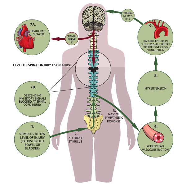
Autonomic dysreflexia:  You get unopposed sympathetic response to some noxious stimuli in a paralyzed patient.   Uti is most common cause.  Fecal impaction or bowel obstruction are the second most common cause.  Treat the cause and manage the BP.

Autonomic Dysreflexia


Wernicke’s syndrome: Thiamine deficiency causes occulomotor dysfunction,  cognitive dysfunction, and ataxia.

EKG’s in the morbidly obese consistently show flattening or T wave inversion in the infero-lateral leads.

Down’s patients have atlantoaxial instability.  Girzadas comment: this makes Down’s patients at greater risk for spinal cord injury in trauma.

Heterotopic ossification can be due to trauma, spinal cord injury, severe neurologic disease, burns and rarely to genetics.   The cause is unknown but alkaline phosphatse is thought to play a role.  Patients will have warmth, tenderness and swelling initially at the site of heterotopic  ossification.  The hip is probably the most common site.  The affected site is always below the level of the cord lesion.

Heterotopic ossification

Garret-Hauser                       Ethics  

Case 1. Trauma pt was driver in MVC with girlfriend who was passenger and also injured.  Male Pt (driver) is HIV positive.   Both he and girlfriend are admitted with injuries.   There was extensive blood at scene.  Patient has asked that his girlfriend not be told about his HIV status.  Treating MD obviously very troubled by this situation.   Strategy:  Discuss with HIV positive patient about his obligation to his girlfriend.  If he refuses to give this information to his girlfriend,  then counsel the girlfriend that she was exposed to unkown blood and she should receive prophylaxis.   Elise comment: The female patient likely has been having sex with the male patient and she needs an HIV  test.   Harwood comment:  Tell the patient we have some information that we can’t pass on to you that makes us strongly advise you to receive HIV prophylaxis.  You can also say you were exposed to HIV positive blood.  Shayla comment: The hospital has to report to the health department that  the male patient is HIV positive.  The health department will eventually notify the patient’s girlfriend.

Case2.  15yo female is pregnant and parent doesn’t know.   Kelly comment: First figure out if there is any issue of safety for the girl if she reveals to family she is pregnant.   Shayla comment: Get the closest responsible female from the family to help in  this situation.   Encourage the girl to disclose the information to the parent while still in the ED.   If the girl refuses, you can tell the parents there are test results that your daughter refuses to allow me to discuss with you.  If you want to discuss this issue with your daughter now, I will be here if you want to discuss.  Also, you can advise parents that they can request the medical records.

Case3. Mom demands drug testing on her son.   If the child agrees to give urine, the results of the drug screen are not protected information and can be given to the parents.   The American Academy of Pediatrics recommends against drug testing outside of the primary care physician’s office.   Attending consensus: Most would not force a catheter or a pelvic exam or draw blood on an actively refusing patient.   There is no medical emergency in a well appearing patient.  If the child agrees to testing then most would do the test for the parents.  If parents request a genital exam to evaluate if a girl had sexual activity, our female faculty all would refuse to inflict this exam on a girl.

Case4.  59yo male found unresponsive after intentional overdose.  Pt has terminal lung cancer.   He has DNR/DNI.   Discussion of whether or not pt should be resuscitated.   Consensus was that you have to intubate patient or try narcan and flumazenil.  But not actively resuscitating the patient would be akin to aiding a suicide.   Although some states have legal physician assisted suicide, the AMA still opposes physician assisted suicide.   Elise comment: not resuscitating this patient places you at very high legal risk and high ethical risk.  You gotta resuscitate the patient.   Christine comment: We are not required to provide medically futile care, so if a patient shots himself in the head you may not need to try to resuscitate that patient if the prognosis is futile.   Girzadas comment: For the patient who overdosed, I would way rather be defending myself on the side of resuscitating him versus letting him die. 

Putman                 M&M

Elderly male with SOB.   Code 44 on arrival.    O2 sat is 76% and  HR=120, normotensive and afebrile.  Lungs are clear.  Pt was incontinent of heme positive stool.   Bipap started.  IV PRBC’s given.  Niece gives history of abdominal pain and diarrhea and coffee ground emesis.  Pt has myelodysplastic syndrome and is on warfarin among other meds. 

Hgb=7.2,  ABG=7.44/19/148/12, Lactate is 7, Bun/cr=117/3.5,  INR result was delayed.   CT was ordered.

Work up proceeds over next 1-2 hours.   Patient at one point arrests.  ROSC obtained with intubation, epi and more IV prbc’s.    Elise comment: LMA may have been sufficient during code.   Harwood countered that intubation in this patient with hx of coffee ground emesis is very reasonable during code.

INR is still pending.   A call to lab revealed that the INR result was not reported because it was too high to be calculated.   When this was learned FEIBA and vitamin K were administered.  Pt received octreotide and pressors as well.   Patient coded multiple times and family withdrew care and patient died.

Lessons:

Coags are a critical piece of info in the GI bleeding patient.   If there are significant delays, we need to investigate what is delaying the result.   Multiple attendees asked why questionable or extreme results can’t be reported to the physician.  We need to know about these time critical results.   Mila comment: there is a system wide initiative to develop a plan for this problem.

Discussion of placement of NG tube in this patient: Consensus that it would have not have helped much.  Harwood comment was that NG can help diagnose UGI bleeding but you already knew the patient had an UGI bleed.  It can also help you quantify UGI bleeding but you were already giving blood and knew the patient had a significant bleed.   Girzadas comment: NG aspirate has a high false negative and false positive rate so it could potentially  have lead you to think the bleeding was less severe than it actually was. 

There was a discussion of the use of bipap in this case.  Bipap can insuflate air in the stomach and exacerbate vomiting.    Group consensus was that bipap was reasonable and helped the patient.   Christine comment: I would have tried bipap in this patient also, but maybe intubation is the better plan of action to avoid gastric distension.   Erik comment: Historically in the past, we used to tube more GI bleeders.  Elise comment: Trust your gut if your feel that a patient is critically ill and secure the airway.   

Kelly comment: You are the managing physician of the patient.  Be clear about what you want a consultant to do.  Lead off your conversation with what you need from your consultant and follow that with further detail  of the case.

 Harwood comment: I am ok with giving FEIBA in this case without the INR result.  The patient was known to be on warfarin with a recent therapeutic INR as an outpt, anemia, and obvious current GI bleed.  You can tell GI or IR that you are giving FEIBA and the INR will likely be reversed in about 30 minutes so they can start coming in to do endoscopy.   Sam Lam comment:  U/S may have been helpful to rule out other intra-abdominal emergencies.

It was discussed that some consultants and other ED’s use gastric lavage as a decision making tool for upper GI bleed.  NG tube placement is safe in the setting of esophageal varicies.

PPI’s have been shown in Cochrane review to not improve mortality, re-bleeding, or surgery in undifferentiated UGI bleed.   It does show that signs of serious bleeding are less on endoscopy following PPI use. SMARTEM podcast did not feel PPI’s were beneficial in UGI bleed.   There is a Cochrane review of PPI’s for  specifically peptic ulcer disease related bleeds that showed no mortality benefit but there was less rebleeding and less surgery required.  

Sangstaken-Blakemore tube. Harwood comment: The only situation that has theoretical benefit with this device  is esophageal variceal bleeding.   There is a problem that you need to keep tension on the tube to keep stomach balloon compressing the upper stomach.  Traditionally people have used foot ball helmets in order to tether the tube to the facemask.   Some attending have tethered the tube to a bed railing.

Activate the massive transfusion protocol in the critically ill GI bleeding patient.

Blachford Scale can be a useful tool to identify low risk and high risk UGI bleeders.


A journal club  on this scoring system within the last 18 months found that with a score of 2 or less and age <70, patients could be considered for possible D/C.   A score higher than 3 likely would need an intervention.

Febbo         Safety Lecture   Diagnosing DKA

24yo female , 13 weeks preg presents with vomiting.  Pt has an insulin pump for DM.  Glucose was 133.   Anion gap=21.   ABG has a PH of 7.54, PCO2 is 19 and HCO3 is 16.  Pt has some respiratory alkalosis and metabolic acidosis.   Ketones in urine was >80.  UA showed signs of UTI.

Pt was treated with IV fluids & IV antibiotics and labs improved with nl anion gap and bicarb up to 19.   Pt was admitted and Insulin pump was turned off per hospital standard practice.   Overnight on the floor, pt did not receive any insulin.   In am next day, anion gap had increased and ph dropped.   DKA was diagnosed and pt was transferred to the ICU.  Harwood comment: There is no mystery here.  A pregnant, diabetic patient with a UTI needs insulin.  With no insulin, she will develop DKA.   Theresa Navarette comment:  In a patient with DKA, turn their pump off because it could be malfunctioning and treat the patient with IV insulin.  In a patient without DKA who has an insulin pump it is reasonable to keep using the pump because it is probably working based on controlled blood sugar.   There was consensus that a shot of Lantus insulin prior to the patient going up to the floor may have prevented the patient going into DKA.   Maggie comment: Endocrine is more than happy to consult on patients with insulin pumps. 

Conference Notes 11-12-13

Much Thanks to Christine and Elise for keeping the Conference Notes going while I was down in Australia!

Joint EM-Peds Conference     M&M

5 week old female.   4 week hx of diarrhea.  PMD tried multiple formula changes.  On arrival to ED, pt not taking po and appears dry and pale/grey.   Also grunting noted.   IV placed expeditiously and fluid bolus given.   Unable to obtain labs initially.   Sign out occurred at this point.   Further attempts at obtaining labs failed.   Finally ,via 4 heel sticks, labs were obtained.    K=7.9 and HCO3<5.    Re-eval of child showed  ill but non-toxic appearance.   Parents refused further attempts at blood draw.    Pt was transferred to the floor at this point.   On floor,  skin was cool/pale,  pt still grunting,  HR=146.   PICU came to evaluate patient on floor and found pt to be lethargic and gray in color.  ABG showed a ph<6.8.    MetHGB=33%.     Pt was intubated, septic work up performed,  methylene blue was given.   Pt did well.   Pt had a congenital food protein related methemoglobinemia.

Issues: There was early recognition of the sick child.  There was difficulty obtaining labs.   Handoffs were considered less than optimal in that severity of illness and management algorithm was not completely clear to receiving teams.   Interdepartmental communication may have been suboptimal.     PICU team was not involved in this case while patient was still in ED.  Bedside handoff may have been helpful to improve the clarity of clinical communication between teams.

Positives: Rapid IV access was obtained.   Multiple re-assessments were made of the patient by the receiving teams in the ED and on the floor.  There was very good communication with the patient’s parents.  Early recognition/treatment of methemoglobinemia and transfer to PICU.

Kettaneh comment: Optimally  PICU could have been consulted prior to the first sign out.  It is helpful to give a management algorithm based on diagnostic testing results to the receiving team.

Roy comment: It is great that the patient was re-evaluated multiple times in ED.   Also very positive that methemoglobinemia was identified and treated rapidly.

Harwood comment: There are multiple system issues. This case involved multiple fatigued docs signing out to other fatigued docs.  ABG was a critical test in this case.  It is good that MetHGB is part of the ABG panel.    If you are facing difficulty getting labs in a seriously ill child, you can get labs from IO draws (T&C, Glucose, Cr, HCO3 are all valid).

Roy comment:  I agree ABG was very important.  Just thinking of alternative diagnoses, if this patient had a high bicarb on ABG it would make CF very likely.

Suggestions for system improvement: C. Kulstad comment: ER physicians should not be defensive toward  Peds Resident suggestions when discussing tranfers of care.   Both teams should discuss options in difficult cases.  Notario comment: Bedside rounding would have been helpful to get fresh eyes on the patient.

Herrmann/Kettaneh           STEMI Conference

Case 1.  Chest pain resolved prior to arrival in ED. Inital EKG was non-specific.  Possibly some Wellens changes anteriorly.   E. Kulstad, Cardiology comments:  Wellen’s is not a criteria for  Code STEMI but cardiology should be consulted for non-emergent cath the same day.  2nd EKG with no pain shows Wellen’s signs.   Cards consulted.  Trop was 0.07.  Pt was cathed later that day.  Pt had an 80% lesion of LAD that was stented.   There was a discussion of the timing of Cath in Wellen’s patients.   General agreement that these patients don’t require emergent catheterization if pain free.  Ok to start ntg and beta blockers and anti-platelet therapy and arrange prompt cath (12-24 hours).  Wellen’s changes are usually noted when pt is pain free.   With chest pain, the changes normalize or evolve to STEMI.  Post-cath, the EKG in this patient normalized.  Al-Kaled comment: Wellen’s syndrome includes chest pain.   If you see these findings in an asymptomatic patient, technically it is not Wellen’s syndrome.

Wellen’s EKG

Case 2.  Ekg showed Afib with anterior Q waves.  Minimal ST elevation noted in lead 3 and minimal ST depression 1 and AVL.   Cardiology consensus was that this was not an acute STEMI.   2nd EKG was similar.  3rd EKG now has minimal ST elevation in AVF and possibly more ST depression in 1 and AVL.  4th EKG has ST elevation in 3 and AVF.  STEMI was called on 4th EKG. Cath showed no coronary artery disease but pt had embolism to distal LAD due to Afib.  There was much disagreement among cardiologists present about the management of this patient.  Most agreed that patient had to go to cath lab with ongoing pain and worsening EKG.  Cardiologists made the point that taking the very elderly to cath is treacherous and if at all possible, attempting a short trial of medical management on elderly  patients without screaming STEMI findings on EKG may be prudent before pulling the trigger on a Code STEMI.    Afib with embolus to the coronary artery is relatively rare. Mechanical valves can also shower emboli to coronary arteries.  Cardiologists note that patient can have concurrent emboli to heart and brain or heart and gut.

Case 3-5.  Multiple rapid fire EKG’s presented.    There was a case of a patient with sob and anterior horizontal ST depression with associated ST depression in AVR.   Most cardiologists would call a STEMI based on that EKG for posterior MI.   Posterior leads showed minimal ST elevation in Post V4 and V5 leads.   Cath showed Circumflex lesion.   

Posterior MI

Last EKG was in a DKA patient.  Silverman comment: Use Echo in a young patient.  If the anterior wall is going like gangbusters it is not going to be a STEMI.

Permar        Trauma Lecture

 60yo Male fell out of a tree.   No sensation below nipples. Could not move upper extremities.  Pt is hypotensive with borderline bradycardia.   Diagnosis: Central cord injury of cervical spinal cord.

Spinal cord injuries are most common in young adult males.  47% are due to MVC’s.  ETOH and underlying spinal disease increase the risk.  Most (55%) Cord injuries involve the cervical spine.

Primary injury= Immediate effect of trauma.   Secondary injury= Progressive injury following initial traumatic insult (ischemia, hypoxia, inflammation, edema, apoptosis)

Backboards can cause pain and their benefit is unproven.  They are the standard of care however in the US.

NEXUS Criteria


Canadian C-Spine Rule


Flexion-Extension views may pick up a ligamentous injury.  30% are non-diagnostic.  Omi comment: For patients with persistent pain this is a worthwhile test to rule out ligamentous injury.   It may save the patient and MRI.  Most useful in a younger, thin patient.  Bigger or more muscular patients will have very limited images and MRI is more useful for these patients.

Unstable Cspine Fractures:   mnemonic: Jefferson Bit  off A Hangman’s Thumb.      Jefferson Fx,  Bilateral Facet dislocation,  Odontoid fractures,   Atlanto-axial dislocation,  Hangman’s fracture, Teardrop fractures.

Back to case: Pt had bilateral C1 arch fractures with central cord syndrome.


Central cord lesion:  Due to forced hyperextension injury.


Anterior Cord Syndrome:  Commonly due to Disc herniation or bone fragmants.  


Brown Sequard: Due to penetrating injury to Cord.


Neurogenic shock: Hypotension and bradycardia. Due to interruption of autonomic pathways causing decreased vascular resistance.  Clinician’s goal is to keep MAP over 85 to maintain cord perfusion.   May require pessors, atropine, pacing. 

The Great Steroid Debate: 2013 American Association of Neurosurgeons state that steroid use is not recommended.    Omi comment: There is no indication to give steroids in spinal cord injured patients.  Many trauma centers have not been giving steroids for over 10 years.   This statement by the Neurosurgery Association helps the emergency physician greatly to not give steroids.

 Prognosis: C1-C3 injuries have highest risk (6.6 fold) of death.    DVT is a common complication of spinal cord injury.

There was discussion of therapeutic hypothermia for cord injury.  Dr. Omi felt that ongoing research would demonstrate that  therapeutic hypothermia will not be helpful for complete cord injuries but for cord contusions or central cord syndrome it may be prove to be useful.   Andrej comment: Recent  Neurosurgical Society statement says there is no current evidence to recommend therapeutic hypothermia for cord injury.    

Omi comment: It is difficult to keep a quadriplegic patient off the ventilator.  They can’t cough forcefully and they have too much respiratory muscle weakness.  Dr. Omi mentioned that some trauma surgeons are placing diaphragmatic pacers to avoid intubation.

Watts      Study Guide        Psychiatric Emergencies  

Phencyclidine related seizures are resistant to phenytoin.   Use benzos first.   PCP is structurally similar to ketamine.

Naltrexone reduces alcohol craving and relapse without unpleasant side effects.

Panic attacks are characterized by discrete episodes of intense fear in absence of real danger.   Patients have both somatic and cognitive symptoms.   Preferred treatment for panic disorder is cognitive behavioral therapy and SSRI’s.   Benzo’s are effective for short term relief.    Harwood comment: Panic attack is a diagnosis of inclusion.  You need 4 of 13 criteria for diagnosis.   There was consensus among faculty the emergency physicians can appropriately to counsel pt’s on how to manage these attacks with cognitive/relaxation stragtegies.   You may need to refer patient for management with SSRI’s.   Symptoms of a panic attack can last for over an hour.

Panic Attack Criteria


Munchausen syndrome: Young to middle aged men  most commonly have this diagnosis.  Munchausen patients create stories about medical illnesses in order to experience medical care.   Malingerers, on the other hand, act in a way to achieve secondary goals and external incentives.   Harwood comment:  The key is what the motivation is for the patient.   If their motivation is to be the center of medical attention then it’s munchausen’s disease.   If the motivation is to get drugs or get out of work then it is not munchausen’s.

Primary gain is related to a patient wanting to improve their internal feelings.   Secondary gain is related to gaining advantage of external factors like getting off work, getting drugs, gaining money.

Dystonic reactions and akathesia  most commonly are caused by any anti-psychotic.  Treat with Benadryl and cogentin.  2nd line treatment is benzos.   Harwood comment: Cogentin (benztropine) is longer acting than Benadryl so you should give the patient some.  Continue Benadryl and cogentin for 2 days.

Risk of suicide: men, older, psychosis, previous suicide attempt, excessive drugs/etoh, lack of social support, serious suicide attempt.  Kelly comment: Get corroborating info from family members or partner/friend. Family/friend history may be significantly different from the patient’s history.

Sad Persons Scale


Venlafaxine (Effexor) is a serotonin-norepi re-uptake inhibitor that can prolong the QT interval.  Methadone can also prolong the QT interval.

Anxiety is the most common class of psychiatric disorders seen by primary care physicians.

Managing the agitated patient:  Physical and chemical restraint are both needed.  Document  that the patient is a risk to patient and staff.   There was a discussion about using chemical restraint to reduce the risk of cardiac arrest from acidosis related to agitated psychosis.   Ketamine was discussed as a tool for chemical restraint.  Kari Tekwani said, in her experience, Ketamin works well  calm patients but some psychiatric facilities will not accept patients in transfer after being given ketamine.  Also there was a discussion  about the differences between geodon and haldol for chemical restraint.  Consensus was that their effectiveness/risk  was largely equivalent.

Treatment of Neuroleptic Malignant Syndrome: Stop anti-psychotic, IV fluids, cooling, benzos, consider dantrolene or bromocriptine.  Classically patients have lead pipe rigidity, hyperthermia, autonomic instability,  and mental status changes.   Differential includes serotonin syndrome.

Ryan      Med Student Review


Conference Notes 11-5-2013

Last guest conference notes, Dan’s back in country soon. This week back to Christine Kulstad.


8:00-9:00- Oral Boards- Felder vs Wise

Case 1- Thyrotoxic paralysis. Critical actions- treat hypokalemia and thyrotoxicosis. This is rare disorder with painless muscle weakness- this is a subcategory of periodic paralysis. Usually in SE Asians, more often men, usually young adults. Respiratory and bulbar muscles are not affected, and arrhythmias are uncommon. Weakness is proximal > distal, legs >arms. All symptoms resolve after treatment.  Treat with potassium, but high dose can cause rebound so give lower initial than you might think. Propranolol will move potassium into serum where it is lacking (total body potassium is fine, just shifted into muscle cells during attacks), dosing is 1 mg q10 to max of 3 mg/kg. Treat hyperthyroidism as usual. Have patients avoid high carbohydrate meals, fasting and heavy exercise as they can precipitate episodes. Chronic potassium supplementation is ineffective.

Case 2- Toxic shock syndrome from skin abscess. Treat sepsis as usual, drain abscess. TSS is caused by either Staph aureus or group A strep. Was associated with tampon use in past, now more with wound-packing or skin infections. Staph will rarely be found in blood cx (GAS does yield + blood cultures). Symptoms arise from exotoxins that staph produce- commonly see fever, hypotension, skin manifestations. May also have chills, malaise, v/d, sore throat, headache. DDx of ill young patient with rash- TSS, RMSF, meningococcemia. Treat with clindamycin and vancomycin. Remember to treat septic oral boards patients with EGDT as you would in the ED.

Case 3- FB aspiration in a child with prolonged cough. Found with air trapping on decubitus x-rays. May also see atelectasis, mediastinal shift, pneumonia.  If FB is left long enough, air in alveoli will be absorbed causing atelectasis. Infection often develops- first pneumonia and then pulmonary abscess and bronchiectasis are possible. Take-home point: do more evaluation (at least xrays) in child with prolonged cough.


9:00-10:00- EMS Study Guide- Motzny (and How to Survive the Zombie Apocalypse)

Triage: When triaging during a disaster, green is walking wounded. Remember triage is dynamic and will require reassessment.

START and SAVE are 2 triage systems. SAVE (secondary assessment of victim endpoint)- determines who will benefit significantly from austere field interventions. Patients who will die if not treated in field (vs those whose fate will not change if treated because too critical or not that bad). START (simple triage and rapid treatment) uses a quick assessment of respirations, perfusion, and mental status. Assess respitations first, if repositioning airway dosen’t fix it they are black/dead. If it does or breathing more than 30- red (immediate rx). If not, check radial pulse. If absent, red. If normal, check MS. If patient can respond they are yellow. Otherwise red.

Disasters: Internal disaster- catastrophic problem in the hospital (power failure and generator failure). External or just a disaster is defined by the capacity of the surrounding hospitals to deal with patients. E.g. 40 victims in a rural area is a disaster while 100 in a large urban area is not.

JCAHO requires a hospital’s disaster plan to be activated twice a year for accreditation.

Communications is consistently identified as a problem during disaster drills. Causes can be failure of equipment and unclear terminology, chains of commands. The first step when you are notified of a disaster is to verify the information (before activating the disaster plan).

Mass gatherings are defined as 1000 people in one site for a common purpose. The most common complaint of patients at a mass gathering is dermal injury (cuts/scrapes).

Medical control from the EMS physician is indirect and direct. Direct is real-time contact with paramedics, whether over the radio or at the scene. SMO (standing medical orders) give indirect guidance for many common problems. Physicians cannot assume control of a scene if unknown to EMS UNLESS you have proof of licensure.

HAZMAT command centers should be set up uphill and upwind. Many toxins are heavier than air (so go uphill), if lighter than air they will soon rise above your head. When dealing with patients with significant chemical exposure, eyes take precedence in decontamination. No procedure (including airway) should be done without decontamination unless you are wearing the appropriate protective gear (HAZMAT suit) and then get decontaminated yourself.

Weapons of mass destruction include chemical, biologic, nuclear, radiologic, and explosive agents. Biological agents classified as Class A agent of concern by CPC are smallpox, anthrax, plague, tularemia, viral hemorrhagic fevers, botulism- all the agents that you think of as really bad. Others are class B if they could be used but not effectively or not that toxic (e.g. salmonella).

Radiation- alpha, beta, and gamma. Alpha particles will not penetrate skin or clothes, only dangerous if ingested. Beta goes a couple of centimeters (lead protects). Gamma goes all the way through (like xrays) and will give whole body irradiation. The good news is you don’t have to decontaminate gamma since its still going. The earliest indicator of radiation injury is lymphopenia. 

Hypoxemia and hypoperfusion are not caused by helicopter EMS transport generally because they don’t fly very high, can factor in fixed wing aircraft transport (planes). Decreased air pressure in planes can 1) increase fluid rates in medications given in glass bottles as gas expands 2) increase pressure of pneumothorax increasing the size 3) tracheal cuff on ETT may pop (use saline instead of air). If you are approaching a helicopter, avoid the rear.

NIMS (National Incident Medical System) standardized terminology and procedures. It states material managment is logistics section responsibility. For more info, www.fema.gov/emergency/nims

10:00-10:30- Discharge Instructions- Kiernicki-Sklar

Don’t make the mistake of thinking discharged patients are not sick or someone’s problem. CRICO 2011 benchmarking report summarized 200k EM malpractice cases, they found #2 cause of malpractice cases related to missed/delayed diagnosis (majority of cases) was development of discharge plan.

Think of d/c instructions as sign-out to the patient. Other functions as a patient care summary, contract with patient (if patient doesn’t follow instructions and suffers harm, care contract is breached).

Patients don’t remember verbal instructions well, make sure you write down important information too.

Mnemonic to remember how to provide good d/c instructions. WTF DR DC?

W- What we did or didn’t find. Include incidental findings (lung nodules, elevated BP).

T- Treatment/tests that are still needed (have BP rechecked, have a stress test in 72 hrs, have sutures removed). Include risks of non-compliance.

F- Follow-up required. Timed f/u when undifferentiated abd pain (24-28 hrs), significant wound checks (1-2 days), chest pain (72 hrs). Stress that patient needs to call to make appointment (or if specific time arranges). Make effort to contact f/u physician in high-risk cases.

D- Drug warnings- legally it is your responsibility to provide warning and check for allergies/interactions.

R- Restrictions. Things that may worsen a current or undiagnosed condition (undifferentiated CP and exertion, seizures and swimming/bathing/driving). Not only could patient sue you, but 3rd party could (driver who seized and hurt someone else).

D- Diagnosis. Don’t feel need to make a diagnosis when it is not clear (abdominal pain vs GERD). Two reasons, easier to sue for misdiagnosis in case 2 and causes anchoring bias in physician who next sees patient.

C- Comeback (what to return for). Highlight specific concerns based on that patient’s problems, don’t assume that they will read all the pages of the pre-formatted d/c instructions. Include contingency plan if f/u cannot be arranged (patient can always return to ED as last resort and should be encouraged to do so).

?- Final checks. Re-check vital signs, reassess pain. Use simple words (6th grade level), and avoid medical terms and abbreviations.

Brief discussion of AMA- think of as informed non-consent. Must discuss and document 1) risk/benefit/alternatives 2) Pt has decision making capacity 3) Patient has understanding of #1 4) patient can ask questions and get answers. Give them good discharge instructions and any treatment they will accept.

10:30- Musculoskeletal Exam- Hands-On Skills Station

You had to be there

Conference Notes 10-29-13

Today's guest for conference notes is Elise Lovell. Don't worry, only one more Tuesday until Dan's back.

Bonus, a tox lecture by alumni and tox fellow Janna Villano.

00 am  Orbital Trauma/Oculoplastics-Dr. Hassan Shah; he’s happy to be involved with any Oculoplastics questions!

Ruptured Globe:  How to tell?  Peaked/irregular pupil, full hyphema, 360 degree subconjunctival hemorrhage, low IOP.

Most important reason to fix orbital fractures?  Prevent diplopia.

What does optho need to know about intraocular foreign body?  High pressure à systemic steroids.  Ruptured or not ruptured.  Visual acuity.

Blunt eye injury: “Blown pupil” actually not helpful to tell if optic nerve injured...can have temporary pupil dilation simply due to blunt injury.  APD however IS a sign of optic nerve dysfunction.  If APD, high pressure, loss of vision, need to perform lateral canthotomy/cantholysis.  Dr. Shah believes cantholysis important; simple canthotomy usually not enough, and most common mistake is to think you’ve relieved the pressure but you haven’t done enough.  He can fix any eyelid laceration that you cause!
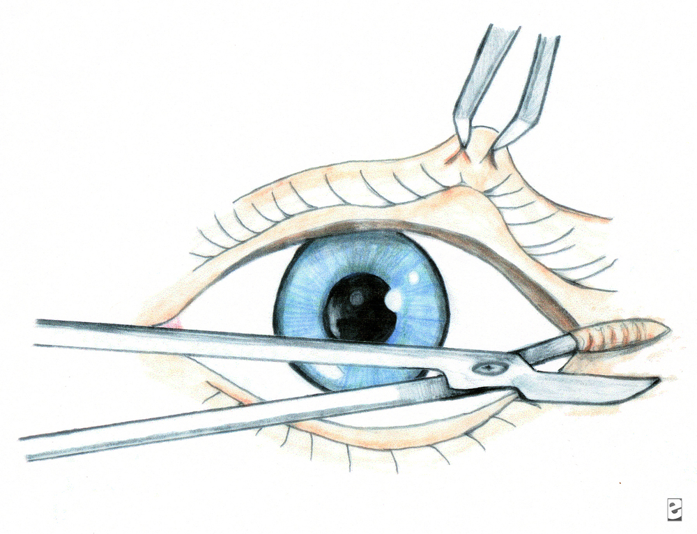
 

This is lateral canthotomy.  You also need to cut up and down (3 total cuts) for complete cantholysis.

Be sure to cover the eye in the setting of eye trauma.  Use ointment, try to bring lid back down over eye.  Best covering for globe is the conjunctiva.

Blow out fractures.  Adults almost never needs to be repaired emergently.  Even if “entrapment”, usually due to soft tissue swelling.  Usually wait for one week before repair to allow swelling to resolve.  Big fractures much less likely to get true entrapment than small fractures.  Big fractures = lots of swelling, but also room for movement.  For kids, they get “greenstick” type fractures of the orbit; softer bones, the fracture is often very small, with mild swelling, but with restricted ROM up and down due to trapped muscle àthis is case for same day surgical repair.  If you don’t have diplopia on Day 1, you will not develop it!

Facial fractures and antibiotics:  He doesn’t usually give antibiotics unless contralateral sinus disease, but there is controversy, not one correct answer.  He has never had an infection.

9:00 am  Janna Villano: - Tox-Uses of Sodium bicarb, and some controversial Antidotes

Most important 4 things in Tox:  Vital signs, Mental Status, Skin and Neuro exam


Many Sodium Channel blockers out there:  TCAs, cocaine, some antiepileptics, dysrhythmics...lots of others.

3 Uses for Sodium Bicarb:  Cardio toxicity, Prevent acidosis, Urinary alkalinization

Bicarb:  Treat cardiotox:  helps with prolonged QRS, wide complex tachycardia, hypotension.  Give 1 to 2 meq/kg bolus over 1-2 minutes.  Boluses better than infusions.   Give boluses and sit there watching for QRS to narrow.

Bicarb:  Prevention of academia:  maintain pH >/= 7.4.  Prevents drug distribution, may be useful peri-intubation of tox patients.

Bicarb: Urinary alkalinization:  enhances elimination.  Maintain pH 7-8.5, ion trapping, must maintain normal K levels for this to work-aggressive K replacement!


Anticholinergics:  antihistamines, TCAs, vertigo meds, antipsychotics, Jimson weed...etc.

Physostigmine:  To treat anticholinergic toxicity.  Will allow history when patient’s mental status clears.  Should see nearly complete resolution of symptoms in order to attribute presentation to anticholinergic  toxicity.  Allows diagnosis, limits workup.  Can cause seizures, GI distress/rest of cholinergic symptoms.  Reports of physostigmine associated with cardiac arrest , but these in patients with TCA OD, also cases of seizure in patients with prior seizure disorder.  If giving physo, watch for bradycardia/heart block,  seizure, cholinergic side effects.  Give 0.5 mg over 2-5 minutes, may repeat up to 3-4  doses.  If necessary can “reverse” physo with atropine.  ***Janna doesn’t give in patients with known seizure disorder, patient who took pro-convulsant, or patients with sodium blocker OD (eg TCA)***


Flumazenil:  Don’t use drug screen to help you decide whether or not to give it-drug screen misses many benzos.  Some studies have shown avoidance of intubation.  Why not to use it in a documented benzo OD?  Inconsistent clinical response, precipitation of benzo withdrawal (rare, and can give benzos), may precipitate seizures (seen in pts who took proconvulsant, or in patients using benzos for sz disorder), maybe risk of complications outweighs harm from OD.

Pick the right patient-no seizure disorder, pro-convulsant use, not if chronic benzo users, not for undifferentiated coma or prophylactically.  Often used in kids, pure benzo OD, procedural sedation.   If patient has seizure after flumazenil, then just flood patient with benzos (competive inhibitor).


10:00 am Dr. Khatiyar-  Chemical Asphyxiants

Fires:  Simple Asphyxiants, Chemical Asphyxiants, Chemical Irritants

Carbon Monoxide:  from partial combustion:  broken furnace, blocked car or boat exhaust, grilling in a garage, heating homes with gas ovens, car idling in garage...etc.  Also from Methylene Chloride, in degreasers/lacqueer thinner, bathrub refinishesàmetabolized to CO.

CO toxicity:  high affinity for hemoglobin forming CO-Hgb, but there are other toxic effects as well.  Also binds to myoglobin (heart tox, rhabdo), direct organ toxicity due to increased NO production and lipid peroxidation.  CO attachment to cytochrome oxidase (inhibits aerobic metabolism)

Think about it in headaches, “flu” in the fall when heaters turned on, kids with “colic”., gastro, viral syndrome, ..great imitator.

Diagnosis depends on a great history.  Ask about recurrent sx, others at home with same sx, time of year, in setting of Headache, flu sx, “hangover” symptoms, chest pain, neuro sx.

Diagnosis:  Breath sampling, CO level:  VENOUS just as good as ARTERIAL blood gas.  Don’t use O2 sat!  Check renal function, pregnancy test as changes management dramatically.  CPK, cardiac monitoring.  CO levels also can be unreliable, depending on treatment with O2, time since ingestion.  Levels:  < 10% no biggie, > 50% likely fatal, everything in between open to discussion.

Delayed neuro sequelae:  days to weeks later, incidence variable (1% - 47%), needs cognitive testing  (think mini mental status) and re-testing on followup.

Red flags:  pregnant women, kids, cardiac patients.

Half life 4-6 hours on room air, 60-90 minutes on 100% O2, 20 minutes with HBO.

Treatment:  HBO controversial:  but if level > 25%, any neuro issues/syncope, coma, Cardiac sx, abnormal cerebellar exam, symptomatic pregnancy or pregnancy with fetal distress à  call HBO chamber and discuss with specialist.  Put patient on 100% O2 as soon as you suspect CO.  Local chambers:  Advocate Lutheran General or Loyola, LCOM, Illinois Masonic, others.

Cyanide:  neuro sx, acidosis, seizures.    House fires, burning plastics, Nipride (sodium nitroprusside), certain foods-cassava, apple seeds, fruit pits, Laetrile.

Toxicity:  Neurotoxin, Enhances NDMA receptor activity, inhibits aerobic energy metabolism (blocks cytochrome oxidase).


Diagnosis:  Symptoms between 3 – 24 hours if oral, few minutes with gas exposure, sicker with premorbid conditions, kids, and dose dependent.

Presentation:  LOC/AMS, metabolic acidosis, GI, SOB, CV instability.  Be worried with rapidly developing coma, lactic acidosis, CV instability, symptomatic industrial worker/chemist.

Treatment:  decontamination (clothing, skin decon), 100% O2.  Old antidote:  Lilly cyanide antidote kit (amyl nitrate, sodium nitrite to cause methemoglobinemia which will bind cyanide, sodium thiosulfate to form thiocyanate which will be excreted in urine).  This kit usually available in ED, and this toxicity very time sensitive.  Cons:  causes hypotension, methemoglobinemia, multiple steps.

New antidote:  Hydroxocobalamin binds with cyanide, conversion to cyanocobalamin (vitamin B12):  safe, no methemoglobinemia, no hypotension, can penetrate to tissues.  Does cause flushing./urine discoloration.  Will mess up color change sensitive testing for hours.  Not available in ED but we have it in pharmacy.


11:00 am : Dr. W.A. Bret Negro - Cold Weather exposure/Hypothermia: 

Core Temp < 35 degrees

Causes:  exposure, EtOH, sepsis, burns, massive resuscitation, metabolic problems (low sugar, low thyroid/adrenal), CNS causes

Mild > 34 degrees:  shivering, “responsive” stage, trying to generate/maintain heat

Mod 30-34 degrees: depressed CV function, stupor, afib with slow ventricular response is classic

Severe < 30 degrees:  low BP, no shivering, coma

Cardiac tox:  sinus tach àsinus bradycardia à slow afib à vfib/asystole.  Heart very sensitive, can cause dysrhythmias with central line placement and tip in heart/rough handling of patient.  Also less responsive to ACLS meds/electricity.  Shock once if T < 30 C, then just remarm until T > 30 before shocking again or giving drugs.  May want to extend intervals between drug dosing.  ECG Osborn wave.
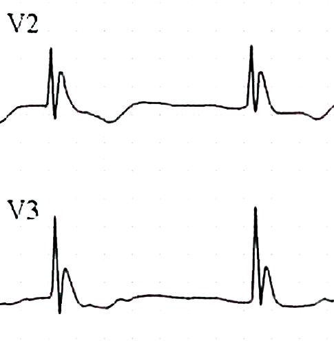


Respiratory depression, messed up ABG values-to keep simple just treat as normothermic when making vent decisions.

Metabolic cold diuresis, rhabdo, ARF, hypokalemia from ion shifts (don’t over correct), pancreatitis, CNS alteration.

Treatment:  remove from environment, dry patient.  Take your time finding a pulse, may be extremely bradycardic.  Don’t start CPR unless absolutely necessary due to cardiac irritability.  Warm fluids (can warm IVF in microwave), warm O2, forced air blanket, Arctic Sun vest/Alsius Icycath central line devices (same devices used to cool patients, but set for warming).  Warm gastric lavage, DPL, Foley fluids, 2 chest tubes with warm fluids, Bypass or thoracotomy as last resorts.

Rewarming shock:  external heat àperipheral dilation, relative hypovolemia and hypotension. 

No firm guidelines on treatment.

Non-freezing cold weather injuries:  Trench Foot in military, direct injury to soft tissues, blistering/anesthesia, treat with heat/dry feet.  Prevent with dry socks/well fitting boots.  Chilblains/pernio:  kids, Raynaud’s, uncomfortable inflammatory lesions of skin by longterm intermittent exposure to damp/nonfreezing ambient temp.  Hands/ears/toes.  Panniculitis:  mild necrosis of subcut fat.  Cold urticaria-sensitivity.

Freezing injuries:  Frostbite, usually < 4 degrees F, frozen tissue, most damage from endothelial damage during thawing.  Clinical diagnosis, warm them up and AVOID RE-FREEZING.  No rubbing-will cause more damage.  Also do not re-warm out of hospital.  Aloe, NSAIDS, tetanus, no antibiotics.  TPA after rewarming to reduce digit amputation.  Heparin/HBO don’t work.  Amputations delayed until final demarcation.


11:30 am:  Dr. Dennis Ryan – MSK Study Guide

Too much fun to take notes

00 am  Orbital Trauma/Oculoplastics-Dr. Hassan Shah; he’s happy to be involved with any Oculoplastics questions!

Ruptured Globe:  How to tell?  Peaked/irregular pupil, full hyphema, 360 degree subconjunctival hemorrhage, low IOP.

Most important reason to fix orbital fractures?  Prevent diplopia.

What does optho need to know about intraocular foreign body?  High pressure à systemic steroids.  Ruptured or not ruptured.  Visual acuity.

Blunt eye injury: “Blown pupil” actually not helpful to tell if optic nerve injured...can have temporary pupil dilation simply due to blunt injury.  APD however IS a sign of optic nerve dysfunction.  If APD, high pressure, loss of vision, need to perform lateral canthotomy/cantholysis.  Dr. Shah believes cantholysis important; simple canthotomy usually not enough, and most common mistake is to think you’ve relieved the pressure but you haven’t done enough.  He can fix any eyelid laceration that you cause!

:756148-799528-584.jpg

This is lateral canthotomy.  You also need to cut up and down (3 total cuts) for complete cantholysis.

Be sure to cover the eye in the setting of eye trauma.  Use ointment, try to bring lid back down over eye.  Best covering for globe is the conjunctiva.

Blow out fractures.  Adults almost never needs to be repaired emergently.  Even if “entrapment”, usually due to soft tissue swelling.  Usually wait for one week before repair to allow swelling to resolve.  Big fractures much less likely to get true entrapment than small fractures.  Big fractures = lots of swelling, but also room for movement.  For kids, they get “greenstick” type fractures of the orbit; softer bones, the fracture is often very small, with mild swelling, but with restricted ROM up and down due to trapped muscle àthis is case for same day surgical repair.  If you don’t have diplopia on Day 1, you will not develop it!

Facial fractures and antibiotics:  He doesn’t usually give antibiotics unless contralateral sinus disease, but there is controversy, not one correct answer.  He has never had an infection.

9:00 am  Janna Villano: - Tox-Uses of Sodium bicarb, and some controversial Antidotes

Most important 4 things in Tox:  Vital signs, Mental Status, Skin and Neuro exam


Many Sodium Channel blockers out there:  TCAs, cocaine, some antiepileptics, dysrhythmics...lots of others.

3 Uses for Sodium Bicarb:  Cardio toxicity, Prevent acidosis, Urinary alkalinization

Bicarb:  Treat cardiotox:  helps with prolonged QRS, wide complex tachycardia, hypotension.  Give 1 to 2 meq/kg bolus over 1-2 minutes.  Boluses better than infusions.   Give boluses and sit there watching for QRS to narrow.

Bicarb:  Prevention of academia:  maintain pH >/= 7.4.  Prevents drug distribution, may be useful peri-intubation of tox patients.

Bicarb: Urinary alkalinization:  enhances elimination.  Maintain pH 7-8.5, ion trapping, must maintain normal K levels for this to work-aggressive K replacement!


Anticholinergics:  antihistamines, TCAs, vertigo meds, antipsychotics, Jimson weed...etc.

Physostigmine:  To treat anticholinergic toxicity.  Will allow history when patient’s mental status clears.  Should see nearly complete resolution of symptoms in order to attribute presentation to anticholinergic  toxicity.  Allows diagnosis, limits workup.  Can cause seizures, GI distress/rest of cholinergic symptoms.  Reports of physostigmine associated with cardiac arrest , but these in patients with TCA OD, also cases of seizure in patients with prior seizure disorder.  If giving physo, watch for bradycardia/heart block,  seizure, cholinergic side effects.  Give 0.5 mg over 2-5 minutes, may repeat up to 3-4  doses.  If necessary can “reverse” physo with atropine.  ***Janna doesn’t give in patients with known seizure disorder, patient who took pro-convulsant, or patients with sodium blocker OD (eg TCA)***


Flumazenil:  Don’t use drug screen to help you decide whether or not to give it-drug screen misses many benzos.  Some studies have shown avoidance of intubation.  Why not to use it in a documented benzo OD?  Inconsistent clinical response, precipitation of benzo withdrawal (rare, and can give benzos), may precipitate seizures (seen in pts who took proconvulsant, or in patients using benzos for sz disorder), maybe risk of complications outweighs harm from OD.

Pick the right patient-no seizure disorder, pro-convulsant use, not if chronic benzo users, not for undifferentiated coma or prophylactically.  Often used in kids, pure benzo OD, procedural sedation.   If patient has seizure after flumazenil, then just flood patient with benzos (competive inhibitor).


10:00 am Dr. Khatiyar-  Chemical Asphyxiants

Fires:  Simple Asphyxiants, Chemical Asphyxiants, Chemical Irritants

Carbon Monoxide:  from partial combustion:  broken furnace, blocked car or boat exhaust, grilling in a garage, heating homes with gas ovens, car idling in garage...etc.  Also from Methylene Chloride, in degreasers/lacqueer thinner, bathrub refinishesàmetabolized to CO.

CO toxicity:  high affinity for hemoglobin forming CO-Hgb, but there are other toxic effects as well.  Also binds to myoglobin (heart tox, rhabdo), direct organ toxicity due to increased NO production and lipid peroxidation.  CO attachment to cytochrome oxidase (inhibits aerobic metabolism)

Think about it in headaches, “flu” in the fall when heaters turned on, kids with “colic”., gastro, viral syndrome, ..great imitator.

Diagnosis depends on a great history.  Ask about recurrent sx, others at home with same sx, time of year, in setting of Headache, flu sx, “hangover” symptoms, chest pain, neuro sx.

Diagnosis:  Breath sampling, CO level:  VENOUS just as good as ARTERIAL blood gas.  Don’t use O2 sat!  Check renal function, pregnancy test as changes management dramatically.  CPK, cardiac monitoring.  CO levels also can be unreliable, depending on treatment with O2, time since ingestion.  Levels:  < 10% no biggie, > 50% likely fatal, everything in between open to discussion.

Delayed neuro sequelae:  days to weeks later, incidence variable (1% - 47%), needs cognitive testing  (think mini mental status) and re-testing on followup.

Red flags:  pregnant women, kids, cardiac patients.

Half life 4-6 hours on room air, 60-90 minutes on 100% O2, 20 minutes with HBO.

Treatment:  HBO controversial:  but if level > 25%, any neuro issues/syncope, coma, Cardiac sx, abnormal cerebellar exam, symptomatic pregnancy or pregnancy with fetal distress à  call HBO chamber and discuss with specialist.  Put patient on 100% O2 as soon as you suspect CO.  Local chambers:  Advocate Lutheran General or Loyola, LCOM, Illinois Masonic, others.

Cyanide:  neuro sx, acidosis, seizures.    House fires, burning plastics, Nipride (sodium nitroprusside), certain foods-cassava, apple seeds, fruit pits, Laetrile.

Toxicity:  Neurotoxin, Enhances NDMA receptor activity, inhibits aerobic energy metabolism (blocks cytochrome oxidase).


Diagnosis:  Symptoms between 3 – 24 hours if oral, few minutes with gas exposure, sicker with premorbid conditions, kids, and dose dependent.

Presentation:  LOC/AMS, metabolic acidosis, GI, SOB, CV instability.  Be worried with rapidly developing coma, lactic acidosis, CV instability, symptomatic industrial worker/chemist.

Treatment:  decontamination (clothing, skin decon), 100% O2.  Old antidote:  Lilly cyanide antidote kit (amyl nitrate, sodium nitrite to cause methemoglobinemia which will bind cyanide, sodium thiosulfate to form thiocyanate which will be excreted in urine).  This kit usually available in ED, and this toxicity very time sensitive.  Cons:  causes hypotension, methemoglobinemia, multiple steps.

New antidote:  Hydroxocobalamin binds with cyanide, conversion to cyanocobalamin (vitamin B12):  safe, no methemoglobinemia, no hypotension, can penetrate to tissues.  Does cause flushing./urine discoloration.  Will mess up color change sensitive testing for hours.  Not available in ED but we have it in pharmacy.


11:00 am : Dr. W.A. Bret Negro - Cold Weather exposure/Hypothermia: 

Core Temp < 35 degrees

Causes:  exposure, EtOH, sepsis, burns, massive resuscitation, metabolic problems (low sugar, low thyroid/adrenal), CNS causes

Mild > 34 degrees:  shivering, “responsive” stage, trying to generate/maintain heat

Mod 30-34 degrees: depressed CV function, stupor, afib with slow ventricular response is classic

Severe < 30 degrees:  low BP, no shivering, coma

Cardiac tox:  sinus tach àsinus bradycardia à slow afib à vfib/asystole.  Heart very sensitive, can cause dysrhythmias with central line placement and tip in heart/rough handling of patient.  Also less responsive to ACLS meds/electricity.  Shock once if T < 30 C, then just remarm until T > 30 before shocking again or giving drugs.  May want to extend intervals between drug dosing.  ECG Osborn wave.


:J-wave-1.jpg

Respiratory depression, messed up ABG values-to keep simple just treat as normothermic when making vent decisions.

Metabolic cold diuresis, rhabdo, ARF, hypokalemia from ion shifts (don’t over correct), pancreatitis, CNS alteration.

Treatment:  remove from environment, dry patient.  Take your time finding a pulse, may be extremely bradycardic.  Don’t start CPR unless absolutely necessary due to cardiac irritability.  Warm fluids (can warm IVF in microwave), warm O2, forced air blanket, Arctic Sun vest/Alsius Icycath central line devices (same devices used to cool patients, but set for warming).  Warm gastric lavage, DPL, Foley fluids, 2 chest tubes with warm fluids, Bypass or thoracotomy as last resorts.

Rewarming shock:  external heat àperipheral dilation, relative hypovolemia and hypotension. 

No firm guidelines on treatment.

Non-freezing cold weather injuries:  Trench Foot in military, direct injury to soft tissues, blistering/anesthesia, treat with heat/dry feet.  Prevent with dry socks/well fitting boots.  Chilblains/pernio:  kids, Raynaud’s, uncomfortable inflammatory lesions of skin by longterm intermittent exposure to damp/nonfreezing ambient temp.  Hands/ears/toes.  Panniculitis:  mild necrosis of subcut fat.  Cold urticaria-sensitivity.

Freezing injuries:  Frostbite, usually < 4 degrees F, frozen tissue, most damage from endothelial damage during thawing.  Clinical diagnosis, warm them up and AVOID RE-FREEZING.  No rubbing-will cause more damage.  Also do not re-warm out of hospital.  Aloe, NSAIDS, tetanus, no antibiotics.  TPA after rewarming to reduce digit amputation.  Heparin/HBO don’t work.  Amputations delayed until final demarcation.


11:30 am:  Dr. Dennis Ryan – MSK Study Guide

Too much fun to take notes

Conference Notes 10-22-13

It seemed a shame to let the conference notes go while Dan took a well deserved break down under. So this week Christine Kulstad attempts to fill his shoes.
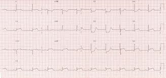
8am: King videoscope, King airway (laryngeal airway), and Ambu aScope training


Ambu aScope- video cable (instead of fiberoptic) with articulating tip and cord to attach to video monitor (all disposable- everything except the monitor is disposable). When starting, keep video cable taut which helps function of articulating tip. Currently there is an oxygen port, next model will also have a suction port. Handle has on/off switch and lever that controls tip. Turn on monitor first- has 3 hrs battery life but should generally be charged. Pre-load ETT on tube stop at top of video cable, can go through nose or mouth. Remember when performing nasotracheal intubation, use a smaller tube (7-0). Consider pre-treatment with nebulized lidocaine and lubrication with lidocaine jelly, oxymetazoline (Afrin) spray or phenylephrine drops if time permits. 

9:00-  Tranexamic acid (TXA) and trauma- Rob Mokszycki (PharmD)

Trauma pts are coagulopathic due to multiple causes. During resuscitation, have to transfuse fluids (crystalloids), blood products (massive transfusion protocol potentially), and pro-coagulant agents (TXA and Factor VII).

TXA works by inhibiting plasmin and preserves platelet function.

First promise of TXA was in CABG- less blood transfusion, platelets functioned better, less blood loss. Same in spinal surgery.

CRASH-2 [Effects of tranexamic acid on death, vascular occlusive events, and blood transfusion in trauma patients with significant haemorrhage (CRASH-2): a randomised, placebo-controlled trial. CRASH-2 trial collaborators. Lancet. 2010 Jul 3;376(9734):23-32. doi: 10.1016/S0140-6736(10)60835-5. Epub 2010 Jun 14.]

- international (except US) study of trauma pts and TXA. Outcome measure was all cause mortality. 2nd meausres- thrombotic complicatiosn, surgical intervation, transfusion. Inclusion- suspicion of bleeding within 8 hrs of injury, age >16. Exclusion- contraindication -active clotting, or allergy. 20,000 pts, groups well matched and low fall out. Injury severity was not measured. All cause mortality was decreased in TXA group. No difference in vascular occlusive events. Dosing not optimized- guesstimate from infusions given during surgery.

Secondary anaylsis of bleeding patients- benefit was greatest when given <1 hr, good if given 1-3 hrs, and negative if given >3 hrs. This was not a pre-specified subgroup.

NNT- 66 for all cause mortality. If this is true, could save 4000 lives in US.

MATTERs  [Arch Surg. 2012 Feb;147(2):113-9. doi: 10.1001/archsurg.2011.287. Epub 2011 Oct 17. Military Application of Tranexamic Acid in Trauma Emergency Resuscitation (MATTERs) Study. Morrison JJ, Dubose JJ, Rasmussen TE, Midwinter MJ.]

- military retrospective trial- primary outcome mortality. 800 patients. TXA group was more severely injured than control, and overall group had more GSW and blast wounds. 48h and in-hosptial mortality were better in TXA group, more pronounced in massive transfusion subgroup. This study did find a higher rate of PE and DVT in the TXA group.  NNT of 15, 7 in massive transfusion group. Time to TXA treatment and TXA dosage was not often recorded.

Summary- use TXA when <3 hrs and life threatening hemorrhage. Don’t use it if known clot. Possible harm in SAH cases (based on other studies).

Given in two 1 g doses, one as 10 min infusion and one as 8 hr infusion.

9:30- Student Loans- Jim Maletich

Medical school debt is increasing rapidly- median private med school debt is $180,000 and only $155,000 for public schools. Our residents owe from $84,000 to $450,000! As accrued interest is based on principal amount borrowed plus last years interest (capitalization), loan amounts continually increase. During residency, can be in negative amoritization- paying less than new interest accruing.

Deferment- grace period where you are not required to make payments. Usually capitalization does not occur during deferment.

Standard payment is your loan divided into 120 payments (generally about $2000/yr). Forbearance- don’t have to make payments but loans are all capitalized). EG- over 3 yrs capitalization adds $3000 to a $200,000 loan vs other fees and interest of $42,000.

Federal Repayment Plan for subsidized loans (IBR- income base repayment)- payments cap at 15% of discretionary income (your income – 150% of poverty limit[$11,490]) which ranges from $0 and $500/month. Government pays your interest so your interest does not capitalize. 3 year limit.

Consolidation- federal option- save 0.25 to 0.5% interest. Private option loses subsidized perks.

Public Service Loan Forgiveness- Must work for tax-exempt 501(c)(3)- federal, state, local, or tribal government for 120 monthly payments (residency counts). Then they repay the rest of your loans. Question if funding will exist by the time you qualify.

10- Admin Study Guide Part 2- Chintan Mistry

Boarding increases LWBS rates, increases length of stay, and diversion rates.

Parental consent is required except for emergency treatment and EMTALA screening, reproductive health, emancipated minors (court order, married, pregnant, minor who is a parent, Armed Forces).

The medical screening exam required by law (EMTALA) checks for an emergency medical condition, does extend to patients in waiting room (no time frame given). Duty ends if patient voluntarily leaves without being seen (LWBS). If an emergency medical condition (EMC) exists, treatment must be provided. EMC means potential serious injury to patient or organ. Transfer the patient when risk of transfer is outweighed by benefits. EMTALA applies to all facilities that accept Medicare payment. EMTALA violation means fines not covered by malpractice insurance, and potential exclusion from Medicare payments.

In all 50 states, child and elder abuse must be reported- can be done by physician, nurse, or social worker. Don’t assume PMD will do it. You are protected legally when good faith reports made (someone may file suit, but they won’t go anywhere). GSWs and stab wounds are also reportable in all states. 

For a malpractice suit to be successful, it must be proven that a breach of duty occurred (patient was in ED usually enough), actual injury did happen, standard of care was violated, and actions led to injury.

On call physicians can refuse transfer of patient with an emergency medical condition when the receiving hospital does not have the capacity to accommodate the patient. Capacity means facilities, expertise, and space.

Leaving against medical advice (AMA)- the treating physician determines if they are competent to make that decision. It’s nice to have a refusal form on file, but it isn’t necessary and doesn’t help much legally. Alternative care should be provided to patients leaving AMA, and discharge instructions should be given. In a teaching setting, the attending should be involved- also don’t let your nurse do it without your involvement. It IS good to have your nurse witness your discussion and document it independently. 

1030- Dialysis- Abraham Thomas

Nephrology update- can do dialysis for ED patients without requiring admission (can be done in ER, SUU, and HD unit). Call nephrologist early for potential discharges who are missing HD so that it can be rescheduled.

Good practice to check access site, ask about dry weights, dialysis schedule, dialysis center.

For HD patients who receive normal amounts of IV contrast for CT, they do not usually need immediate HD. It will increase their fluid balance by about 0.5 L. No good evidence it worsens anything, but caution may be beneficial for patients with temporary HD or improving renal function.

Gadolinium is contraindicated, but evidence of fibrosis was in patients who received multiple doses. If need, patients will need daily HD for 3 days afterwards.

BNP is not an accurate marker of volume status in HD patients- study shows some correlation with wide amounts of variation when studied in HD patients. However, a low BNP is probably accurate. Better to use clinical judgment to determine volume overload (dyspnea, RR, CXR, hypoxia).

Troponin can be elevated, but don’t ignore an elevated troponin. Change in troponin is more meaningful. Baseline elevation is not that concerning.

Please take down all bandages to look at access sites- especially intravascular catheters. Catheters are tunnels into IJ even though it looks subclavian. For AV grafts and fistulas- feel for thrill and listen for bruit. If it is not there, contact nephrology.

For bleeding vascular access (fistula or graft)- occluded artery for a minute or two. Then place mattress suture through skin ONLY  at bleeding site. You can also use gelfoam or hemcon or similar. Bleeding is a marker of a problem with the fistula/graft so let nephrologist know even when bleeding is controlled.

Aneurysms can form at fistula/graft site. If you see firmness, skin turning white, or ulceration nearby be very concerned. This aneurysm is at high risk for rupture and vascular & nephrology should be contacted.

Acute kidney injury- creatinine increased by 0.5. ATN, obstruction, and pre-renal common causes. Always ensure pt is voiding (consider Foley), image kidneys with U/S or CT, check UA. Avoid NSAIDs, ACE/ARBs, contrast. When you have a patient with hyperkalemia, order repeat lytes in 4-6 hours.

Indications for HD- fluid overload; hyperkalemia, esp. with EKG changes; refractory acidosis; overdose- aspirin, lithium, toxic alcohols; uremia with encephalopathy/pericarditis

Chronic kidney disease- GFR <60 for at least 3 months. Usually from DM and HTN. These patients likely to present with volume overload, anemia, infection, hyperkalemia. Try to avoid pRBC transfusion in someone waiting for transplant as it will make a match harder. Don’t give these patients NSAIDs, gentamycin, or IV contrast if possible. If contrast necessary, give normal saline 1 ml/kg/hr for 6 hours pre/post. No benefit from bicarbonate or Mucomyst. Give instructions for creatinine check in 2-3 days.

Expect to see more kidney transplants at ACMC, we do them here. Call Dr. Chet Desai or Dr. Darshika Chabbra (417100), who are transplant nephrologists, surgeon is Dr. Mittal. Any change in baseline creatinine is critical. Any obstruction is potentially catastrophic. Evaluation after surgery- hematoma or urine leak. Afterwards worry about rejection and infection, drug toxicity. Make sure you check UA, BMP and levels of immunosuppresants which should be back in 2 hours. U/S likely helpful- specify that patient has a transplant.

Many causes of hyperkalemia- final common pathway through aldosterone which opens Na/K pump in tubule. Urgency of treatment depends on cause and clinical condition of patient- ie hydrating a patient who can urinate will lower K by itself.

Calcium-onset is immediate and duration is 30-60 minutes. Give 1 g of CaCl or Cagluconate over 2-3 minutes. Stabilize cardiac membranes.

Insulin/D50- insulin will push K into muscle cells, will drop 0.5-1.2 mEq. Onset in 20-30 minutes.

Bicarbonate- does not do very much. You can use it if pt is very acidotic (7.2 or lower) and you are putting them on a bicarb gtt.

Albuterol- safe and very effective. Must use 10-20 mg over 10 minutes. Effect seen in 90 minutes.

Kayexalate- exchange resin that may bind potassium but there have been cases of bowel necrosis. Do not use in pts with ileus, recent surgery, opioid use, bowel obstruction. Avoid multiple doses or large doses (30 mg OK). Onset in 90 minutes, lasts 4-6 hours.

If patients make urine, treat with IVF (saline) and loop diuretic (furosemide).

Peritoneal dialysis- most common ED presentation is peritonitis- happens about 1x/year/patient. May have all or only one of abdominal pain, n/v, cloudy dialysate. Work up as usual plus add 1 L of 1.5% dextrose dialysate, let it dwell for at least 20 minutes but ideally 60 minutes. Send for cell count and cultures. Peritonitis – 100 cells with 50% PMNs. Give vancomycin 1 g IP if possible, IV otherwise PLUS ceftazidime 1 g IV/IP  or 500 mg ciprofloxacin bid. Can be discharged if not toxic and can be seen the next 1-2 days.

Hypercalcemia- treat levels 12-14 if symptomatic, all over 14.  First, 200-300 ml/hr of normal saline. Do not had furosemide unless volume overload. Add calcitonin (4-6 IU/kg sq over 6-12 hrs x 48 hrs), bisphosphonate (ie zoledronic acid 4 mg iv over 15 min- slow onset but lasts for weeks). 

1130- Amphetamines- Andrea Carlson

Pharmaceutical amphetamines-for ADHD, obesity (short term use), depression, enuresis, parkinsons (selegeline), alcoholism, narcolepsy.

Majority of drug abuse is misuse of drugs prescribed to patients, but illegal sales are rising. Check IDPH website before prescription refills of Adderall, etc.

Mild-moderate effects- agitation, hallucinations, HTN, tachycardia. Unlikely to have serious side effects from small, accidental ingestions.

Methamphetamines: 2nd only to cannabis in use. Significant geographical variation, seems to be increasing in Midwest.  Highest rate of admission in all drug abusers, mostly psych. Crank is dirty, smelly meth. Yaba is meth + caffeine, big in Thailand.

Meth works by releasing norepinephrine and dopamine. Binds to serotonin receptors and effects brain glutamate. Renally excreted, large Vd. Causes neuroexcitation, tachycardia, HTN, mydriasis, hyperthermia, diaphoresis. Like cocaine but less dysrhythmias and seizure, lasts longer, more psychosis.

Meth- get horrible teeth. “Meth bugs”- from tweaking and picking at skin. Gross.  


Meth- increases child neglect and violence, HIV transmission. Montana meth project (now in 7 other states including Illinois) has been very effective at reducing use.

Ecstasy (aka Adam, XTC, Molly)- often not in the pill (can be caffeine, designer drug, decongestants). Lower dose- trouble speaking, agitation, repetitive behaviors. Higher doses- more repetitive/compulsive behavior (bruxism), chorea, hyponatremia. Less cardiac effects. Chronic use- necrotizing vasculitis, psych problems.

Bath salts- cathinones- are newer congeners.

Drug screen- positive for a couple of days. Lots of false +- decongestants, buproprion, selegiline. MDMA and designer drugs false negative. Treat clinically.

Overall treatment- cool, calm room. Watch HR and core temp. Check BMP, CPK, EKG. CT head if seizure or significant CNS depression. Generous chemical sedation- up to 100 mg Valium (10 mg q 10 min). Can add anti-psychotics (newer generation). If sedated but still hypertensive, give phentolamine or nicardipine. 

Conference Notes 10-15-2013

Another Great week of conference!   

I will be taking three weeks off from doing Conference Notes due to a trip “Down Under”.    Conference Notes will start back up the week of November 12th.

In the meantime, be safe out there, and be careful with sharps.

Walchuk/Urumov                       Oral Boards

Case 1. 37 yo woman who is 35 weeks pregnant presents with RUQ abdominal pain, hypertension, headache, and emesis. Patient had a prolonged seizure after initial evaluation.  Diagnosis was Ecclampsia. Critical actions: IV Magnesium and Ativan to initially manage the seizure,   IV hydralazine or labetalol for BP control, left lateral decubitus positioning to improve IVC flow, set up fetal monitoring, arrange for delivery.   

Case2. 30 yo male hemophiliac presents with headache due to head injury from MVC.  Diagnosis was hemorrhagic cerebral contusion . Critical actions: Give factor 8 prior to CT head, correct to 100% of factor 8 level (50u/kg of factor 8), Get CT head, consult hematology/neurosurgery, ICU admission. Andrej teaching point:  You can also give FEIBA in this situation especially for patients with Factor 8 antibodies.  More Andrej teaching points: Don’t delay administering factor 8 for any reason if a head bleed is in the differential. Gotta give the Factor 8 prior to sending patient for CT head.   Easy way to remember Factor 8 dosing:  Head bleeds need to correct to 100% so give 50 IU/kg.  All other bleeds correct to 50% so give 25IU/kg.  

 Case3. 38yo male fell from ladder and injured left wrist.  Diagnosis was  perilunate dislocation.  Critical actions: Consult ortho for reduction, do complete neurovascular exam, administer pain medication.  Andrej teaching points: One way to help differentiate a scapho-lunate dissociation and a perilunate dislocation is that in the scapho-lunate dissociation there will be no overlapping bones on the AP view.  The perilunate and lunate dislocations will have overlapping bones on the AP view.  Also use the lateral view to help figure out the radiographic diagnosis.

Scapholunate disociation (Terry Thomas sign/No overlapping bones, Arcs of Gilula are intact)


Perilunate dislocation (AP and Lateral Views, Gilula's Arcs not intact)


Girzadas                Selection  Committee Meeting

The Breakfast with the RLT lecture was discussed.  Unfortunately no breakfast was provided at this lecture.  :-) My Bad.

Lambert                     Aortic U/S

First thing to look for is the spine shadow.  It’s your easiest landmark to identify in the transverse  view.   Aorta is anterior and left of spine.  IVC is right and anterior to the spine.     First branch off the aorta is the celiac trunk.   Second branch off the aorta is the SMA.

You will need to apply slow steady pressure with the probe onto the abdomen to move away bowel gas .  Angle the probe inferiorly slightly.  Keep the indicator on the probe oriented to the right.

Differentiating the Aorta from the IVC; get transverse images first. Make sure the indicator on the probe is toward the patient’s right side.  Look for arotic branch vessels.  Look for color and pulse waves.  Non-compressibility and thick walls favor aorta. Compressibility and thin walls favor IVC.

Transverse images will pick up both fusiform and saccular aneurysms.   The longitudinal image can miss saccular aneurysms.

Hypodense round area within spine shadow is the spinal canal.  Don’t confuse that with aorta.

Measure the aortic diameter from outer wall to outer wall using a transverse view.  Longitudinal views may underestimate the true diameter of the aorta.

Mike said he never found a case where someone was looking for AAA and missed it.  These usually are not subtle.

Iliac artery aneurysms can present as groin pain.  All the iliac aneurysms Mike has found have been in octogenarians.  If an older patient has groin pain, he scans down below the aortic bifurcation.

Bilateral iliac artery aneurysms

Tips for more difficult patients:  Use liver as an acoustic window for proximal aorta.  Apply firm steady pressure to compress bowel gas.  You can try the decubitus position to displace bowel. 

Lambert                      Cardiac U/S  

Mike presented a case of a young adult patient presenting with weakness and hypotension.  Echo showed pericardial effusion and plump aortic root (3.4cm).  Maybe had some RV collapse.   Pt responded to IV fluids. Pt went to CT and was found to have aortic dissection.  Pt never had any pain.   As noted in last week’s Conference Notes, Diagnosing aortic dissection is a bitch!

Bedside Echo changes the management of many patients.

Indications: cardiac arrest, near arrest, call an arrest, PE, pericardial effusion,  tamponade, severe LV dysfunction, valvular heart disease.

Use the probe with a small footprint to get a sound beam between the ribs.   The phase array probe has 2.5-3.5 frequency and gives you better contrast and has a high frame rate so you can see motion better.

B mode is brightness mode.    M mode is motion mode.  Basically, M mode drops an ice pick image through the bright mode image and shows that specific area repeatedly over time.  With M mode you can measure fetal heart tones or mitral valve motion.

Standard Bedside Echo  Images: Subcostal, parasternal, apical.   Apical is the least important.

In subcostal view, start looking right at the liver under the right costal margin and then sweep medially. When Mike says sweep he doesn’t move the probe on the abdomen but angles the probe toward the left shoulder.

Parasternal Long view:  Keep probe in the plane between the right shoulder and the left elbow.  Mike said he doesn’t care if the indicator is oriented to the right shoulder or left elbow.   In a heart with good contractility the LV wall and the septum should move pretty close together mid chamber and the mitral valve should come close to slapping the septum.

 Parasternal long view probe positioning

Parasternal Short view: keep probe in the plane between the right shoulder and left elbow. Orient the probe indicator to the left shoulder.

PE on echo will have a proportionately larger RV than LV. 

 PE in a Parasternal Short Window.  Note large RV in relation to normal size LV


Lambert /Chastain/Chan/Villalba                    Aorta and Echo Scanning Workshop

Have a great week everyone!

Conference Notes 10-8-2013

Another Conference chock full of high quality presentations!

Urumov     Study Guide   Orthopedics

Maisonneuve Fracture: Fracture of  proximal fibula with a tear in the tibiofibular syndesmosis. There is frequently a boney injury of the distal tibia or rupture of the deltoid ligament as well.

    J

Bohler’s angle:  Normal is 20-40 degrees.  If less, suspect fracture.


Pittsburgh knee rules :  Get xrays in a patient that fell or had blunt injury to knee AND  pt is over 50 or under 12 or pt cannot take 4 steps in ED.


Lateral Tibial plateau fractures are associated with ACL and MCL ligamentous injuries.  Medial tibial plateau fractures are associated with PCL and LCL ligament injuries.  

Lateral knee xrays are most sensitive for fracture.  Lipohemarthrosis (blood/fat interface seen on lateral film, or fat globules in blood seen on arthrocentesis) is another sign of knee fracture.

Knee exam: collateral ligaments should be tested with the knee in 30 degrees flexion.  >1cm of laxity is significant.  If you test the knee in full extension and there is laxity that suggests that the ACL is also injured.   Harwood comment: compare the pt’s injured knee exam  to the un-injured knee and look for assymetrical laxity.  That is probably a better marker than the classic 1cm rule.

Osteo chondritis dessicans: Specifically, OCD is a localized lesion in which a segment of subchondral bone and articular cartilage separates from the underlying bone, leaving either a stable or unstable fragment (up to date)  Stages range from a small compressed/non-displaced fragment to a completely detached and displaced boney foreign body


 Chopart Joint (yellow squigly line) and Lis Franc Joint (red squigly line)


Delta Pressure in patient with concern for compartment syndrome: Diastolic pressure  minus   compartment pressure should be greater than 30mm hg.  In one study noted in Tintinalli no patients had serious outcomes from compartment syndrome with a delta pressure greater than 30.

Risk factors for Achilles tendon rupture:  Age over 40, quinolone use, prior steroid injection.   Also more common in weekend warriors.  Diagnose with thompson’s test.

Pylon Fracture: Severe axial load fracture to ankle mortis.


Lis Franc Fracture:  Complications include compartment syndrome of foot, chronic arthritis, nonunion.  Tips to diagnose the injury is plantar ecchymoses, fx of proximal 2nd metatarsal, and wide space between proximal first and second metatarsal.  2 examples below:


Jones and Pseudo Jones fracture:  Jones fracture involves the metaphysic of the 5th mt.  Pseudojones is an avulsion fracture of the tuberosity of the 5th mt.   Jones fractures require post mold and non-weight bearing.   Psuedojones can be treated with an ace wrap/cast shoe and weight bearing as tolerated.


Jones Fracture


Pseudojones Fx

Kessen   

  EKG changes with elevated ICP are giant T-wave inversions.

Sodium Channel Blockers like TCA’s will cause prolonged QRS, right axis deviation and prominent r wave in AVR.  Herrmann comment: If you have a TCA overdose with a wide complex rhythm and tall R wave in AVR, give repeated bolus doses of sodium bicarb until the QRS narrows down.   Elise comment:  Bicarb drip will not work initially, you have to give bolus dose bicarb.


TCA EKG


Hyperkalemic changes of ekg include: Tall, narrow, peaked T waves,  p wave will flatten and eventually be lost,  qrs can progressively widen, bradycardia, sine wave.    Putman coment: Many of the critically ill hyperkalemic patients  I have seen had a wide, bradycardic rhythm.  So when you see bradycardia especially with a wide complex think hyperkalemia.

Hyperkalemia ECG


Gore       Tachyarrythmias

Consideration 1: Is the patient stable or unstable.   Place zoll pads on patient either way

Consideration 2: Are there P waves?

Consideration 3: Regular or Irregular

Consideration 4: QRS wide or narrow.

Categories:

Narrow/Reg: If there are p waves, sinus tach.  If no p waves: 1. SVT  TX=adenosine. 2. Aflutter with consistent block Tx=diltiazem.  3. Orthodromic WPW  Tx=adenosine or procainamide.   Elise comment: Give 0.5 mg of propofol prior to giving adenosine to prevent the very uncomfortable chest pressure/impending doom  feeling related to adenosine.    Elise comment:  In a patient with Orthodromic SVT from WPW  treat with adenosine.   It will look initially like the run-of-the-mill SVT.  There is no danger in treating orthodromic, narrow complex SVT from WPW with adenosine.  On initial presentation, you won’t know that it is WPW.

Narrow/Irreg:  If P waves present think aflutter or MAT.  If no P waves think afib.   You can use diltiazem for all of these.  Magnesium is another option, as is Amiodarone.   If the patient is unstable with a narrow/irregular tachycardia consider cardioversion.   Afib usually takes higher doses of electricity to convert.   Harwood and Elise comment: Ibutalide, procainamide, and propafenone are also options for conversin of afib.   Elise comment: Check for hypomagnesiemia. Harwood  comment: I give magnesium prior to giving ibutilide. It may decrease the risk of torsades with ibutilide.   Tekwani comment: Digoxin is a nice option for the borderline hypotensive patient with rapid afib.

Wide/Reg: Vtach.   Unstable Cardiovert.   Stable: amiodarone, procainamide.   If you can prove by previous ekg that it is SVT with bundle branch block then adenosine is ok.  Any doubt, treat as V-tach. 

Wide/Irreg: Afib with Abberancy (WPW)  Tx with procainomide.  

Case discussed of pt with WPW and presented with antidromic  SVT.

Chastain                      OB Ultrasound

To document that a pregnancy is in the uterus the image must include bladder, vaginal stripe and uterus.   Gestational sac/yolk salk/fetus must be within the uterus.

Ovaries lay medial and anterior to iliac vessels.   Ovaries look like chocolate chip cookies because of the follicles.

Ovary on ultrasound

You should see a gestational sac with TVOB at a B-hcg level of 1000.

Fetal heart rate should be 150-170 at 7-9 weeks.  The heart rate  will be a little lower before and after this period.

In second trimester, measure the biparietal diameter (inner skull to outer skull) to get accurate gestational age.  In third trimester you can estimate gestational age with femur length.

Free fluid in pouch of douglas or morrison’s pouch in the setting of pregnancy with abdominal pain and no definite intrauterine pregnancy indicates very high risk of ruptured ectopic pregnancy.  

Michelle presented multiple OB ultrasounds for the residents to interpret.

Postive fetal heart tones does not mean that the baby is in the uterus.   Mike Lambert knows a person who was a full term ectopic pregnancy in the peritoneal cavity.    Lam Socratic Question:  Can you have an IUP with an empty uterus on U/S and a quant-hcg over 2000?  Yes it is possible.  These patients need close follow up and repeat beta-hcg and U/S in 48 hours.   Also should return for pain, lightheadedness, vaginal bleeding.    Girzadas comment: You should obviously be consulting with OB in this situation.  This clinical situation is very high risk.

Permar    Safety Lecture      Sharps Safety

800,000 needlestick injuries per year in US.  Most are un-reported.

Sharps injury risk exposure to HIV, HBV, HCV.

40% occur during procedure.   40% occur during clean up.  20% occur during recapping, passing needles, improper disposal.    25% are inflicted by a co-worker.

Trale presented the data on all our EM resident sharps injuries.   Girzadas comment: I have been notified by Hospital Safety that we have a high number of  sharps injuries in our department.

Harwood comment:  Straight needles seem to be a common cause of needle punctures during central lines.    We should consider getting the straight needles out of the central line kit.   Trale comment: In the ICU, they don’t allow you to suture in the lines.    Harwood comment: We need to  change our central line kit to have curved needles and a needle driver.  Harwood comment: Consider dermabond to attach line to skin.   Walchuck comment: We should invest in thimbles for all the residents.  J

No recapping of needles.   Elise comment: Just don’t recap the needle! Just don’t do it!

Include sharp safety during time out.   Vocalize when you are moving sharps on the field or tray.

Double gloving has multiple advantages.   It reduces perforations to inner glove, makes visualization of out glove perforations easier and reduces the amount of blood that is transmitted into the injured caregiver by 95%!  The first glove “wipes” the blood off the needle before it gets through the second glove.

If you get stuck: wash hand thoroughly and  immediately.  Go to the ER to get medical attention and to report the injury.

28 day anti-retroviral therapy for HIV has a lot of side effects.  

Girzadas comment:  We will have thicker ,first layer gloves (green indicator gloves) available in central line kits and in the charting rooms.  Use a sterile glove over these thicker green gloves.  Please double glove for invasive procedures.  Discuss sharps safety during timeouts.  Be careful out there!

Den Ouden      An ER Hobit’s Tale

Case 1. Pt felt Imminent delivery of child.    Multiple checks and U/S showed a second trimester baby who was not engaged in the pelvis.   Pt had a large BM and felt much better.  Lesson: Trust your exam.

Case2. 21yo male with headache for 2 weeks prior to ED visit.  CT head shows 5cm mass of right thalamus.  Pt became anxious and his BP went up in the ED after hearing this news.    Pt then began posturing and blew right pupil.  Pt was treated with intubation/hyperventilation, mannitol, IV lido, and surgery.   Pt survived but had neuro deficits.   Lesson: Be prepared for rare emergent  events like herniation,  crics, peds resuscitations. 

Case3. 49yo male with Down’s.   Severe constipation for 2 weeks.  Christian disimpacted the patient.   5 minutes later patient became unresponsive.   Pt was bradycardic and hypotensive.  IV fluids started.  Pt went to the unit on pressors.   Pt was dc’d home with diagnosis of presumed prolonged vaso-vagal response.    Lesson: Be aware of possible vagal response to disimpaction and even rectal exam.  Christian discussed an abstract that described using rectal exam to convert SVT.    Probably don’t do rectal exam in patients with bradycardia.

Case4. 29yo female, 20weeks pregnant with worse headache of life.  Pt had central venous thrombosis.  Lesson: Beware pregnant and post-partum complaints.

Case5. 25 yo female 13 weeks pregnant with 2 days of left eye pain with eye movement.   Diagnosis with mri showed optic neuritis and MS.  Lesson: See Lesson 4.

Case 6. 35yo male with chest wall pain. Tender lump on exam.   CRP was 17.  Osteo/abscess at sterno-clavicular joint.    Lesson: Bad shit can happen with minimal exam findings.

Case 7. 40 yo male with Pharyngitis on amox.   Pt had thrush.   Do wet mount to check for candida.   Rapid HIV was positive.   Treated with nystatin and anti-retrovirals.   Lesson: Adult with oral candidiasis, think HIV.

Case 8. Pt had Infer-Posterior STEMI on EKg.  On exam, pt has acute left side paralysis.  History of acute chest pain.   Pt got CT on way to Cath Lab.   Wife later arrived and said stroke findings were chronic.   CT’s showed old stroke and no aortic dissection.  Lesson: Be alert for possible aortic dissection with chest pain and neuro findings/complaints.

Case9. 45 yo female with septic cholangitis.  Lactate=13, hypotensive.   Know how to give your own pressors without the pharmacist’s help to get through a tight spot when the pharmacist is not available.  1mg of any pressor in 1 liter of NS= 1mcg/ml.   Epi give 1mcg/min.  Norepi (levo) 10mcg/min.  Phenylepherine (Neo) 100mcg/min.   (1:10:100 rule)

Case10. 31yo male altered mental status,  in restraints.  Na was 106. Pt on multiple psych meds.  Pt received bolus NS by nursing staff.  Pt developed massive dieresis and rapidly corrected NA.  Chrisitian consulted nephrology and started D5 and DDAVP.  Hypertonic saline dosage if you are going to give it is 50-100ml. (maybe only for seizures) Harwood comment:  Psychogenic polydispia patients frequently don’t need any treatment.  If they have normal kidneys they will correct themselves with water restriction.   Lessson: Altered mental status in psychiatric patients can have serious organic causes.

Case 11. 1am, patient wants to leave. 33 male with hx of HTN.  Pt has chest pain. EKG, 2 trops,  and PERC all negative.   Pt came back to ED with severe leg pain.   Pt had ischemic right leg on second visit.  Chrisitian started esmolol/ntg.   5 min later pt becomes unresponsive.   Pt intubated.  Pt’s ekg shows ST elevation.   Pt gets tachy then bradycardic and arrests.   Labs come back with trop#3 negative.  Dimer was elevated to 3. Suspected diagnosis was aortic dissection.   Lesson: Diagnosing aortic dissection is a bitch.   The standard of care is to miss the diagnosis.

Case12. 35yo morbidly obese male.  Ruq and periumbilical pain. Couldn’t  scan him due to size.  Surgery wouldn’t OBS him without scan.   Brookfield Zoo CT is an urban legend, they don’t have CT’s for large animals.  Other hospital  had a CT that could handle a patient up to 550lbs. Pt was transferred for CT only and then back to same hospital for admission. Lesson: It is good to be aware of both your hospital and nearby institutions’ resources.

Case13. 55 you female with severe agitation.  Ativan, etomidate, succinylcholine all not working!!   IV access was the problem.   The first IV was not working.  Second IV placed and pt went down quickly with IV meds.   Lesson: If drugs aren’t working, check your patient’s IV.

Iannitelli       Acute Pancreatitis

Case1. Psych patient with ETOH abuse.  Pt had markedly elevated lipase.

Case2.  Severe epigastric pain.  Pt had ruq tenderness.  RUQ U/S showed acute cholecystitis.   Pt had gallstone pancreatitis.

Case3. 42 yo male with abdominal pain for one month.  Etoh abuse due to PTSD.   Lipase was normal but CT showed edema of pancreas.

20% of pancreatitis cases progress to SIRS and multi-organ failure.

Difficult diagnosis because presentation is protean and there is no diagnostic gold standard.

Etiology:  Gallstones, ETOH (heavy etoh abuse for more than 5 years), medications, infections, hypertriglyceridemia (increasing incidence, level should be >1000 to cause pancreatitis).

  Serum lipase is more specific and remains elevated longer than amylase.  Only downside to Lipase is that it can be elevated in other GI diseases.  To make the diagnosis of pancreatitis the lipase should be   3X normal.   All patients that you diagnose pancreatitis should get RUQ U/S to look for stones.   You don’t need to get a CT on every patient.   Get CT for uncertainty of diagnosis or concern for complications.

Most severe pancreatitis cases present initially as mild run-of-the-mill pancreatitis.  Probably safest to assume all initial cases have the potential to become severe in 48 hours.  

Cornerstone of treatment is early aggressive IV hydration in the first 24 hours.   250-500ml/hour in the first 12-24 hours.  LR is preferred due to lower rate of SIRS development.      

Give antibiotics if infection is suspected. Don’t give antibiotics for straightforward pancreatitis.

Consult GI and General Surgery as needed.

Hypoxemia, tachycardia, or confusion are subtle signs of early organ failure.   1 or 2 Ranson criteria should suggest a sicker patient.


Have a great week everybody! 

Conference Notes 10-1-2013

Conference Notes   10-1-2013

Be safe out there!  Be careful with sharps.

Maletich/Anderson                        Oral Boards

Case 1. Adult Epiglottitis; pt presented with severe sore throat and forward leaning posturing.  Lateral neck xray showed enlarged epiglottis. Pt maintained a patent airway and did not require intubation/cricothyrotomy  Critical actions:  IV antibiotics,  ENT consultation, prepare for any airway compromise by having both advanced intubation options (direct and video laryngoscopy) and a surgical airway option ready.  Racemic epinephrine is another option to reduce epiglottis edema.

Case 2.  Pt presented with hypoglycemic seizure and altered mental status.  Diagnosis was panhypopituitarism due to previous surgery of pituitary gland.  Patient was on vacation and was not taking her usual medications. Critical actions: IV fluids, treat hypoglycemia,  IV hydrocortisone ,  management of hyperkalemia.  

Case 3.  38yo male struck in mouth with baseball bat while coaching kids little league.  Tooth was completely avulsed. Critical actions: Place tooth in Hank’s solution (Save-a-Tooth) until reimplantation,  attempt reimplantation of tooth, consult Oral-Maxillofacial Surgery.   Get tooth in the socket as soon as possible.  Do not scrub tooth.  Only rinse tooth gently  with saline.  Alternative mediums to transport an avulsed tooth include chilled milk, and saliva.

Elise and Harwood comments: You have other options to lower fever besides oral or rectal acetaminophen.  You can use IM/IV ketorolac or in kids, you can use IV acetaminophen.

Elise comment: appropriate local anesthesia to replace an avulsed tooth requires injection of anesthetic around maxillary ridge (supraperiostial injection).  Infraorbital nerve block does not provide adequate anesthesia of maxilla or tooth socket.

Roberts and Hedges’ Text:

The most common technique for intraoral local anesthesia of individual teeth is supraperiosteal infiltration (Fig. 30-5). This technique can provide complete relief of a toothache. Select the area to be anesthetized and dry it with gauze. Apply a topical anesthetic, such as 20% benzocaine or 5% lidocaine ointment, as described. Ask the patient to close the jaw slightly and relax the facial musculature. Grasp the mucous membrane of the area with a piece of gauze. Pull the gauze outward and downward in the maxilla and outward and upward in the mandible to extend the mucosa fully and to delineate the mucobuccal fold. Puncture the mucobuccal fold with the bevel of the needle facing the tooth. Aspirate the area and deposit approximately 1 to 2 mL of local anesthetic (2% lidocaine should be used here) at the apex (area of the root tip) of the involved tooth (see Fig. 30-5). It is helpful to place a finger against the outer aspect of the lip overlying the injection site. Apply firm and steady pressure against the lip and slowly inject the local anesthetic into the supraperiosteal site. This helps prevent ballooning of the lip.

The purpose of the injection is to deposit the anesthetic near the bone that supports the tooth. The anesthetic must penetrate the cortex of bone to reach the nerve of the individual tooth, so the injection may fail if the solution is deposited too far from the periosteum, if the needle is passed too far above the roots of the teeth, or if the bone in the area is unusually thick or dense. If anesthesia is unsuccessful, inject the palatal side. It may take 5 to 10 minutes to achieve full anesthesia with this technique, however, and the procedure may not be as effective for the posterior molars. Infiltration of the area around the maxillary canine and the first premolars will anesthetize the MSA and ASA nerves; lacerations of the upper lip can be treated by bilateral injection in the canine fossa areas.


Balogun    Trauma Lecture

Blunt poly-traumatized patient due to motorcycle accident transferred to ACMC.  Pt had LMA placed in outside hospital and had a cardiac arrest at outside hospital prior to transfer to ACMC.  Pt had massive free fluid in peritoneal cavity on FAST exam.  Pt  had cardiac arrest in ED.  Pt was intubated and bilateral chest tubes were placed.  PRBC’s were transfused.  Pt had severe facial trauma and had nose packed multiple times for severe epistaxis.    OR was not immediately available due to 2 other trauma  cases going at the time.   Massive transfusion protocol was initiated.   Patient arrested again and was resuscitated with ACLS.  Pt develops a massive neck hematoma.    Patient continued to bleed profusely from head/facial injuries.  Pt had pelvic binder placed for widely diastatic pelvic fracture.

Elise comment: At some point you have to decide if utilizing massive resources to further attempt resuscitation is futile.  

Sola’s response:  That is exactly correct.  The trauma attending evaluated the patient and when pt again arrested the decision was made that further resuscitative efforts were futile.  

Altered mental status or agitation can be a sign of shock.

(Up to Date) The Advanced Trauma Life Support (ATLS) manual describes four classes of hemorrhage to emphasize the early signs of the shock state [3]. Clinicians should note that significant drops in blood pressure are generally not manifested until class III hemorrhage develops, and up to 30 percent of a patient's blood volume can be lost before this occurs:


Girzadas comment:  Class 1=10% (500ml) blood loss (no change in vitals),  Class2=20% (1000ml)blood loss (tachycardia),  Class 3=30% (1500mL) blood loss (hypotension/mental status change), Class 4= 40% (2000ml) blood loss (hypotension/altered mental status/minimal urine output).

No evidence favoring either NS or LR for resuscitation.

Massive Transfusion Protocol: 1:1:1 transfusion ratio of of PRBC’s: FFP: Platelets.   Here at ACMC the massive transfusion protocol can be activated by a Trauma attending or an ED attending.   Elise comment: as a resident you have to really push to get the blood products into the patient.  You have to place order in care connection and call blood bank at (41-5581) Nurse must send a “Blood Release Request” in order for blood to be released.   Christine comment: Have a tech run this form down to blood bank and bring the blood products back.  Massive transfusion protocol gives the patient 10 units of prbcs, 6 units of FFP, 1 unit of apheresis platelets, 2 units of cryoprecipitate. The first 4 units of prbc’s of the massive transfusion protocol are O-neg prbc’s that are sent to ED immediately.  Use the level 1 rapid warmer/infuser to give fluids and blood rapidly.

No human studies support the use of vasopressors in the resuscitation of trauma patients.   Elise comment: If patients with hemorrhagic shock are on pressors, stop the pressors.  If they aren’t on pressors, don’t start them.  Give blood.   

There was a discussion of using IO lines for prbc transfusion.  Consensus was that IO lines are an ok way to give prbc’s but not as effective as large bore iv lines.   Roberts and Hedges reference a rate of 21 ml per minute for prbc transfusions via IO versus 35 ml per minute transfusion rate via large bore IV.

Tranexamic acid: CRASH-2 showed lower mortality overall in trauma patients and lower death from hemorrhage.   Best if given in the first 3 hours.  1g IVPB over 10 min followed by 1g IVPB over 8 hours.  You can’t give this in the same line as blood products.   TXA is not indicated for isolated head bleeds. Elise: Give this in the first three hours in patients who are hemorrhaging or who you expect to need blood transfusion.    Harwood comment: TXA can also be used for gynecologic bleeding.  Kelly comment: I had a patient who requested TXA for gyne bleeding. She had received TXA for severe vaginal bleeding previously   Elise comment: OB attendings here feel there are other options preferable to TXA for vaginal bleeding.

Opportunities for improvement indentified by Sola:

Get control of scalp lacs and epistaxis.   Use a staple gun to close scalp wounds.   Harwood comment: consider using hemecon  or some other material promote coagulation to control severe nasal bleeding.  Christine comment: You may consider posterior balloon to stop epistaxis.  Harwood comment: be careful with placing objects in the nose when you suspect there are mid face fractures so that you aren’t placing a catheter into the brain.

Be diligent with placing the pelvic binder and check it regularly to make sure it is in proper position.  Activate the massive transfusion protocol early (as soon as patient arrives in ED).  Control hypothermia with arctic sun device or bear hugger.   Get help  from ED resources to help manage these difficult cases. Elise comment: Please ask the ED attending for guidance and help with these resuscitation if the Trauma attending is in the OR.  Harwood comment:  Nice job intubatiing this patient with massive facial injuries.  Some brain injuries are associated with coagulopathies and that may have been a factor in his hemorrhage.   IR may have been an option to embolize his pelvic vessels or his spleen.  Trushar comment: If you are at a non-level 1 ED, get a patient such as this out of the ED rapidly.  Maybe intubate them and get blood stated and get them out.  Erik comment:  Don’t do any work up at the transferring hospital, just get them out to the receiving hospital ASAP.

Mistry        Study Guide        Administration   

Occurrence-based malpractice insurance policies cover you for events during the policy period regardless of when the claim is made.    Claims-made policies cover you only when the policy is in effect.  So with claims-made policies you need to purchase a tail to cover you for any claims made after the policy ends.  A Claims-made policy plus tail coverage effectively becomes an occurrence policy.

 Insurance distributes risk over a large group.

If you are sued more often than the average physician you are flagged by insurance companies.

A simple made up word to remember charting requirements: “Fourtwoteneight” (pronounced 4-2-10-8 say it fast)    Four HPI elements,  two past med/ family/surg/ social history components,   Ten ROS systems, eight physical exam components are what is needed for a level 5 chart.

Boarding increases the rate of patients who leave without being seen.    Boarding does not affect the waiting times of critically ill patients but it does affect the waiting time of less ill patients.

Criteria for deiversion: No inpatient telemetry beds open.    ED is overwhelmed by patients or other problems such as electrical failure or water main breakage.    If all the hospitals in a region go on diversion, then all the hospitals in that region have to go off diversion.

Consent for minors: Attempt to contact the parents but if the patient’s health is at risk do the right thing and treat the patient while attempting to contact the parents. In fact, most routine, indicated care can be started while you are attempting to contact the parent.   You definitely can perform the EMTALA screening exam prior to getting parental consent.     Emancipated minors are those that are married, pregnant, minor who is a parent, minor who is in the military.  Emancipated minors do not need parental consent to begin treatment.

Chiefs/Faculty              Joint Aspiration and Reduction Lab

5 Stations:  1. Shoulder, 2. elbow/knee, 3. wrist, 4. Hip, 5. Ankle


Have a good week everybody!

Conference Notes 9-24-2013

Hayward    Study Guide   Hematology 

Case of elderly female with melena and dark bloody emesis with a hgb of 9.  Teaching point was to transfuse prbc’s  based on the patient’s condition rather than just the number.   Harwood and Elise comments:  We have had it beaten into our heads to not transfuse unless the hgb is <7.  This question makes the point that you also need to consider the patient’s clinical status.  If the patient has life threatening bleeding, it is reasonable to initiate transfusion prior to a hgb of 7.

Petechiae is the most common sign of ITP.   Acute ITP is more common in kids.   Chronic ITP more common in adults.

Differentiating severe liver disease from DIC is difficult but the d-dimer should be normal or only minimally elevated with severe liver disease.

There is debate regarding doing a paracentesis with a platelet count less than 20 or an INR >2.  Elise comment: There is controversy on this topic.  Elise would be ok with doing a paracentesis with an INR <4.  Christine agreed.     Elise comment:  Elise found a  study of over 1000 paracentesis and many patients had an INR of around 2 or platelet count less than 50 and all did well.

Quick medline search also found this abstract:


Most commonly observed abnormality with DIC is thrombocytopenia.   Most common cause of DIC is gram negative sepsis.

Hemophilia patient with a head injury needs factor 8 replacement prior to any imaging.  Elise comment: this is critical knowledge for both real life and the boards.   Girzadas comment: Patients will often come in with their own factor 8.

Von Willebrand DZ with gingival bleeding can be treated with DDAVP intranasally.   Avoid NSAIDs in patients with von willebrand dz.

Acute chest syndrome is the leading cause of death in patients with sickle cell disease in the US.   Hydroxyurea can decrease the incidence of painful crises in adults but is not approved for pediatric patients.

Clopidagrel inhibits the ADP receptor on the platelet.

ABO transfusion reaction results in hemoglobinemia and hemoglobinuriauria,  decreased serum haptoglobin, elevated ldh.   In one series of reports, the combination of an increased serum LDH and a reduced haptoglobin was 90 percent specific for diagnosing hemolysis, while the combination of a normal serum LDH and a serum haptoglobin >25 mg/dL was 92 percent sensitive for ruling out hemolysis. Haptoglobin binds to Hgb released during intravascular or extravascular hemolysis.   (Up to Date)  

Febrile neutropenic patients:  You should avoid rectal exam if you can.   Harwood comment: the only time I would do a rectal in such a patient is if the patient has a fever and complains of rectal pain.  Any other complaints, I don’t do a rectal.  Elise comment: I totally agree. 

Finger tip laceration on a patient with INR of 6:    Harwood comment:  Stitch it up, apply pressure dressing and give oral vitamin K (2.5mg).  Girzadas comment: For skin avulsion of finger tip, apply heme-con and give oral vitamin K.   Elise comment:  New recommendations from American College of  Chest Surgeons  says no oral vitamin K in the non-bleeding patient unless the INR is 10 or higher. 

Up to Date Chart for Management of Supratheraputic INR.  They lean toward the 2008 guidelines in that they still suggest vitamin k for non-bleeding patient with INR between 5 and 10.  Again the ACCP 2012 guidelines recommend no vitamin k for non-bleeding patients with INR’s less than 10.


Treatment of hypercalcemia:  Initial treatment is normal saline IV to restore euvolemia.    Aggressive hydration and lasix has fallen out of favor because it does not lower calcium more than saline alone. Next step is calcitonin. Steroids may be useful with some tumors.

Managing sickle cell pain crises:  Elise comment: new thinking is avoid fluid boluses and just give maintenance fluids.   Excess fluids may result in atelectasis or acute chest syndrome.   Consider using D5.45 because normal saline may increase sickling.   Also no benefit to O2 unless patient is hypoxic.  Oral rehydration is also acceptable.

Treatment for TTP is plasma exchange.   

Wise        Morbidity and Mortality   (some details changed so specific case is not identifiable)

Teenage  female  was hit by an auto.  Obvious head trauma with altered mental status.  Pt is intubated on arrival.    After tracheal intubation, pt desaturates and becomes bradycardic.   Pt had left chest needle thoracostomy  placed with rush of air from the left chest.  Subsequently vitals improved.  Presumed diagnosis was tension pneumothorax.    Team was next concerned about head injury, pulmonary contusions, possible intra-abdominal injury, and open right humerus fracture.

Pt was sent directly to the PICU following  CT imaging.  This was done based on neurosurgery concerns about intracranial injury.  Neurosurgery wanted the patient up in the ICU to have a intracranial pressure monitoring device placed.   Harwood comment: I don’t feel this was the right decision by the ED to let this patient go up to the PICU at this point in the workup. 

When patient gets to PICU, the results of the abdomen/pelvis CT shows severe pelvic fx with pelvic hematoma.   Pt becomes hypotensive in PICU.   She is basically an unstable peds trauma patient in the PICU.   Patient was started on dopamine and IV fluids.   Hgb was 6.  PRBC transfusion was started.  Patient was too unstable to go to IR.  Peds Ortho Attending took patient to OR to place ex-fix of pelvis. 

Opportunity for improvement:  Don’t let a patient out of the ED environment until the patient is stabilized even if a consultant is saying to send the patient up to the PICU now.  We should review critical tests prior to sending patient from ED to another unit.

No strong evidence that intracranial pressure monitoring improves outcomes in kids.

Harwood:   A type and cross and PRBC’s generally should be ordered in ED.  Pelvic fx can be identified and management begun in ED by wrapping  pelvis.   Prioritizing the head injury over the torso injuries risks missing life-threatening injuries.  The torso injuries are what are going to kill a polytraumatized young person.  Dopamine is generally not indicated to manage hypotension in trauma patients.   PRBC’s and IV crystalloid are standard management of hypotension in trauma patients .  Kids/teenagers can maintain a normal range bp until they crash.  You gotta watch the pulse because that will show early hypotension before the blood pressure will.   Central lines are generally not critical to trauma resuscitations.  Large bore peripheral IV’s and Intraosseous lines can provide crystalloid and blood products probably faster.  Elise comment: Trauma resuscitations are different than medical resuscitations and in general should be done in the ED not the ICU.

Adjust your trauma algorithm for kids/teenagers.   Speak calmly and softly to them.  Do not give direct orders to them.

Thoracic trauma accounts for 20% of pediatric trauma deaths.  Kids have a compliant chest wall and can have serious injuries of chest and abdomen without fx.   Ribs provide less protection for liver and spleen.  Use broslow tape to determine chest tube size in younger children.

Abdominal wall is less developed and less protective.  Kidneys more prone to fracture.  Spleen has a strong capsule and is less likely to rupture.

Pediatric Trauma score: (higher the better/less than 8 has 20% mortality and needs transfer to trauma center)   Our patient had a score of 4.

Pediatric Trauma Score

Components

+2

+1

-1

SCORE

Weight

>20 kg (44 lbs)

10-20 kg (22-44 lbs)

<10 kg (22 lbs)


Airway

Patent

Maintainable

Unmaintainable


Systolic BP

Pulses

> 90

Radial

50 – 90

Carotid

< 50

Nonpalpable


CNS

Awake

+LOC (responsive)

Unresponsive


Fractures

None

Closed or suspected

Multiple closed or open


Wounds

None

Minor

Major, penetrating or Burns > 10%


TOTAL SCORE


9 –12 Minor Trauma Use local guidelines/protocols

6 –8 Potentially Life Threatening Suggests need for Trauma Center

0 –5 Life Threatening Need for Trauma Center

<0 Usually Fatal Transport to Nearest Facility


Mckean comment: Sometimes with trauma and non-trauma patients, the ER is the best place for them for awhile.   We have the specialized knowledge/skill and access to rapid diagnostic testing that is not available in any other area of the hospital.   Tekwani comment:  We need to overcome our own anxiety as physicians with these traumatized kids/teenagers and understand we are the best people to manage them in the hospital.   Harwood comments: If you see tire marks on the torso, you have to proceed under the assumption that there are internal injuries.   However, at times you will be amazed how kids and adults can have body parts get run over by cars and not have any injury.  Every patient has an end point when it is reasonable for them to go up to the unit. Key thing is to identify the life threatening injuries and begin addressing their management in the ED.  After that, the ICU can continue management.     

 McKean   CPC Case Presentation  for the National Championship at ACEP in Seatle

4yo child with altered mental status.   Developed on a plane ride home from Walt Disney World.  Pt was noted to have left side weakness.  Ct showed right sided hypodense lesions.   Angiogram shows carotid dissection on right.   It was thought child suffered dissection from roller coaster ride.  Pt was treated with ASA and did well.

Kettaneh    My right leg is cold ( some details altered so specific case is not identifiable)

  79 yo male with cold right leg and abdominal pain.  Pt has RLQ tenderness and absent pulses in the right leg below femoral pulse (femoral pulses present bilat).   Vascular surgery consulted immediately.  Heparin started.  Dissection was clinically ruled out by the presence of  bilaterally equal femoral pulses.  Harwood comment: Heparin is problematic prior to evaluating for AAA.  Kettaneh: Pt had an ultrasound done at the bedside showing a normal abdominal aorta.  Treatment for arterial occlusion is heparin, asa, iv fluids, pain control, dependent positioning of affected limb.  Labs in this case showed acutely decreased gfr, elevated cpk and elevated lactate, and  leukocytosis.   ED physicians ordered CT abdomen to evaluate abdominal pain/ abnormal labs.  Prior to CT aabd/pelvis however,  pt went to the OR for vascular surgery thrombectomy.  The night after surgery, pt developed peritonitis.   CTA showed ischemic bowel, renal infarcts and thrombus in the aorta.  There was some note of an intimal flap in the aorta.  Patient died.   Although this patient was likely not salvageable even at ED presentation, again this case brings up the issue that the ED has the global picture of the patient and needs to identify critical diagnoses before giving the patient up to the management of the consultant.

Knight      FEIBA for the Rapid Reversal of Bleeding in Patients with Warfarin Induced Coagulopathy

FFP for reversal has the limitations of large volume, incomplete reversal, and need for type and cross match.

FEIBA is basically a 4 factor prothrombin complex concentrates

We gave FEIBA and IV vitamin K to reverse warfarin coagulopathy in patients with major hemorrhage (life/limb threat, intracranial bleed, drop of 2 HGB).

Time to INR  of 1.5 or less was 127 minutes.  4 patients died (19%).   4 patients had thrombotic complications after day 3  following FEIBA for a rate of 14%.   This rate of thrombotic events is higher than previous studies.  FEIBA’s duration of action is 72 hours so these thromboses may be due to the patient no longer being anticoagulated rather than the effect of FEIBA.    Elise and Harwood comments: there is risk of thromboses from underlying pathology and being in the ICU.  Those risks are likely more the cause of thrombotic events in these patients rather than FEIBA

Katiyar    McGurk   Sayger                   Documentation/Coding for Medical Decision Making

Medical decision making section: Provides a capsule summary of what is going on with this patient and the direction of management.

3 categories that make up medical decision making: # of possible diagnoses, amount of data/diagnostic testing  that was analyzed, risk of significant morbidity/mortality.

It is not enough to say I reviewed a test.  You have to say what your interpretation is of the test.

Document for example: pt presents with fever, cough and sob.  Will evaluate with cxr, cbc, chemistries  abg and blood cultures.  Will initiate treatment with nebulized albuterol. 

Document what you learned from review of prior records.

Document your discussions with consultants and pmd.

Document your discussion with family members.

Document re-evaluation of patients.    Include if you utilized a specific guideline like PERC or Heart Score.

Ryan   Med Student  Review


Conference Notes 9-10-2013

Please consider donating to our EM Foundation to benefit resident education/development.  Thanks for your consideration/generosity.
/em-foundation/

*EMF Golf outing October 8.  All faculty and Alumni welcome!

*Congratulations to Andrej Urumov for winning the International CPC Competition in Marseilles, France!

Jakubowicz/Carlson       Oral Boards

Case 1.   Rocky mountain spotted fever.  62 yo male with fever and headache for 2 days followed by new rash on distal extremities for the last 12-24 hours.  Critical actions: Recognize the rash, treat hypotension with fluids, start abx (doxycycline is first line, chloramphenicol  can be used for the doxy allergic patient and pregnant patients.  doxy is ok in kids)  , report the illness to health department and get confirmatory testing.   The frequency of diagnosis of RMSF  has tripled in the last 10 years but the mortality has dropped.  RMSF is also know as “tick typhus” because of it’s clinical similarities with typhus.  Infectious agent is ricketsia ricketsii.  Vectors are dog, wood, and lone star ticks.  Incidence increases with age.  Rash starts 2-6 days after onset of illness.  Labs show anemia, low platelets, low sodium, and ARF.  Test with ELISA.   Gore comment:  Can this look like TTP or meningitis?   Andrea: Yes, this is a difficult diagnosis but you have to look at the context of the presentation.

RMSF Rash


Case 2.   Phenobarbitol  overdose in a child.  Child presented with altered mental status, hypothermia, and hypoglycemia.   Mom was poisoning child.     Critical actions:  Intubate, treat hypoglycemia,  correct hypothermia, Multi-dose  charcoal, Alkalinize the urine, identify child abuse and contact child protective services.   Andrea comment: get an alcohol level as alcohol can  cause hypoglycemia and sedation as well.   On CXR, the patient had non-cardiogenic pulmonary edema.  Andrea gave the differential of toxin caused non-cardiogenic pulmonary edema :  Salicilates, opioids, phenobarb, ethclorvynol, calcium channel blockers were the ones I caught.

Case 3.  Spontaneous  pneumomediastinum in a teenager due to laughing while doing situps.       Critical actions:   Don’t do a CT chest.  Treat pain.   Don’t let patient fly on a plane or run in track meet until pneumomediastinum resolves.    Both can increase the volume of pneumomediastinum.   Boerhaave syndrome can have a similar CXR appearance to spontaneous pneumomediastinum but  is usually related to vomiting and this patient wasn’t vomiting.  Boerhaave’s is due to esophageal rupture and is a serious risk of mediastinitis.   Spontaneous pneumomediastinum is not a risk for mediastinitis.  Spinnaker sign:  Elevation of thymus due to pneumomediastinum.

Bad Pneumomediastinum                         


Spinnaker Sign


Knight      Facial Trauma

Case 1. 21 yo male in a high speed mvc.  Pt had a seizure in the field.  Pt also had serious facial trauma.   On arrival to ED pt was intubated using etomidate/rocuronium.  Cricothyrotomy tray was the planned back up maneuver if intubation failed.  Another option is intubation with ketamine sedation and topical anesthetic in oropharynx without neuromuscular blockers.   Don’t nasally intubate patients with severe facial trauma.  

LeFort  Fractures

Treatment for LeFort fxs: airway management, nasal packing, IV antibiotics, plan for surgical repair, ophthalmology evaluation.

Mandible fractures:  Order of frequency  body>angle>condyle.  Tongue blade test 95% sensitive/68% specific.   Check the ears for tm rupture/hemotympanum/ear canal injury.   Open fractures need IV pcn or clinda, admission and ORIF.   Closed fractures can be dc’d home on pain meds and soft diet.

Case 2. 38yo female assaulted by cousin.  Pt was punched in the head and bit in the nose.    Pt’s nose was largely amputated by bite.    Human bite wounds have 10% risk of infection.  Infections are polymicrobial with concern for eikenella corodens.    Pt required complex reconstruction including a forehead flap and cartilage graft from ribs.

Case 3. 40 yo male transferred to ACMC for multiple gsw’s to chest and face.  One gsw went through left orbit.   Orbital blowout fractures are more commonly caused by blunt trauma to orbital area.   Patients with orbital blowout fractures may have infraorbital anesthesia and diplopia on upward gaze.   Dispo home with instructions to not blow nose, smoke, or drink through straw.  Need follow up with plastics or max-face surgery.    Globe rupture:  Use bent paperclips to retract eyelids to see eye.  Patients may have irregular or tear shaped pupil.    Konicki Comment:  In an eye that is not obviously ruptured, you can do a very gentle U/S with a lot of gel.  If the probe just touches the gel, you wil get an image of the eyeball.   Girzadas comment: Have a low threshold for getting a CT for possible orbital fractures.  I have been impressed with how many orbital fractures I have found with relatively mild ecchymoses around orbit after blunt trauma.  Don’t “diss” the paperclips.  Bent paper clips are very effective in retracting swollen eyelids. 

Harwood comments: You can anesthetize the airway by injecting lido through cricothyroid membrane.  Knowing the different types of LeFort fractures is not that clinically relevant.  Most facial fractures are complex and don’t follow the strict classifications.

Levato      ACMC Recommended  Management of Infections

Uncomplicated UTI: Macrobid BID X 5 days,  Cephalexin TID X7 days,  last choice is Cipro X3 days.    Cipro should be avoided unless you can’t use the first two.   GFR cutoff for using macrobid is 50.     Harwood comment: You have to be careful with macrobid in the elderly with low GFR.   Definitely need to use cephalexin or cipro in patients with GFR less than 50.  

ACMC is really trying to not use Cipro for uncomplicated uti’s.    Bactrim is not a good choice for empiric treatment of uti.  Resistance to bactrim is around 31%.

Cipro has growing resistance from pseudomonas and e coli.  Both bacteria has 25-30% resistance to cipro.    Cipro and other quinolones  are associated with NAP-1 hyper-toxin producing C-diff strain.

Use cipro/flagyl combo only for the pcn allergic patients.

Question by Adam Wise: What do we use for nursing home patients with  pcn allergy who are diagnosed with UTI?    Answer:  Cipro or Gent.   Aztreonam also suffers from a lot of resistance from e coli.  Similar resistance pattern as cipro.   You have to pick on a 3 not great choices in this situation.   Gent probably has the best microbial coverage but you have to be aware of potential nephrotoxicity.

For intra-abdominal infections that are uncomplicated like simple diverticulitis give Ceftriaxone/flagyll.  If patient has a complicated intra-abdominal infection such as post-surgical, hospital acquired,  or abscess formation give Zosyn.  In the last 10 years, Cefoxitin has had less than 80% effectiveness against anaerobes.  That’s why we use it less often.  We have been using it less over the last few years so it’s effectiveness may have improved. Christine comment: She feels she has had surgery request cefoxitin being given for appendicitis.   Should I push back and recommend Ceftriaxone/flagyll?  Levato answer: Cefoxitin is ok for that situation. 

Harwood comment:  If you have a nursing home patient with ED diagnoses of both pneumonia and uti.  Just treat the pneumonia.  You will get decent coverage of most likely uti pathogens.  When you get urine culture back in 2 days you can narrow your antibiotic choice.

Diabetic lower extremity cellulitis limited to 2cm around ulcer : Treat strep and staph with Ancef.      For legit diabetic foot (foot is red/swollen/painful and at risk for amputation) give Vanco and Zosyn.    Interesting discussion about the common presentation we see which is diabetics with cellulitis involving a larger area of the lower leg but don’t have foot at risk for amputation.   Consensus was to consider the overall picture of the patient, level of blood sugar control, vascular status of affected leg, and rapidity of progression of infection when deciding between ancef and Vanco/Zosyn combination.

This Diabetic Foot would get Vanco/Zosyn based on our discussion


Two grading scales for diabetic foot


General rule at ACMC  for all infections is go to a beta lactam first and move to other classes for specific reasons.  One exception is macrobid for uncomplicated uti.

Herrmann question: Where do we find the ACMC recommendations for antibiotic treatment of infections.    Levato answer: We are working on an online bug and drug section on the ACMC website.

Zosyn, Unasyn, Imipenem, cefoxitin all have adequate anaerobic coverage and when using these, you do not need to add flagyl.

Febrile neutropenia: Cefipime or Imipenem.   Add vanco for HCAP, cultures  gram positive organisms,  suspect MRSA, line infection, neutropenic shock,or  pcn allergy.

C-Diff treatment: Treating with flagyl (500mg po TID) alone is only appropriate for mild first presentations  or first recurrances   (WBC<15K, Cr<1.5).   Anything else treat with oral vanco (125mg qid).    Severe C-Diff infections give 500mg Vanco enterally QID + IV Flagyll 500mg QID.

Katiyar     Study Guide   Ortho

Sorry I missed the majority of this excellent lecture.

Traumatic knee dislocation: This injury needs emergent ortho referral.   Popliteal artery and common peroneal nerve are at risk.    50% of  these dislocations spontaneously reduce prior to arrival to ED.  Do ABI, if <0.9 do arteriogram or CT angio.

Knee Dislocation

Harwood comment:  You can have an intimal injury of popliteal artery that clinical looks ok with nl pulses initially. I would not trust ABI’s to decide on doing a CT angio.  If the patient has a knee dislocation do a ct angio and admit.  Group discussion ensued,  One of our residents had a knee dislocation personally, had an angio and was discharged with a knee immobilizer.   All faculty present felt they would admit all knee dislocations.   We are talking knee dislocations, not patellar dislocations.

Tibial plateau fractures: Can be subtle.   Harwood and Girzadas comments:  these patients have a lot of pain, and have effusions.  Pay close attention to any depression or fracture lines through the lateral tibial plateau. 

Positive birefringent crystals is pseudogout (PP;positive=pseudo).  Negative birefringent crystals is gout (NG; negative=gout).


Fort         EKG Basics

Professor Chamberlain’s 10 rules of normal ekg

PR=120-200 ms

QRS should be less than 120

QRS should be up in I and II

Qrs and T’s should be concordant

All waves are negative in AVr

Normal R wave and S wave progression


St segments should be isoelectric except V2 and V3

No abnormal q waves.

T waves must be upright except avr


Weinckeback (progressive prolongation of pr interval)


Mobitz 2 Block


Accelerated ideoventricular rhythm: Rate 50-100, has fusion and capture beats.   Common Boards Question: In the setting of TPA reperfusion, what is the  management  for Accelerated Ideoventricular Rhythm?    Answer: Observation


Accelerated junctional rhythm :  Rate of 60-130.  Retrograde p waves can be present.    Harwood comments: When you see this rhythm, think  congenital problem, medication effect,  rheumatic fever, post surgery, anorexia, CHF, caffeine, isoproterenol, and brain injury. 


 WPW 


Brian covered the common myocardial infarct patterns.


Wellen’s syndrome:  Biphasic T wave inversions in the antero-septal leads.  Associated with critical stenosis of proximal LAD.


Left main coronary artery occlusion: Widespread horizontal ST depression, most prominent in leads I, II and V4-6.  (6 or more leads with ST depression)

  • ·  ST elevation in aVR ≥ 1mm
  • ·  ST elevation in aVR ≥ V1


This is considered a STEMI equivilent in the new ACC guidelines.  AVR ST elevation should be alittle more than ST elevation in V1.  In this EKG they are pretty close.  Bottom line if you see more than 6 leads of ST depression and AVR and V1 are elevated and the patient is having chest pain, call a STEMI or Speak with the cardiologist.

Patient Safety/Doctor Safety     Be Safe out there.   Be careful with Sharps.


Conference Notes 9-3-2013

Reminder  Emergency Medicine Foundation Golf Outing Tuesday afternoon/evening, October 8th.      

Gupta     Study Guide     Trauma

If you suspect tension pneumothorax, do immediate needle thoracostomy.  Do not wait for CXR or a clinical response to IV fluids.

If you are seeing this CXR you are late in needling the chest:

CT is more sensitive/specific than ultrasound for diagnosing solid organ injury.   It is more sensitive than ultrasound for picking up small amounts of intraperitoneal blood. CT is not great for picking up hollow viscous injuries.    Harwood comment: Use serial exams to diagnose hollow viscous injuries if you are suspicious and CT is non-diagnostic.

Kelly Williamson comment:  In adults with blunt flank trauma who don’t have gross hematuria and who are not hypotensive, you don’t need to work up the patient for renal injury.   You don’t need to send the urine to the lab.   You can visually inspect the urine to check for gross hematuria.  If the urine is not grossly bloody, you don’t need to work patient up for renal injury.

In contrast to adults, microscopic hematuria in pediatric patients who suffered blunt trauma still requires further evaluation.  Harwood comment:  You won’t find a lot of treatable injuries using microscopic hematuria as your only criteria to do a IV contrast CT.    Gupta comment: For the boards,  microscopic hematuria in adults requires no further urinary tract evaluation.   Microscopic hematuria in kids,  requires a CT abdomen/pelvis with iv contrast.  Elise comment: The injury you would be looking for is at the junction between the kidney and the ureter.  This area is very mobile in kids and is prone to injury.    Harwood/Elise/Erik/Christine comment: They wouldn’t CT a kid for just a few RBC’s in their urine.   The kid would need in addition some history or sign of serious trauma to proceed with CT.  There was then a discussion about the recent publication of the PECARN decision instrument to decide on work up of the abdomen in kids with blunt trauma.


Elise comments: Strengths-as always with PECARN, really big study, well done, reliable results. Weakness- they don't incorporate US in their workup (during EMRAP interview with an author he stated that some centers weren't using US routinely).  Doesn't specifically discuss microscopic hematuria, but simple 7 point instrument to help decrease CT use in these kids.  Decision instrument missed 2 kidney injuries:  one kid had gross hematuria, the other had distracting injury (rib fracture) and microscopic hematuria (10 RBC per hpf).

Harwood comments: This is a great rule. It has a 99.9% (-)  predictive value & doesn't use micro urine or LFT's (in old rules). It did missed 7/203 injuries (requiring treatment). However, of the 7 misses, 2 kids were 2 y/o's. 1 did indeed have gross hematuria & was treated without an operation (did get a blood transfusion). 5 were 16-17 y/o's. All would have gotten ED CT's based on presentation. This includes a 17 y/o MVA had rib injury, blood in the belly (& 10 RBC's in urine). This pt got got embolization (spleen? most likely).


Intraperitoneal bladder rupture usually results from a compressive force to abdomen causing rupture of dome of the bladder.   Gross hematuria is present 98% of the time.  


Extraperitoneal bladder rupture is caused usually by sharp boney edges of pelvic fracture.


Large myocardial contusions can result in cardiogenic shock.   Harwood and Girzadas comment: There is no good diagnostic test for myocardial contusion and there is no accepted treatment for myocardial contusion so there is no point expending resources looking for this.   If the patient is in shock you gotta think hemorrhage, hemorrhage, hemorrhage.  But if hemorrhage is ruled out and the pt is hypotensive or has an arrhythmia it could possibly be from myocardial contusion but still is more likely to be due to hemorrhage.

The spleen is the most common  organ injured in blunt trauma . Blunt trauma; organs most frequently injured in decreasing order: Spleen>liver>kidney>small bowel .  Penetrating trauma; organs most frequently injured in decreasing order:  Small intestine>liver>colon.      

Harwood comment:  In elderly patients with multiple rib fractures (3-4 broken ribs) , they have a surprisingly high mortality.   Admit them for observation.  Christine comment: Mortality in the elderly increases by 19% for every rib broken above 2.

Left sided diaphragmatic hernias are three times more likely than right side.   The liver is thought to protect the right diaphragm.  Diaphragm injuries can be subtle initially and pt may become symptomatic years later.

Treat patients with pelvic fractures and hypotension with  pelvic binding, prbc/ffp transfusion,  and angiography with embolization.

Jakubovich     Pediatric Trauma   M&M

Pediatric GSW victim(5yo) transferred from outside hospital to ACMC.  Intubation was attempted at initial hospital but was either unsuccessful or tube became dislodged.    Upon arrival at ACMC,  pt could not be intubated using laryngoscopy.   Resident and attending were also unable to intubate with glidescope.   Patient, however, was able to be bag-valve-mask ventilated.    ED/Surg team proceeded to perform cricothyrotomy on pt.  PRBC transfusion started.

Pt went to OR and had multiple intra-abdominal injuries repaired.   In the OR, the cricothyrotomy developed an air leak and was removed.  Peds annesthesia was able to intubate the patient.

Patient was discharged home.  He suffered from PTSD.

Airway issues:   In infants with prominent occiput, you can put a towel under the shoulders to line up the airway.    In kids age 5 or older, it is recommended to put a towel under the head to line up the airway.  Harwood comment:  Place a towel under the shoulders first, if that doesn’t work to line up the airway, move the towel to behind the head and look again. 

Pediatric bougie will fit through a 4 ET tube.   Adult bougie will fit through a 5.5 ET tube.

Since we were able to bag-valve-mask this patient, did we need to cric this patient?  Elise comment: A pediatric LMA may have been a reasonable option in this situation.  Our pediatric difficult airway box should have a pediatric sized LMA. Erik comment:  Maybe a retrograde intubation over a wire passed through the cric would be a nice technique.  Kelly comment: Don’t beat yourself up over doing the cric, you need an airway and getting the airway anyway you can is OK.   Christine/Elise comment: If you can bag the patient successfully, you usually have time to consult pediatric anesthesia emergently.   There was some consensus among attendings present that an LMA would have been the optimal bridge device until pediatric anesthesia could be consulted.

Should we have done an open cric or a needle cric?  Age recommendations go down to 5yo for open cric.  However at age 5 and below it will be more difficult due to anatomic size.   In this case Trauma Surgery had no difficulty passing a shiley through the cricothyroid membrane.   Harwood: It is best to use a commercial Melker percutaneous seldinger technique cricothyrotomy kit.   Other jerry-rigged needle cricothyrotomy techniques and jerry-rigged jet insuflation are prone to failure.  Elise: I agree the Melker kit is the best option.  But again, if you can ventilate a patient it is better to continue bagging or place an LMA and get help from anesthesia.   Christine comment: Your respiratory therapists will have no experience with needle crics.   Erik comment: It may be helpful to  remember that in many places around the world, they do entire surgical procedures without intubation and just bag-valve-mask the patient.

Elise follow up comment: We currently stock size 3 LMA in the PED.  This is sized for patients weighing 30 - 50 kg, and would work for average sized kids age 8 to small adult.

Plan is to add smaller sizes (2 and 2.5) which will cover kids 1 year old and up.  There are smaller LMAs, but my thought is to keep this simple, and little babies are usually less challenging intubations.  

Getting blood:  You can make an MR# for a DOE name prior to arrival of the patient to order blood from Blood Bank.

Kmetuk        Cardiac Trauma

Case 1.    Penetrating left chest trauma.   CXR discussed by Harwood shows hemothorax despite left chest tube, heart is enlarged and is very suspicious in a young patient for hemopericardium.   FAST exam identified hemopericardium.   Pt went to OR for repair of RV.  Pt did well.

Cardiac Box  

RV is the most commonly injured cardiac structure due to penetrating trauma.  Penetrating injury to the chest can definitely injure abdominal organs as well.   Left ventricle is more likely to self seal than right ventricle.   Stab to the heart is 17X more likely to survive than GSW to the heart.

Salzman comment: Patients with stab wounds to the heart  are the one’s who can survive an ED thoracotomy.    If you do an ED thoracotomy, push the ET tube down the right mainstem bronchus to deflate the left lung and improve your visualization of the heart.

Salzman comment:  If you have pressors infusing in a young trauma patient it means they need blood Dammit!   Young trauma patients should never get pressors (except possibly neurogenic shock) they need blood.

Salzman comment: You can close wound in the RV with foley catheter, stitches, or staples, or just having someone put a finger in the hole.

Salzman comment:  In a 20something patient with penetrating chest trauma, and FAST shows some pericardial fluid, that is blood.    Also in a patient who is arrested or peri-arrested don’t hesitate, just get started on the thoracotomy.  ( As an aside, when breaking bad news to families, in a similar fashion get to the fact that their loved one is dead with little preamble. )  There is no role for needle pericardiocentesis in penetrating cardiac injury.   The blood in the pericardial sac is usually clotted and can’t be aspirated with a needle.

Case 2.  51yo male victim of motorcycle vs. truck collision. Blunt chest trauma.  Intubated, hypotensive.   CXR shows wide mediastinum.    CT chest shows aortic injury.   PT was started on esmolol but BP dropped and esmolol was stopped.  Pt’s aorta was repaired in the OR with a graft.   Pt walked out of hospital 20 days later.

90% of patients with aortic injury will die at the scene.  50% of the survivors die in the next 24 hours.

High risk historical factors for aortic injury: rapid deceleration injury, t strikemvc, fall more than 3 stories.

Febbo question:  Do you need to CT scan an asymptomatic patient who has one of the above mechanisms?    Salzman comment: Yes, he has seen pt’s who have intimal injuries or pseudoanuerysms that have been relatively assymptomatic. 

Walchuk       Mandibular Dislocation

Anterior dislocation is the more common dislocation and is reducible in the ED.   Posterior dislocations are rare and need specialty consultation and likely OR reduction/surgery.

Pt’s with anterior dislocations can’t close their mouths.  There will be a preauricular depression in the skin.   Check cranial nerves 5 and 7.   Unilateral dislocations will have jaw deviation to the side opposite the dislocation.

Posterior dislocations can result in external auditory canal injury.

Intra TMJ lidocaine can give significant pain relief and even allow reduction.

You can try to elicit a gag response.  The gag response blocks contraction of the masseter and can result in reduction of the dislocation.

Dan Gromis method of masseter massage was presented on video.    Basically, an assistant massages the masseters on both sides while the operator applies inferior and posterior pressure on the mandible while at the same time tilting the chin upward.   The video demonstrated very easy and non-painful reductions.

After reduction, pt should be on a soft diet for a couple of days.

Harwood comment: Main thing is to apply some downward traction on the mandible and get the chin elevated relative to the angle of the mandible and then move the mandible posteriorly.

Lam       Fever in infants 0-90 days

Fever/age cutoffs to initiate work up in kids:  0-90 days=38C    3mo-36mo=39C

10% of kids 0-90 days with 38C or higher temp will have serious bacterial illness.

Clear cut national consensus to do sepsis workup and admit/give IV antibiotics in kids 0-28 days.   Give Amp and Cefotaxime.   Sam said he has the experience of regularly giving  ceftriaxone in kids 0-28 days with no complications during his fellowship.

Well appearing infants 29-90 days old with Temp 38 or higher is the difficult group to make decisions on.

 Rochester criteria: WBC 5-15K, UA with less than 10wbc/hpf, less than 5wbc in the stool.  No LP criteria.  Also kids can’t have any significant pmh or clinical exam findings of serious infection.  These low risk criteria miss 15-45% of serious bacterial illness.. 

Boston criteria: WBC under 20k, CSF with less than 10wbc, UA with less than 10 WBC or neg urine dip. All kids got 50mg/kg ceftriaxone IM.  All had re-check and second dose of ceftriaxone in 24 hours.   These criteria missed 6% of serious bacterial illnesses.

Philadelphia criteria: Non-toxic, WBC <15K, low bands, urine with <10wbc /hpf and no bacteria on gram stain, CSF<8wbc/hpf, CXR with no infiltrate,  Stool with not a lot of WBC’s.  High risk kids were admitted with antibiotics.  Low risk kids did not receive antibiotics and were sent home.  This strategy missed the least amount of serious bacterial illnesses.

In this age group there is a 10% chance of viral meningitis.

If you are going to treat a febrile child in this age group with ceftriaxone, you need to do a LP prior to giving ceftriaxone.   There is national consensus on this management point as well.

OM does not make a child in this age group low risk.  They still need an LP.

Bloody taps require admission and antibiotics because the results are uninterpretable. 

Sam uses the Philadelphia criteria in kids less than 8 weeks.  He does a full septic workup on them.  If results fit low risk criteria, he sends them home.   Scott Altman and Sam both said they will give kids antibiotics (ceftriaxone) prior to sending them home.  

There was some practice variability voiced in the group.   Some faculty will do full septic workup, give antibiotics, and admit for observation in all these febrile kids 0-8 weeks. 

Occult bacteremia in kids 3 months to 36 months in the era of pneumo and HIB vaccinations is basically dead.  The incidence of contaminated blood cultures is approximately 10X the incidence of positive blood culture. So getting blood cultures in these patients leads to many more false positives than true positives.   In Immunized kids 3-36 months with high fever, instead of getting blood cultures, look for uti.  A kid with 2-3 doses of vaccine can probably be considered immunized.

In kids in this  age group, if you get a CBC for whatever reason and the WBC is over 20K get UA/UCX and get a CXR.  Kids with a WBC >20 are at higher risk for both.

Kids who have a clear cut diagnosis of a viral illness(positive rsv, chicken pox, croup, bronchiolitis,etc), meningitis is very unlikely.   

Thanks for reading!  Stay safe out there.   Double glove and be cautious/take your time with procedures to avoid Sharps Injuries.  


Conference Notes 8-20-2013

Some Awesome Pearls at Conference this week!  But first gotta promote the Foundation:

SAVE THE DATE for the ACMC EM Foundation Golf Outing

What: Emergency Medicine Golf Outing – October 1, 2013

 Location: TBD

Time: Following the Emergency Medicine Conference/Early Dept Meeting and Vice-Chair Vote

Cost: $150/golfer (includes golf, dinner & drinks)

Purpose: To raise awareness for the Emergency Medicine Endowment which provides funding for residents to further their education

Contact: Jason Keene, Director of Development 708-684-2012 or jason.keene@advocatehealth.com

*Proceeds will benefit the Emergency Medicine Endowment

*Electronic Invitation to follow


McKean/C. Kulstad     Oral Boards

Case1.   TTP    Critical Actions: plasmapheresis (the same thing as plasma exchange) is the key treatment, CT head, No LP, Admit to ICU, get Hematology Consult.  Optimal care: DIC panel, discuss FFP transfusion, assess for life threatening bleed,  discuss steroids with hematology.   Classic TTP Pentad:  microangiopathic hemolytic anemia (MAHA), neurologic abnormalities, fever, renal disease, thrombocytopenia.  Frequently the pentad is not present.  The cornerstones of the diagnosis of TTP are thrombocytopenia and hemolytic anemia.  Most patients have some neurologic and renal abnormalities but the severity of the neuro and renal abnormalities is highly variable.     This disease is more common in middle age persons. Females more commonly affected than males.  TTP has high mortality  90% untreated and 20% treated.   Don’t give platelets.  TTP and HUS are very similar disease processes in adults.

Group discussion about management of TTP in regard to LP.   Most agreed when there is diagnostic uncertainty between TTP and meningitis give antibiotics and steroids prior to getting labs back and prior to CT/ LP.  If platelet count is less than 50K (Harwood’s cutoff) don’t do LP.  If platelets above 50K probably safe to do LP.  Girzadas: Some case series have shown no bleeding complications with very low platelet counts.    Erik comment: If you do a LP for possible meningitis and the platelet count comes back below 50K, the patient will likely be fine.   Everyone agreed though that Harwood’s cut off at 50K was prudent.

Up to Date reference:  It is traditionally stated that a platelet count greater than 40,000 to 50,000/microL provides safety for interventional procedures such as lumbar puncture [11-13]. In certain procedures where bleeding risks are greater, or the risk of complications from minor bleeding is high, a platelet count greater than 80,000 to 100,000/microL is often required by surgeons (especially neurosurgeons) or anesthesiologists.

A quick Abstract on this topic:

The risk of spinal haematoma following neuraxial anaesthesia or lumbar puncture in thrombocytopenic individuals.

van Veen JJ, Nokes TJ, Makris M.

Source

Sheffield Haemophilia and Thrombosis Centre, Royal Hallamshire Hospital, Sheffield, UK. joost.vanveen@ukgateway.net

Abstract

Neuraxial anaesthesia is increasingly performed in thrombocytopenic patients at the time of delivery of pregnancy. There is a lack of data regarding the optimum platelet count at which spinal procedures can be safely performed. Reports are often confounded by the presence of other risk factors for spinal haematomata, such as anticoagulants, antiplatelet agents and other acquired or congenital coagulopathies/platelet function defects or rapidly falling platelet counts. In the absence of these additional risk factors, a platelet count of 80 x 10(9)/l is a 'safe' count for placing an epidural or spinal anaesthetic and 40 x 10(9)/l is a 'safe' count for lumbar puncture. It is likely that lower platelet counts may also be safe but there is insufficient published evidence to make recommendations for lower levels at this stage. For patients with platelet counts of 50-80 x 10(9)/l requiring epidural or spinal anaesthesia and patients with a platelet count 20-40 x 10(9)/l requiring a lumbar puncture, an individual decision based on assessment of risks and benefits should be made.

Case2.   Achilles tendon rupture    Critical actions: Complete extremity exam,  consult ortho, splint in moderate  plantar flexion.  Tear is usually just above the calcaneus.   Surgical repair has less risk of re-rupture but of course carries surgical complications.   Plantaris tendon rupture causes whip like pain in calf.  Treatment of plantaris rupture is pain control  and ace wrap.  Medial gastrocnemius tear also can cause sudden calf pain and is treated with conservative measures.

Thompson's Test: Right leg in photo is abnormal and doesn't plantar flex with squeezing the calf (positive test=tendon rupture). You can also do the test with the patient prone and knee flexed.

Discussion between Harwood and Elise about best position to splint ankle with Achilles tendon rupture.  Harwood basically said it is not that important to splint in plantar flexion.  Elise felt plantar flexion de-stressed the tendon and was  preferred over  splinting  at 90 degrees.

Case3.   Perforated Ulcer    Critical actions: IV antibiotics (cover gram negatives & anaerobes)  and emergent surgical consult.    Optimal care: PPI, NG, foley. 

Free air under the diaphragm on CXR:


C. Kulstad    Study Guide Trauma

Nexus

The NLC decision instrument stipulates that radiography is not necessary if patients satisfy ALL five of the following low-risk criteria:

  • §  Absence of posterior midline cervical tenderness
  • §  Normal level of alertness
  • §  No evidence of intoxication
  • §  No abnormal neurologic findings
  • §  No painful distracting injuries


Canadian C-spine rules

The CCR involves the following steps:

  • §  Condition One: Perform radiography in patients with any of the following:
    • ·         Age 65 years or older
    • ·         Dangerous mechanism of injury: fall from 1 m (3 ft) or five stairs; axial load to the head, such as diving accident; motor vehicle crash at high speed (>100 km/hour [>62 mph]); motorized recreational vehicle accident; ejection from a vehicle; bicycle collision with an immovable object, such as tree or parked car
    • ·         Paresthesias in the extremities
  • § 
    • ·         Simple rear end motor vehicle accident; excludes: pushed into oncoming traffic; hit by bus or large truck; rollover; hit by high speed (>100 km/hour [>62 mph]) vehicle
    • ·         Sitting position in emergency department
    • ·         Ambulatory at any time
    • ·         Delayed onset of neck pain
    • ·         Absence of midline cervical spine tenderness

Patients without any of the low-risk factors listed here are NOT suitable for range of motion testing; they must be assessed with radiographs.

If a patient does exhibit any of the low-risk factors, perform range of motion testing, as described in Condition Three below.

  • §  not

Head injury on Coumadin: Check INR,  observe in ED for a few hours   Observe  in hospital if INR is very high. 

 Pregnant patient with chest trauma and pneumothorax: Place tube in the 3rd intercostal space in the mid-axillary line.   It’s alittle higher placement than in a non-pregnant patient.

In contrast to orbital floor “blow out fx’s”,  Orbital roof fractures and fractures through the naso-ethmoid bone risk injury or infection to the brain.  These roof and naso-ethomid fractures need admission and consult of neurosurgery.

Stable C-spine fractures: compression less than 25%, spinous process fracture, transverse process fracture.

 Fractures of spinous and transverse processes are stable.


Treatment of severe blunt laryngeal injury (laryngeal fx).   Group discussion on this one:  Start with RSI but be prepared to have great difficulty with the airway.  You want to paralyze the patient to give you an optimal look but loss of muscle tone may cause the airway to collapse.  There is a high risk of creating a false passage with the ET tube.  Your rescue approach is problematic as well.  Cricothyrotomy is not recommended due to disruption of the normal cricothyroid anatomy.   Harwood and Kelly Williamson suggested being prepared to do a seldinger cric through the trachea in this situation.   Everyone cringed at facing this difficult situation.

Zones and Triangles of the neck


Girzadas comment: Way to remember that zone 1 is at the bottom and zone 3 is at the top. Big Red 1= Aorta is in zone 1 and Zone 3 is up toward the Third ventricle. 

Open mandible fractures require admission for IV antibiotics.   Open fracture is identified by blood between teeth.   Tongue blade test is 96% sensitive for identifying mandible fracture.   Closed  mandible fractures can go home.

NG and foley sizes for kids are 2X the ET tube size.   Chest tubes are 4X the ET tube size.  ET tube size is age/4 + 4.

Evaluate for incomplete cord injuries with anal wink, cremasteric reflex, and bubocavernosis reflex.

Chest tube output to prompt OR in Kids: 15ml/kg initially and 4ml/kg/hr ongoing.  For adults: 1500ml initial and 200ml/hr x 4 hours

Central cord syndrome: Upper extremities weaker than lower extremities and proximal weakness more pronounced than distal weakness.  Man in a barrel is the classic analogy for this neuro deficit. 

Brown Sequard syndrome : Ipsilateral paralysis and loss of pain and temperature on the contralateral side.


Subdural/Epidural/SAH Bleeds


Williamson/Patel/Tekwani         Post-Partum Emergencies

Case 1: 36 yo female G2P2 s/p c-section one week ago.  Pt had headache.  She then developed some chest pain.   BP 132/80  P 52  P/O 98%.    Broad ddx of head and chest emergencies was considered.    Kari was thinking pt had predominantly a headache problem.  Pt improved with treatment.   D-dimer was 17.  CXR was pretty much pristine.   CT-PE study showed aortic dissection extending to innominate and carotid arteries.    There was debate between radiology and CT surgery about whether the CT findings were artifact.    Kari called in Cardiology to do transesophageal echo.  TEE showed/confirmed  dissection.  Pt went to OR.   Discussion about CT evaluation of PE vs. Dissection.   If you are specifically looking for one disorder (Dissection or PE) get the appropriate test.  If both are in the differential, get the CT PE. You are probably more likely to see a dissection on the PE study than see a PE on the dissection study.

Kari comment:  The presentation of dissection can be extremely protean or subtle.

Case 2: 38yo female with chest pain 9days after c-section.     DDX was PE, dissection, musculoskeletal pain,  pre-ecclampsia, nstemi, cardiomyopathy.  EKG showed subtle inferior depression and high lateral minimal ST elevation.  Most people in the room read it as nonspecific.  Nobody would have called a STEMI on this EKG.  Troponin was elevated.   Pt got cathed and was found to have LAD dissection.  There are multiple case reports of post-partum woman having spontaneous coronary artery dissections. 

Coronary artery dissection or thrombosis is more common in the post-partum patient than acute coronary syndrome.  These patients don’t usually have underlying atherosclerosis.

Case 3.   31 yo female 1 week after delivery.   Pt has sob.  Went to an urgent care center the day before.   Pt had nl ekg and neg d-dimer, trop from previous visit at urgent care center.   DDX=PE, post-partum cardiomyopathy, pericardial effusion, anemia, pre-ecclampsia.  EKG did not show any acute ischemia.  CXR is pristine.  Trops are negative.  Hgb is 11.6. VQ is low prob.  Venous dopplers are negative.   Ambulatory pulse ox was nl.  Pt had no protein in urine.  BP was 156/83.  Pt was dc’d home…..    Pt develops a headache 5 days later and she collapses.  Pt was severely  hypertensive.   She arrests in the ED. Difficult intubation.   CT head was neg.  Pt was placed on magnesium and labetalol.   She remained in a coma and support was withdrawn.  Diagnosis was pre-ecclampsia.

20% of patients present with htn alone (>140/90) and no proteinuria.  Ecclampsia can present up to 8 weeks after delivery.   It is critical to get a urine protein/creatinine ratio.  This can be ordered as a single urine test from lab.  Anything above 0.19 suggest pre-ecclampsia.  Also get CBC and CMP.  Treat pre-ecclampsia with magnesium.      

Lecturers comments:  The post-partum patient should be considered very carefully and deserves a more detailed work up.

#1 risk factor for endometritis is C-section.   Harwood comment: There is a sub population that can be treated as an outpt.    Kelly’s comment: If patients receive IV antibiotics in the hospital, when they clinically improve and defervesce they no longer need any antibiotics IV or oral.

Peripartum cardiomyopathy can go out to 5 months after delivery.  

PE is more likely post-partum than during pregnancy.

Harwood comment:  Pregnancy is a stress test.  Some women fail this stress test and get head bleeds, aneurysms, post partum cardiomyopathy, pe’s , and dissections.

Frazer       Safety Lecture

Sign out: Admit to MICU for ativan overdose.  Pt is somnolent but stable and is boarding in the ER waiting for the ICU to open up.   Pt has been in ED for several hours.   He becomes more alert.  He gets downgraded to step down unit.   Pt gets transferred up to floor.  Although the ICU attending was contacted, no one ever contacted the floor attending that the patient was going to the floor.  The patient spent several hours on the floor with no orders.

All attending should be contacted when there is an upgrade or downgrade of admission destination.

There was a trouble shooting discussion and the group felt that  putting the medical team as a consult with the reason as MICU admit.    At sign out we should be asking each other is this patient downgradable?   If so who needs to be contacted.

Felder       CV Workshop

It should be a “map” of your career and interests.  

Put  information lines under each heading  in reverse chronological order.

Keep it concise.    If you have a lot of pubs just list topics and the number of references under each topic.  Then add at the bottom: Detailed list of publications on request.

Make a top 5 list of achievements/strengths/interests.   Be sure your CV highlights these strengths.

Girzadas comment:  Use the decision points in residency to differentiate yourself on your CV.   Lectures, elective rotations, resident project, and leadership positions all can be used strategically to demonstrate a focus in your residency training. 

5 quick fixes:

  •  
    1. Creative editing like bolding should be used to highlight your strengths.
    2. Make  your career timeline easy to follow.
    3. List your publications but if you have many make it more conscise as described above.
    4. Make the CV easy on the eyes
    5. Make formatting consistent.

You may want to customize your CV to different jobs you are applying to.

Before you interview for your dream job, think about applying and interviewing for a job you are not super-interested in to give yourself a chance to practice.

Cover letters should give alittle more definition to who you are and what kind of doctor you are beyond the objective work milestones on your CV.    Christine comment:  It should give the employer some info about why you are interested in a particular geographic area or hospital.  

Altman comment:  Personal connections such as phone calls and networking are the most powerful way to get jobs.    Say,  “Dr. X told me to call you”


Ryan                       Medical Student Review

Conference Notes 8-13-2013

Another Outstanding Week in Conference!  but first an important word from our sponsor: 

SAVE THE DATE for the ACMC EM Foundation Golf Outing

What: Emergency Medicine Golf Outing – October 1, 2013

 Location: TBD

Time: Immediately Following the Emergency Medicine Conference

Cost: $150/golfer (includes golf, dinner & drinks)

Purpose: To raise awareness for the Emergency Medicine Endowment which provides funding for residents to further their education

Contact: Jason Keene, Director of Development 708-684-2012 or jason.keene@advocatehealth.com

*Proceeds will benefit the Emergency Medicine Endowment

*Electronic Invitation to follow


Lotsa pics in these notes if you don't see them scroll to the bottom and click "read in browser"

Coghlan      Study Guide   ENT

Most common source of bleeding in posterior nose bleeds is the sphenopalatine artery.

Compications of posterior nasal packs: eustacian tube dysfunction, nasopulmonary reflex  causing  apnea/bradycardia/hypoxia/bronchoconstriction/cardiac arrest,  necrosis of the columella. 

Best nerve block for complex ear laceration is the auriculotemporal block.


Diabetic with signs of otitis externa:  Consider malignant OE due to pseudomonas.   These patients are ill-appearing, diabetic, febrile, elderly and have granulation tissue in their external ear canal.


Malignant OE


OE with TM perforation:  Use Floxin because it is the only topical antibiotic approved by FDA for use with an open middle ear.    If cost is an issue, use Cortisporin otic suspension as an alternative .

Tooth avulsion: never re-implant primary teeth.  Adult tooth that is avulsed hold by crown,  irrigate with saline and re-implant in the socket ASAP.   Beyond 2 hours the chance of success of re-mplantation is quite low.   Best transport mediums for an avulsed tooth is Hank’s solution.   Saliva, milk, saline are 2nd, 3rd, 4th best..

ANUG (acute necrotizing gingivitis) :  Due to oral anaerobes,  pt’s have “punched out inter-dental lesions of the gingival”.    Treat with antibiotics that cover anaerobes.

    ANUG


Lemierres syndrome:  Infectious thrombophelbitis of the IJ due to a parapharyngeal space infection.  Pt’s can get septic pulmonary emboli.


Diagram of Lemierre's Syndrome


Aphthous stomatitis: Cellular immune mediated response.   Treat with pain medication and steroids.

Pt  gets hit in the mouth with a baseball.  Pt has dental injury.  Dental mobility but not displacement is called subluxation.    Dental mobility and displacement=luxation.    There can be intrusive, extrusive, posterior and anterior luxations

For burns, don’t use silvadene above the clavicles as it can permanaently alter the color of facial skin.

There was a discussion about complications from OM.   Harwood comment: This is a bogus question.  It is extremely rare for a patient to develop meningitis from otitis media.   If the infection gets severe, the TM ruptures before they get meningitis.   Joan’s humorous response: Well Tintinalli says you’re wrong.

We discussed parotitis and parotid gland/other salivary gland abscess due to a stone in the salivary duct.  Joan’s comment:  It is analogous to pyelonephritis complicated by an obstructing ureteral  stone.  You gotta get that stone out of the duct.  Although the textbooks say you can milk the stone out of the duct, it can be rather difficult to remove and ENT may need to surgically extract the obstructing stone.  Treat with anti-MRSA antibiotics but the infection won’t resolve until you remove the obstructing stone.

 Sickle cell patient with spontaneous hyphema and elevated IOP:  Avoid carbonic anhydrase inhibitors because they can cause sickling of RBC’s.

Recurrent jaw dislocation should raise suspicion for ehler-danlos syndrome or marfan’s syndrome.   Most mandiblar dislocations are anterior.


Adult epiglottitis:  Most common organisms are strep and staph but 25% are still hemophilus.    Think about this diagnosis in the patient with severe symptoms but a relatively benign appearing pharynx.   They may also have anterior laryngeal tenderness.   Girzadas comment:  Every patient I see with a sore throat has severe symptoms and anterior neck pain so those symptoms I feel are nonspecific.   Joan felt that most patients with severe symptoms have pharyngeal findings to go along with the symptoms.  If they don’t, then get a soft tissue neck xray and look for epiglotitis.    If you make this diagnosis, put patient in the ICU because there is some chance of airway obstruction.


Xray of Epiglotitis with Thumb print sign and Valecula not extending to hyoid bone


Herrmann/Kettaneh    STEMI Conference

Case 1: Elderly female with previous CABG and current chest pain and non-diagnostic EKG with non-specific interventricular conduction delay. Pt had a ph of 7.01, cxr showed CHF and she was tubed. Pt was admitted to the ICU and was cathed the next day.   Pt ended up having an ostial vein graft 99% occlusion.   All cardiologists agreed the management was appropriate.  They felt pt did not meet criteria to go to the cath lab emergently from the ED.    They all agreed that vein grafts don’t recanulate once occluded, they only get worse.    So this patient never had a complete occlusion of the vein graft.     New 2013 guidelines state: Pt with ischemic chest pain and LBBB and a sgarbossa score of 3 or higher may indicate AMI.   Low sgarbossa score however, does not rule out AMI.    Dr. Al-Khaled comment:  Low pH greatly increases risks  related to angiogram/plasty.    She may have died if she went to cath.

Case 2.  78yo male with chest pain, jaw pain for 2-3 days. No hx of CAD.  EKG shows LBBB.   Pt became bradycardic down to 30 transiently in ED.    Labs show Trop of 15.    2nd EKG shows no evolution, still with LBBB.  On cath pt had 100% occlusion of mid-RCA.   Silverman Comment:  The LBBB had nothing to do with his acute RCA occlusion.  It is a confounding factor.   Harwood comment: first EKG has a 1st degree heart block.  Al-Kaled:  If pt has typical ischemic chest pain with BBB (old or new) they go to the cath lab.   Robust discussion about LBBB with chest pain.   If patient has Typical ischemic pain and LBBB you should activate the cath lab.   Trevedi comment: Acute MI causing LBBB should make patient pretty sick.  LBBB and first degree AV block puts patient at risk for 3rd degree block and should have standby transcutaneous pacing available.

2012 JACC study by Neeland: Low incidence of LBBB and AMI.  No culprit lesion on cath.  Early PCI not helpful.   If you have sgarbossa score of 3 or higher increases the chance of AMI.  Other markers are ongoing chest pain and elevated troponin.   Echo showing wall motion abnormality is also helpful.

Algorithm:   Chest pain and LBBB>> if unstable go to cath>> sgarbossa score>If 3 go to cath>>get echo looking for wall motion abnormalities.   Siverman comment: Echo looking for wall motion abnormality is the most useful modality to help figure out this situation.   Elise comment: You have to look at the patient like the LBBB isn’t there.  LBBB shouldn’t sway you either way for or against cath.  Look at the patient, if they have typical pain, if they look at all sick, or have abnormal contractility on bedside echo activate cath.

Case 3.  31yo male with chest pain.  EKG with Infero-Lateral  ST elevation with no reciprocal changes.   Silverman: This is either pericarditis or big wrap-around LAD infarct.   Bedside echo looking for wall motion abnormality would be very helpful in this situation.    Second ekg looked a little worse.  Code STEMI called.  Bedside U/S showed subtle anterior wall motion abnormality.   Dr. Silverman comment: don’t delay cath for the echo.   Cath showed wrap-around LAD lesion and trop peaked at 45.  Silverman comment: I am seeing more and more young persons in their 30’s who have stemi’s.  Don’t let age dissuade you from going to Cath Lab.     Harwood comment:  Young people  don’t have collateral flow and are at higher risk of large/serious infarct secondary to vessel occlusion from plaque rupture than older patients who are more likely to have some collateral flow.   

Algorithm for STEMI vs. Pericarditis

If EKG shows any st depression, st segments are convex up, or ST elevation in lead  III > lead II then call a Code STEMI.  If none of the above,  then if pt has multiple PR depression and history fits it may be more likely to be pericarditis.  Dia comment: If ST elevation in lead III> lead II in setting of STEMI more likely to be RCA.   If ST elevation in lead  II> lead III it is more likely to be circumflex.    Silverman comment: Don’t hang your hat on the height of ST elevation in leads III vs. II to decide between pericarditis and AMI.  These leads are highly variable and he has seen many cases that don’t follow the rules.  

Chiefs/Faculty   ENT Workshop 


Conference Notes 8-6-2013
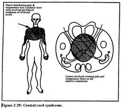
Conference Notes 8-6-2013.  A little late this week,  but   Another Great Day of Learning at Conference!  

There are images in this document.  If you don't see them, scroll to the bottom and click "View in Browser" 
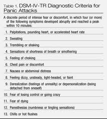
 Please consider donating to our EM Foundation to benefit resident education/development.  Thanks for your consideration/generosity.
/em-foundation/

Chastain   U/S

I am sorry I missed this excellent lecture. 

Campanella    Code 30 (Acute Stroke)

Dizziness is a completely non-specific quasi-medical term and should be avoided.  Try to get to a more specific term like vertigo, lightheaded, or ataxia.

New recommendation: The only labs you need prior to giving TPA for stroke is a finger stick glucose and an INR if the patient is on warfarin.    The lower limit for glucose to give TPA is 50.  Hypoglycemia below 50 is more likely to be the cause of neuro symptoms/findings than stroke.  The upper limit of blood sugar is not defined.  You can give TPA in the setting of hyperglycemia but also treat hyperglycemia with insulin.

Key HPI point is the last time patient was seen normal.   That time starts the clock for the 3 hr or 4.5 hr windows for TPA.

Don’t delay CT scan for anything even physical exam, lab draws or xrays.

Dysarthria can result from multiple causes other than stroke.   However, if someone is dysarthric,  you should also worry about dysphagia.  Must do swallow screening prior to oral intake in all stroke patients.  Swallow screen is : If Aphasia is present then they should be npo.  Next have them try sips of water, then drink a half cup of water.  If they cough or struggle with these tests, then they should be npo.

Blood pressure control is another key management issue in stroke patients.

Door to CT scan completion time optimally should be 25min.    Door to CT interpretation time should be 45min.  Door to TPA administration time should be 60 min.    Door to stroke unit time should be 3 hours.

Risk of serious hemorrhage (CNS or GI) from TPA is 6.5% and usually occurs within 48 hours.   However, morbidity and mortality at 3months has been shown to be better than placebo. 

If pt has signs of cortical stroke (hemi-neglect, visual field cut, global aphasia, dense hemiparesis)  endovascular treatment may be indicated because likely there is a large vessel involved.   Lovell comment: Can you comment on the negative outcomes in the endovascular trials in NEJM recently?   Dr. Campanella said the findings are disappointing.  He said they may not be valid because they used old technology.  Campanella has seen many anecdotal successes.   Lovell comment:  What about all the patients who didn’t do well.   Campanella: If patients have not improved with the standard of care TPA treatment  then endovascular management may be indicated.  The recent international stroke conference spent the majority of its time debating the use of endovascular results.   Parker comment: The studies showed good reperfusion with the old device so why would new devices show benefit.   Campanella response: There are mechanical differences between devices that may affect outcomes.   Lovell: Future endovascular procedures should all be performed within the confines of an IRB approved study.   Campanella:  It is a problematic situation.  Careful patient selection is critical for endovascular treatment of strokes.

Kelly Wiliamson comment: Would you ever activate a code stroke for suspected central vertigo?  Campanella response: Without focality, no do not activate code stroke for vertigo.  There is no category for vertigo on the NIH stroke scale.    Harwood comment:  Isolated aphasia with an overall low stroke score does uniformly well.  Do you use TPA?   Campanella: Depends on the severity of aphasia.   Mild word finding problem does not warrant TPA but severe expressive aphasia does.  

There are imaging modalities that can define the age of a stroke.    There has been a study of “wake up strokes”  (strokes first noted in morning when pt woke up) and they have found that the majority occur just prior to waking.     Campanella has concerns about giving tpa for mild strokes.  Difficult judgment calls.

Kettaneh/Girzadas     Study Guide

 Case 1  Infective Endocarditis.  Critical actions: Get  blood cultures (preferably 3) prior to antibiotics. Give IV antibiotic coverage for acute endocarditis   (Vanc and Gent).  Consult  Cardiology and ID.  Echo showed mitral vegetation.  Mitral valve is the most common site to embolize.  Pt had multiple signs of endocarditis (embolic phenomena, janeway lesions, osler nodes, glomerulonephritis, roth spots on fundi)

Case2  Iron poisoning in a pediatric patient. Critical Actions: IV Fluid Bolus, Serum Iron level, IV Deferoxamine.   Optimal Care: Identify anion gap, Identify abnormal liver labs, Figure out history of iron overdose, Consult Toxicology/Poison Control,  Admit to PICU.   Serum Iron levels >500 are associated with serious toxicity.  Pills but not liquid Iron may be visible on abdominal radiographs.  Deferoxamine IV at 15-35mg/kg for 24 hours  (ARDS is a potential complication).   Classic presentation of severe overdose: Bloody diarrhea, Shock, Wide anion gap acidosis, Hepatic dysfunction, Leukocytosis, Lung Injury, hyperglycemia.    Good mnemonic for anion gap acidosis: METAL ACID GAP   Methanol/metformin/massive OD, Ethylene glycol, Toluene, AKA, Lactate, APAP, Cyanide/CO/colchicine, Iron/Isoniazid/Ibuprofen, DKA, Generalized seizure producing toxins, ASA, Paraldehyde/phenformin. 

Case3 High Pressure Injection Injury. Critical Actions: Treat Pain, Identify High Pressure Injection      Injury , Start IV Antibiotics,  Consult Hand Surgeon for urgent surgical debridement.  Optimal management: Get x-rays of hand, Update tetanus shot, Elevate affected hand, Keep Johnny NPO.   High pressure injection injuries have  30% Amputation Rate.  Amputation more likely if debridement is delayed >6 hours.    Digital blocks are contraindicated.  They can increase compartment pressure.   Left index finger is most common location.

Google Image of Hi pressure injection injury and resultant surgical dissection.


 Erickson    Heat Emergencies

Prickly heat: Blockage of sweat pores leading to inflammation of sweat ducts.   No specific treatment.  Can use antihistamines or chlorhexadine lotion.

 Heat cramps:   Sweating leads to volume loss and hyponatremia resulting in cramping.   Most commonly patients have been drinking hypotonic fluids.   Treat with po Gatorade or IV NS.

Heat tetany:   Hot environment stimulates hyperventilation leading to respiratory alkalosis and tetany of hands.   Get patient to a cool environment. 

Heat syncope:  Variant of postural hypotension (volume depletion and vasodilation).   Get to a cool environment and hydrate. 

Heat Exhaustion:   Due to dehydration and electrolyte depletion.  Pt subjectively doesn’t feel right but still has normal mental status.   Treat with rest in cool environment and IV or PO fluids/electrolyte repletion

Heat Stroke:  True medical emergency.  Classic heat stroke: Mostly affects elderly and chronically ill.  Occurs in periods of high environmental heat stress.   Exertional heat stroke:  Healthy person who is exerting themselves in hot environment.      Patients have altered mental status or seizures.   Cerebellum is highly sensitive to  heat and ataxia may be an early sign of heat stroke.   LFT’s are usually elevated with heat stroke.  Patients can get ATN and rabdomyolysis from heat stroke.  Treatment of choice is evaporative cooling.  Spray lukewarm water on skin and direct a fan at the patient.  This is an effective cooling method pioneered in Mecca.   You need to prevent shivering with benzo’s.   Another effective cooling method is ice water immersion cooling.   This is a method that is harder to perform.   Another method is to use the device you use for therapeutic hypothermia (coolguard, arctic sun).  

Girzadas comment:  How do elite athletes complete the Badwater race (running 100+ miles through the desert)  Brian Fort response:  They drink specialized electrolyte drinks.  Brian has drank a glass of liquid with 1400 calories and a ton of electrolytes.  It tastes terrible.

Febbo    Anaphylaxis

Diagnostic Criterion 1 — Acute onset of an illness (minutes to several hours) involving the skin, mucosal tissue, or both (eg, generalized hives, pruritus or flushing, swollen lips-tongue-uvula) and at least one of the following:

 Respiratory compromise (eg, dyspnea, wheeze-bronchospasm, stridor, reduced peak expiratory flow, hypoxemia).

OR

  • Reduced blood pressure (BP) or associated symptoms and signs of end-organ dysfunction (eg, hypotonia [collapse] syncope, incontinence). 

Note: Skin symptoms and signs are present in up to 90 percent of anaphylactic episodes. This criterion will therefore frequently be helpful in making the diagnosis.

Criterion 2 — Two or more of the following that occur rapidly after exposure to a LIKELY allergen for that patient (minutes to several hours):

  •  Involvement of the skin-mucosal tissue (eg, generalized hives, itch-flush, swollen lips-tongue-uvula).

  Respiratory compromise (eg, dyspnea, wheeze-bronchospasm, stridor, reduced peak expiratory flow, hypoxemia).

  •   Reduced BP or associated symptoms and signs (eg, hypotonia [collapse], syncope, incontinence).
  •   Persistent gastrointestinal symptoms and signs (eg, crampy abdominal pain, vomiting).

Note: Skin symptoms or signs are absent or unrecognized in up to 20 percent of anaphylactic episodes. Criterion 2 incorporates symptoms and signs in other organ systems and is applied to patients with exposure to a substance that is a likely allergen for them.

Criterion 3 — Reduced BP after exposure to a KNOWN allergen for that patient (minutes to several hours):

  •  Reduced BP in adults is defined as a systolic BP of less than 90 mmHg or greater than 30 percent decrease from that person's baseline   (Up to Date)

Skin/mucosal findings can be absent 10% of the time.   Respiratory signs are present  in up to 70% of patients.  GI signs are present in 45% of patients and can be a sign of impending cardiovascular collapse. 

PCN and NSAID’s are the two most common drugs causing anaphylaxis.

Immunologic (anaphylactic)and non-immunologic reactions (anaphylactoid)are treated in an identical manner.

Mechanism of shock is vasodilation, fluid shifts and myocardial depression.

Anaphylaxis onset is within 30 minutes of exposure. 

80% of reactions are uniphasic.   Biphasic reactions most commonly occur within 10 hours but can occur out to 72 hours.

Treatment: Epinepherine/IV fluids/Airway management.   No contraindications to epi in the setting of anaphylaxis.   Just use IM Epipen or Epipen jr (kids <30kg).   No need to remember the dose of epi just use the epi pen.

Harwood comment: You have to leave the epipen in the muscle for 5-10 seconds to allow it to auto-inject.  Elise comment: I was in the PED recently and the nurse pulled the epipen out right after sticking it in the muscle.   We had to administer a second epipen and make sure it was in the muscle for 5-10 seconds.

Empty ventricle syndrome :  Pt’s treated for anaphylactic shock in the ED who get up to walk to bathroom  while they are still vasodilated and tachycardic develop an empty ventricle and go into PEA.  There are case reports of this.   Brian recommended keeping pt’s supine for 30-60 minutes after treatment for anaphylaxis.

Anaphylactic shock should be treated with iv epi.   Criteria is marked hypotension or patient is still symptomatic after two IM epipen injections.    Give infusion of epi :  1mg in 1 liter of saline and infuse at 2ml/min.  Nick Kettaneh comment:  http://academiclifeinem.com/dirtyepi/  describes the making of this drip. This reference says you can run the drip wide open.   Each ml of this drip has 1 microgram of epi.   Girzadas advice:  probably start  drip somewhere between 2-10 ml per minute and titrate up if needed.   Bolus dosing not recommended.  Vasopressin is probably best choice for refractory shock if epi  fails.

Adjuvant therapy: Steroids,  diphenhydramine or zyrtec (second generation antihistamines are effective and less sedating),  ranitidine or pepcid, albuterol or racemic epi nebs for wheezing.   Glucagon is indicated  if patient is on a beta blocker.   Everyone gets a 6 hour observation.   Selected patients should be observed for 24 hours (severe symptoms, hx of asthma, use of betablockers, extremes of age, barriers to care)

Give everyone a referral to an allergist.

Bradykinin induced angioedema:  ACE-I and Hereditary angioedema.    HAE may have intense abdominal pain and can mimic a surgical abdomen.   Treat HAE with FFP.   Ecallantide and Icatibant are new drugs for this disease but we don’t have them at ACMC.

ACE-I angioedema:  Lip edema rarely progresses to involve the airway.  Edema of tongue/soft palate/larynx have higher risk of needing airway intervention.  

Conference Notes 7-23-2013

Conference Notes      7-23-2013     Another Great Conference Day!

If you can't see the pictures scroll to the bottom and click on "Read in Browser"

Please consider donating to our EM Foundation to benefit resident education/development.  Thanks for your consideration/generosity.
/em-foundation/
 

Putman/Lovell           Oral Boards

Case 1    5 day old infant with malrotation and midgut volvulus.   Recognize bilious emesis in an infant.  Malrotation with midgut volvulus usually presents in the first week of life.   Duodenal atresia usually occurs in first day of life.   Get a KUB then follow that with upper GI and emergent surgery consult.   This is an ischemic condition that can evolve to death. 

Case2     Jones fx of 5th MT.  

This is a diaphyseal FX of the 5th MT that is distinct from an avulsion FX of the tuberosity of the 5th MT.  Jones fractures occur in an area of relatively poor blood supply and are at risk of nonunion if the patient continues to weight bear on this Fx.   One way to identify a Jone’s FX is to see if the FX line extends into the space between the 5th and 4th metatarsals.

Case3  Cryptococcal Meningoencephalitis      Get a CT in HIV patients prior to doing an LP.  Get Crypto AG of bood and CSF.   Treat with amphotericin and flucytosine.   High opening pressure carries a poor prognosis and requires hi volume taps and repeated LP’s to drain CSF fuid.

Elise was Gracious enough to give me all her notes:

Malrotation with midgut volvulus:  In patients with midgut malrotation, the usual gut rotation ceases after the first 90 degrees, and the duodenum and ascending colon are juxtaposed around the superior mesenteric vessels, with the entire midgut suspended from this narrow axis. The malrotated bowel itself does not cause any significant problem. However, because of the narrow axis, the midgut can at any time twist around the axis.  The tighter the twist, the more the midgut suffers from obstruction of the lumen, obstruction of venous and lymphatic return from the midgut, and obstruction of arterial inflow, thus threatening midgut viability. Unless it is treated in a timely manner, bowel strangulation results in an ischemic loss of extensive bowel, causing death if not treated.


In neonates, malrotation with midgut volvulus classically presents with bilious vomiting and high intestinal obstruction. While most neonates with bilious vomiting do not have midgut volvulus, this diagnosis must be ruled out. Older children with malrotation may manifest a failure to thrive, chronic recurrent abdominal pain, malabsorption, or other vague presentations.

Malrotation and volvulus should be suspected in all cases of proximal small-bowel obstruction, especially in infants.  Most patients with midgut malrotation develop volvulus within the first week of life.  Besides the cardinal clinical manifestation of bilious vomiting, clinical features may include pain (colicky at first, then steady), anorexia, blood and mucus in the stool, abdominal tenderness, and, eventually, shock.


In early cases, patients may appear well, and abdominal examination findings may be normal. In fact, normal findings on abdominal examination have been reported in as many as 50% of patients. Because the obstruction is very proximal, abdominal distention is not usually present. 

An upper GI series is the preferred diagnostic test for malrotation with midgut volvulus and must be performed. Upper GI series sensitivity is 85-95%, with a higher specificity. In malrotation with midgut volvulus, upper GI à dilated, fluid-filled duodenum, a proximal small bowel obstruction, a "corkscrew" pattern of the jejunum.  Malrotation without volvulus on upper GI seriesà duodenal jejunal junction is displaced downward and to the right on frontal view.  KUB of malro without volvulus is often nml.   KUB  of malro with volvulusà classic finding is a partial duodenal obstruction (dilation of both the stomach and proximal duodenum, with a small amount of distal bowel gas).


Double bubble sign.   S=stomach     D=duodenum


UGI showing midgut volvulus

Ultrasonography and CT may suggest the diagnosis of malrotation; however, their sensitivities and specificities are low compared with those of an upper GI series.

Duodenal obstruction, seen in midgut volvulus and malrotation, is also seen with duodenal stenosis, duodenal web, annular pancreas, preduodenal portal vein, and duodenal atresia (atresia presents within first hours of life, double bubble, may delay surgery 24-48 hours, assoc. with Down’s). 

Labs:  may have initial hypochloremic metabolic alkalosis.  As they get sicker, think metabolic acidosis,  gut ischemia, necrosis,  perforation/peritonitis, sepsis/death.  Mortality up to 15%.   Long term complication short gut syndrome.


Jones fracture:

The diaphyseal bone, where the fracture occurs, is an area of poor blood supply; a watershed area between two blood supplies. This makes healing difficult. In addition, there are various tendons, including the peroneus brevis and fibularis tertius, and two small muscles attached to the bone. These may pull the fracture apart and prevent healing.

If the fracture enters the intermetatarsal joint, it is a Jones fracture. If, however, it enters the tarsometarsal joint, then it is an avulsion fracture caused by pull from the peroneus brevis. An avulsion fracture is sometimes called a Pseudo-Jones fracture or a Dancer's fracture.  Avulsion fracture can be treated with walking cast, Jones must be non-weight bearing.

Jones fx is located at least 1.5 cm distal to tuberosity of 5th metatarsal & should not be confused with the more common avulsion fracture of the fifth metatarsal styloid which is usually proximal to the metatarsal cuboid joint and treated with walking cast or cast shoe.


 Jones FX    Fracture line extends to 4-5 intermetatarsal space

Cryptococcosis is the most common fungal disease in HIV -infected persons, and it is the AIDS-defining illness for 60-70% of HIV-infected patients.


In addition to invading the lung and CNS, cryptococci also invade the skin, bone, and genitourinary tract, but meninges appear to be the preferred site.


Cryptococcal disease usually develops only when CD4+ lymphocyte counts fall below 100 cells/microL.


Disease onset is usually insidious; time from symptom onset to diagnosis is, on average, 30 days or more. The delay also may be due to the waxing-and-waning course and the nonspecificity of symptoms.  Headache, fever, malaise, nonspecific mental status sx, uncommonly CN palsies, seizures, focal neuro sx.  Also uncommon to see meningismus (no immune system).


CSF analysis may yield normal (ie, reference) results in 25% of patients and may be minimally abnormal in as many as 50%; therefore, identifying the organism via India Ink and serology is crucial.


Increased intracranial pressure (>200 mm H2 O) occurs in over half of all patients with AIDS who have cryptococcal CNS infection, probably because of obstruction of the basal meninges or impaired CSF absorption. Since increased intracranial pressure is a poor prognostic factor (increased mortality) whose correction leads to symptomatic improvement, this must be managed aggressively.

In the absence of obstructive hydrocephalus or risk of herniation, increased pressure (>250 mm H2 O) can be relieved by serial spinal taps or a lumbar-peritoneal shunt.  Daily lumbar puncture removing approximately 30 mL of CSF until the pressure has decreased 50% is recommended. 


Close to 100% of CSF culture results are positive for Cryptococcus neoformans, whereas 66-80% of blood culture results are positive. India ink stain is positive in 74-88% of infected patients.

CrAg in the serum usually is indicative of systemic disease and correlates with fungal burden. A localized cryptococcal infection, such as pulmonary cryptococcosis without lymph node involvement, usually is not associated with a positive serum CrAg; a positive result warrants a search for disseminated disease. Detection of CrAg in either serum or CSF has >95% sensitivity and specificity in the diagnosis of true invasive cryptococcal infection. Cryptococcal capsular polysaccharide antigen in spinal fluid is now the method of choice for diagnosing patients with cryptococcal meningitis.


Twenty to thirty percent of patients show meningeal enhancement, parenchymal solid mass lesion without hemorrhage (granuloma), atrophy, cerebral edema, or hydrocephalus on CT/MRI.   If the imaging studies show a cryptococcal mass lesion (ie, cryptococcoma), toxoplasmosis and lymphoma must be considered in the differential diagnosis; brain biopsy may be indicated.


If left untreated, cryptococcal CNS infections are fatal.  Mortality rate of 6%, despite aggressive therapy, has been reported.

Current guidelines from the Infectious Diseases Society of America (IDSA) for primary therapy of cryptococcal meningitis in HIV-infected patients recommend amphotericin B deoxycholate (0.7-1.0 mg/kg/day IV) plus flucytosine (100 mg/kg/day orally in 4 divided doses) for at least 2 weeks. This is followed by fluconazole (400 mg [6 mg/kg] per day orally) for a minimum of 8 weeks).


Lifelong secondary prevention may be required. Relapses occur if secondary prevention is stopped or becomes ineffectual. Relapse rates without prevention range from 15-27%; this drops to 0-7% with prophylactic antifungal agents.


Predicting CD4 count by absolute lymph count.  If ALC < 1,000, CD4 likely < 200.

Acad Emerg Med. 2011 Apr;18(4):385-9.

Absolute lymphocyte count in the emergency department predicts a low CD4 count in admitted HIV-positive patients.

Napoli AM, Fischer CM, Pines JM, Soe-lin H, Goyal M, Milzman D.

866 patients known to be HIV+ and admitted from the ED, who had an ALC measured in the ED and a CD4 count measured within 24 hours of admission. An ALC of <950 × 10(6) cells/μL has a sensitivity of 76% (95% CI = 73% to 79%), specificity of 93% (95% CI = 87% to 96%), and positive likelihood ratio of 10.1 (95% CI = 8.2 to 14) for a CD4 count of <200 × 10(6) cells/μL.


Prophylaxis (another way to guess CD4 count, based on antibiotics patient is taking)

PCP CD4 <200, bactrim

Toxo CD4 < 100, bactrim

MAC CD4 < 50, Zithromax

Erik comment: When doing an opening pressure during an LP, the patient has to be laying down in the lateral decubitus position.  Measuring the opening pressure with the patient sitting up will be inaccurate.

Harwood comments:  HIV patients with headaches should get CT brain with IV contrast.   You can get a closing pressure instead of an opening pressure.   So if you want to not risk your ability to get CSF from an LP, you can get the fluid first and then lay them down and measure the pressure.  Removing 8ml of CSF will not completely resolve  the increased  pressure.


Kettaneh     M and M     Case F/U’s

 Case 1   Missed distal femur buckle fx in a young child.   Elise comment: Gotta carefully palpate the entire extremity.  If the child won’t bear weight you have to xray the entire extremity if you can’t find palpable tenderness. 

 Case 2    55 yo female with exertional near syncope and blurry vision.    In hospital, it was found patient had central retinal artery occlusion.   Nick felt that he could have communicated with the consultants better in the ED because in their note the mentioned possible CRAO but he did not speak with them.

 Nick recommends that you follow up the hospital course on most of his patients.  You learn a lot of stuff by doing this.   Elise comment:  I call some patients, especially the one’s I am worried about.   Patients invariably really appreciate this.

 Case 3    49yo male bunt trauma  to face and amnestic to events.   HGB noted to be 8.7.   Pt was observed  in ER.   Pt developed epistaxis in the ED.  Nick asked the nurse to put some packing in the patient’s nose. Pt was not re-assessed for hours.  Pt became unresponsive.   He was successfully resuscitated.  Repeat labs and multiple CT’s after pt was resuscitated showed no clear cut etiology of what happened to patient.    The suspected etiology was that nasal packing stimulated the nasopulmonary reflex causing  apnea/bradycardia/hypoxia/bronchoconstriction.   At Dr. Lovell’s suggestion, pt was cooled using our therapeutic hypothermia protocol.   Pt gradually improved over weeks.

Consider the risk of nasopulmonary reflex whenever you a pack pt’s nose.    

Girzadas comment:  this patient’s risk of the nasopulmonary reflex may have been higher because the patient was head injured, had elevated ammonia level, was older and had pulmonary comorbidities.   Lovell comment: I am more conscious of this potential complication and really think about sending home a patient who is elderly or has multiple comorbidites with a nasal pack.  McKean comment: You will never regret personally going to re-assess a patient.  You may regret not going to re-assess a patient.  Chastain comment: When you are on an off-service, don’t forget you are an ER doc.  Think globally about your patients.  Harwood comment: Is there a bright line to decide which trauma patients would benefit from therapeutic hypothermia?   Nick, if it is a non-traumatic arrest with no active bleeding and no brain bleed then therapeutic hypothermia is indicated.

Girzadas    Neuro Study Guide

 I was giving the lecture so couldn't take the notes. 

Gore      Safety Lecture

Case 1     40 yo Pt presented with multiple physical complaints related to severe hypothyroidism.   

Case 2     35yo female presented with headache similar to previous migraines.

Kasia ordered migraine cocktail accidentally on patient  1.   She did not switch out of the electronic record of patient 1 prior to ordering what she intended for patient 2.

Safety strategies:  Limiting distraction, EMR blocks to ordering on the wrong patient,  Doing  time outs prior to placing orders.    Easiest to rapidly implement: Do a mental checklist or “safety check” while waiting for orders tab to load.   Check patient, allergies, and intended orders.  

Multiple persons commented that they have made this same mistake.   Multiple people asked that chief complaint be added to the top bar.   Robbie suggested that you can place orders while you are in the patient’s note.  It keeps you from accidentally placing orders into the wrong chart. 

Collander                             Meningitis/Encephalitis

Bacterial Meningitis: Strep pneumo 61%,  Neiseria 16%, others are less common.


Gram negative intracellular diplococci N. Meningitidis

Gram positive rods in chains  Strep pneumo


Just a reminder for Boards what the gram stain colors are in case they give you a slide on the test

Purple= positive      Red=negative


Predisposing factors: otitis media, immunosupression, etohism, dm, endocarditis, extremes of age, liver disease, many more.

Brudzinski:  Neck flexion by examiner causes pt to flex hip and knees

Kernig’s  sign:   With patient’s hip flexed,  examiner cannot extend knee  fully due to pain in posterior leg.

Criteria for doing a CT prior to LP:  altered mental status, focal neuro deficit, papilledema, seizure within the last week, immunocompromised state, history of CNS disease, age over 60, malignancy, concern for a CNS mass lesion.   If patient has none of these, you an do an LP without a prior CT.

Steroids:  Give before or at the time of first antibiotics.   Reduces morbidity in  H flu and S Pneum inkids.  50% reduction of M & M in adults with S. Pneumo.  Equivocal results in adults with N. Meningitidis.    Dose is 0.15mg/kg Q 6 hours or 10mg Q6 hours for adults.   Lovell: Reason to not start steroids after antibiotics is that it doesn’t work.    Harwood comment: The reason steroids don’t work after antibiotics is that the bacteria have already started to be killed and the steroids are less effective in halting the inflammatory cascade at that point .     I give steroids if I have a strong suspicion of meningitis or  abnormal csf on visual inspection.   Elise comment: Steroid work best for strep pneumo but because you won’t know the gram stain or culture results at the time of first antibiotics, base your decision to give steroids on visual inspection of csf during tap. 

Normal opening pressure is <170 mm of H2O.  Bacterial meningitis is usually >300 mm of H2O.   Viral  between 170 and 300 mm of H2O.    Girzadas comment: As a practical point the manometer in our LP kits measures the column of CSF in centimeters.  So 17 cm =170mm of H2O, and 30cm=300mm of H2O.

Antibiotics: 

0-1 month  Ampicillin and Cefotaxime, or Ampicillin and Gentamycin

1-3 months Amp and Ceftriaxine

3mo-50 year   Cetriaxone and Vanco

>50yr  Ceftiraxone, Vanco, and Ampicillin 

Chemoprophylaxis:  Rifampin 600mg Q12 for 4 doses.  Single 500mg Cipro dose is another option.  Single dose of IM ceftriaxone is another option.

Viral meningitis:   If CSF study not clearly bacterial or viral, cover with antibiotics and do CSF viral studies.  CMV,EBV,VZV,HSV  are in the standard panel.  West nile is a separate IGM study.    If you have suspicion for HSV give acyclovir 10mg/kg in the ED.  

*Great teaching point*Andrea comment: Most common drug induced cause of aseptic meningitis is NSAID’s.  Ibuprofen is the most common.   NSAID induced aseptic meningitis can look exactly like viral meningitis.  It is an idiosyncratic reaction to an NSAID.