Conference Notes 3-8-2017

Wellness Retreat at Lake Katherine

We had our 2nd annual Wellness Retreat this last Wednesday.  Multiple aspects of wellness were covered including Financial Wellness (Special thanks to Nick Kettaneh), Yoga, Meditation/Spirituality, Healthy Eating and Team Building.  And..... Therapy Dogs!  

Snip20170310_5.png

Conference Notes 3-1-2017

Lovell     Procedural SedationJeopardy

Key Principles of Pain Management

1. Assess Severity 2. Use appropriate doses of analgesics 3. Titrate your pain meds 4. Monitor the patient's status.

Local anesthetics can be divided into amides and esters.  Amides are longer acting.  Amides have lower incidence of allergic reactions than esters.   Amides all have two i's in the name (lidocaine, bupivicaine).   Esters only have one i in the name (procaine).   If a patient has an allergy to local anesthetics it is usually due to the preservative.  Cardiac lidocane has no preservative so is usually considered safe in regard to allergies.  Subcutaneous diphenhydramine can also be considered as a substitute for local anesthetic in the setting of severe local anesthetic allergy.

*ASA classification of patients for sedation and anesthesia

The risk of using ketamine in kids with an active uri is laryngospasm.  If this develops you can bag the patient through this complication most of the time.  Ketamine is OK in head injury.   You can use ketamine in children down to 3 months of age for sedation.  For patients younger than age 3 months ketamine is not approved and there may be a higher risk of laryngospasm.

3 drugs for treatment of regional complex pain syndrome: corticosteroids, calcitonin, and bisphosphonates.  These patients present with burning pain, edema, warmth to skin, local sweating, and allodynia (pain to light touch).   To treat, get the patient's affected limb re-mobilized (take off splint or cast) and start oral prednisone.  They need follow up with PMR.  If you don't identify and treat these patients early they can develop severe chronic pain. 

Healthcare Disparity:  A large study showed that African American pediatric patients had a 60% chance of getting pain medications for abdominal pain compared to white pediatric patients. In other words African American children were 40% less likely to get analgesia for abdominal pain compared to white patients.

Ways to reduce the pain of local anesthetics: 1. inject thru the open margins of the wound. 2. buffer with sodium bicarb 3. Inject slowly4. Warm the anesthetic5. Use a small (25 or 27 guage) needle 5.  Reassure or distract the patient

Non-ASA NSAID's are noted by the FDA to cause heart attacks and CHF.  Elise avoids giving NSAID's in elderly patients, patients with heart disease, HTN, or renal disease.

Benzocaine and prilocaine are the two local anesthetics that can cause methemoglobinemia.

Max dose of lidocaine is 4mg/kg without epinephrine or 7mg/kg with epinephrine.  For bupivicaine, the max is 3mg/kg plain or 5 mg/kg with epinephrine.   Girzadas comment: Draw up your local anesthetic before you enter the child's room so you never enter the room with a potentially toxic dose of anesthetic and the child does not see the needle.

Toxicity from local anesthetics initially causes dizziness, facial or extremity paresthesias, ringing in the ears.  Patients may progress to seizures and then cardiovascular collapse (V-tach).  Bupivicaine has the highest risk of cardiovascular toxicity.  Treatment for local anesthetic toxicity is lipid emulsion therapy.

* Levels of procedural sedation

Recent large study in NEJM shows that if emergency physicians are high intensity opioid prescribers (prescribed opioids to 24% of their patients) the patients receiving opioids have a 30% higher relative risk of having long term dependence on opioids. Overall absolute risk of about 2% of becoming addicted.  Elise comment: the pendulum has swung away from opioid use so be cautious of prescribing opioids to patients. Discuss with patients that there is a 2% risk of becoming addicted.  Abhi comment: Warn patients about nausea and constipation to better inform them and encourage them to limit their use of narcotics. 

Scoring system to determine who can be safely discharged after procedural sedation

Scoring system to determine who can be safely discharged after procedural sedation

KatiyarEM Billing and Coding

Optimizing your RVU's requires optimizing your documentation.  Medical decison-making documentation is critical to your charting.    Also make sure you write a procedure note for any procedures.  Another key documentation item is to document your plan of care for a fracture or sprain.  Also document a re-exam after a splint was placed to show neuro vascualar status is intact and the splint is not too tight.  Ortho cases in general have high RVU's.  Appropriate fracture and joint reductions have very high RVU values.

If you incise an abscess, probe, break down loculations and pack, that is considered a complex abscess.

Any wound checks following abscess drainage can be billed as a level 2 or 3.  If you have to re-pack or give antibiotics, it becomes a level 3 chart.

Measure the length of the lacerations you repair.  There is a ruler printed on the paper wrapping of a tongue blade that you can use to measure the wound.

If you make management changes with oxygen for a low pulse ox, document your thought process regarding the pulse ox and oxygen therapy.  This is important for the medical decision making and for the RVU documentation.

The diagnosis you place on the chart is critical for determining the ED's case mix index.  If you document acute STEMI rather than just Chest Pain it better characterizes the acuity your ED is seeing.  Try to be as specific as you can in the ED.  If the patient has an nSTEMI, document that diagnosis rather than Chest Pain.  If the patient has DKA document that rather than hyperglycemia.  If the patient has Pyelonephritis, document that instead of uti. 

When you are describing patient behavior use objective terms as much as possible.  For example, instead of writing the patient is "beligerent", write the patient was screaming obscenities at staff, violently rocking the cart and throwing punches at staff or other specific actions.

Review all chart documentation including what the nurses and the techs and EMS personel wrote.  I any court case, the lawyers will go thru all charting with a fine tooth comb.

Marshalla      Patient Safety LectureMassive Transfusion Protocol

We now have refrigerated blood in the ED for any patient in hemorrhagic shock.  In that refrigerator there are 2 units of O pos blood for all males and for females over age 49.  There are 2 units of O neg blood for females under age 49.

ED Attending physicians and Trauma Attending physicians can order blood from the ED blood supply for all causes of hemorrhagic shock.  Only nurses can physically access the blood from the ED refrigerator.

Einstein       ED EKG's

Noah discussed strategies to improve the information flow of EKG's in the ED.

Sedation Workshop

We broke into small groups and discussed different sedation scenarios. 


Conference Notes 2-8-2017

McKean     Resuscitation Procedures

Unfortunately I missed this excellent lecture

Girzadas    Zebras Lecture

I gave this lecture and did not write up notes. I did send out a PDF of the lecture to all the residents. If you would like a copy, please send me an email separately.

Critical Care Device Workshop   

Much Thanks to our awesome ED Nurses for teaching us abut the devices commonly used in resuscitation situations in the ED.  Special shout outs to Kristen, Monika,  Danielle , and Nick!

Conference Notes 1-25-2017

Airway Day

Samir Patel     Airway Disasters


5% of intubations in the ED are considered difficult

2% of intubations in the ED are failed (Defined by first 3 attempts fail)

You have to be prepared for this.  It eventually will happen to you.

Treat every airway as a difficult airway

Prior planning before starting intubation is the key to success.


* Algorithm for Difficult Airway


Indications for Intubation:

Hypoxic or hypercapnic respiratory failure

Airway protection

Anticipated deteriorating course of illness

Work of Breathing (tachypneic, septic patients are using 30% of their cardiac output for diaphragm contraction)


*MOANS    Predictors of Difficult BVM


*Lemon Law    Predictors of Difficult Laryngoscopy


*3-3-2 Rule


*Mallampati Score     Class 1 and Class 2 predict reasonable airway visualization.  Class and 3 and Class4 predict poor airway visualization.


A Major Rule is: Don’t have any pride when working with a difficult airway.  Get help from ICU, anesthesia, surgery and other EM physicians.  Whoever you need.  There is no shame in getting help.


Sedated Nasal fiber -optic Intubation

Start with 4% nebulized lidocaine

Afrin and glycopyrrolate can help dry secretions

Ketamine for sedation

Warm the ET tube in warm water.  It makes the tube more malleable

When you visualize the cords with the fiberoptic scope have someone spray the cords with lidocaine so you don’t get laryngospasm


Apneic Oxygenation (basically a nasal cannula running at 15 liters per minute or more during intubation) prolongs your safe apnea time.  High flow nasal oxygen devices are even better than nasal cannula but it is more bulky.


*Delayed Sequence Intubation.  Give Ketamine slowly to avoid apnea.  The ketamine will calm an agitated patient allowing you to better pre-oxygenate and prepare.


Avoid IV Ativan in patients with respiratory distress. It reduces their respiratory drive and it may force you to intubate before you are ready.


Lovell        Airway Devices


To lessen your anxiety during a difficult intubation, it is useful to have familiarity with the airway tools you are using.   So practice with different devices so you are comfortable using them.


You always need to have a supraglottic device and a surgical option in your armamentarium as rescue devices.


Elise’sMinimum List of Devices every Intubating physician needs to have available and be comfortable using:

Bougie

LMA

Direct laryngoscope

Video laryngoscope

Fiberoptic device (Really CMOS/digital camera technology.  It is more durable than fiberoptic technology)

Cricothyrotomy kit


The difficulty with the Glidescope is properly passing the tube once you get a great view.   You need to pop back the stylet once you have the ET tube at the glottis to appropriately position the ET tube thru the cords.


Elise discussed many advanced airway devices available on the market.


The biggest error with cricothyrotomy is waiting too long to start doing it.


If the O2 sat is dropping and you have a failed intubation, stabilize with an LMA.  Based on your ability to oxygenate with an LMA you either must rapidly do a cricothyroidotomy or if you can oxygenate OK, attempt intubation thru the LMA.


If the patient is in a HALO, initial intubation attempt should be with video laryngoscopy.


Airway Workshop

Conference Notes 1-18-2017

Joint EM-Pediatric Conference    Pediatric Sepsis


Pediatric sepsis is culture negative in 25-60% of cases.


About half of children with sepsis will have a low cardiac index and high SVR (cold shock) where 90% of adults will have high cardiac output and low SVR (warm shock)


*Pediatric Sepsis Definitions


Cornerstones of Sepsis Therapy in Children

Early recognition is the key to treating sepsis effectively.

A key resuscitation goal is 60ml/kg of normal saline infused in the first hour.

Vascular access in the septic child should escalate to IO after 2 attempts at IV.

IO placement is not more painful than IV placement but IO infusions are painful.  So give lidocaine 0.5 mg/kg, not to exceed 40 mg thru the IO line. (Lovell reference)

You need to use a pressure bag, rapid infuser, or push-pull method to give 60ml/kg within one hour.


There was a discussion of which patients should get 60ml/kg in the first hour.    The strong consensus was that all pediatric sepsis patients should receive 60ml/kg of NS. 

You should be cautious giving 60ml/kg to kids with cardiac disease, history of abnormal kidney function, and neonates. 

The panel felt that, in general, septic kids need more rather than less fluid. 

The panel felt that it would be reasonable to re-assess the patient after every 20ml/kg.

Neonates are a high-risk group for large volume fluids and should be bloused in no more than 10ml/kg aliquots at a time.  You should carefully re-assess the neonate after any bolus to determine if more fluids are required.


Early antibiotics are another cornerstone of sepsis management.  A 3 hours delay increases the likelihood of need for PICU admission.  Ampicillin & Cefotaxime or Vancomycin &Ceftriaxone are the basic empiric antibiotic combos. But there are many variations based on age, allergies, and source of infection.


If you need a pressor for sepsis in a pediatric patient use peripheral epinephrine.  It has lower mortality compared to dopamine.


Children who are on chronic steroids (asthma, cancer) are at risk for adrenal suppression.  Give hydrocortisone (2mg/kg) Q 6 hours in septic kids who are at risk.


All kids in septic shock should receive hi-flow nasal cannula O2.

To avoid a hemodynamic crash during or after intubation, give a fluid bolus prior to intubation. Consider an epinepherine drip prior to intubation.

The Panel suggested avoidingetommidate for RSI in septic children.  I assume that is based on concern for adrenal suppression and maybe a risk of hypotension.


Lactate levels are unreliable in kids for identifying sepsis. 


Basically in the first hour after you suspect sepsis:

Give 60ml/kg of NS, Start IV antibiotics, Start high flow O2 via nasal cannula. If you need a pressor give IV epinephrine.


Kerwin/Denk     Oral Boards


Case 1. 25yo female with severe dyspnea.  Patient has a history of asthma and is severely wheezing.  Despite therapy with nebulizers, magnesium, and subcutaneous epinepherine,  the patient was still in severe distress and the ABG shows respiratory acidosis.   Patient was then intubated using a sedated look with ketamine for sedation and topical lidocaine. 


*Use peak flows to determine severity of asthma.  


Give 10-15mg of albuterol nebs every hour for severe asthma.   Give steroids and IV magnesium. You can also try high flow nasal cannula, heliox, subcutaneous terbutaline or IM epipepherine.   Sub-dissociative dosing (0.1mg/kg)etamine may be helpful in the anxious/agitatedpatient to help them tolerate bipap and nebs to stave off intubation. Bipap should be tried but the data in asthma for bipap is limited.


Case 2. 68yo male presents in Cardiac arrest.  Patient had V-tach as his initial rhythm.  In the ED, patient was in V-fib .   ACLS protocol was initiated.   ROSC was obtained.  EKG post-arrest showed STEMI.  Asa and heparin were given and patient was taken to the cath lab.  Therapeutic hypothermia was initiated as cath lab was being activated.


Chris advised that if repeated shocks for V-fib are not working, try double shocking with two defibrillators at the same time.  There are a few small case series showing some efficacy to this method.


*Double defibrillation method for persistent V-fib


*2015 ACLS Guidelines recommend Amiodarone and epinephrine for treating ventricular fibrillation that is resistant to first shock.

Therapuetic hypothermia is indicated after V-fib arrest either with or without STEMI.  


*Some criteria that make therapeutic hypothermia less likely to have a postive outcome.  My brief review of the literature would add: unwitnessed arrests, asystole/PEA arrests, significant delays to starting CPR and ACLS care, intracranial hemorrhage, subarachnoid hemorrhage, pre-arrest inability to perform ADL's, and arrest due to sepsis.


Case 3. 56 yo male with altered mental status and vomiting.  Patient had a headache earlier in the day.  Patient has a history of HTN and Afib.  Patient is on xarelto and aspirin.  Head CT shows ICH.   Treatment is with FEIBA or PCC’s.


The data shows that lowering the BP to less than 140 systolic has worse outcomes.  Goal should be a BP just above 140 mm/hg systolic.


Yasser Said     Observation Medicine


Kelly comment: The phrase I use for documenting chest pain patients in the OBS unit is: Patient placed on OBS service for further cardiac risk stratification.


Elderly and frail patients should be considered for inpatient management over OBS management. 

AARP has advised people to refuse OBS stays because OBS stays are more expensive.  This is not always true. It really varies on a case-to-case basis. 


Average OBS stay is 20-22 hours


OBS service has a policy of Dilaudid restriction.  Please inform patients that this medication will be restricted in OBS.


Physicians can be criminally prosecuted for prescribing opioids to a patient who has an overdose or bad outcome.


Pharmacy Lecture    Status Epilepticus Management


1st line IV lorazepam (2-4mg) or IM midazolam (5-10mg) or IV diazepam

Rectal diazepam (0.2mg/kg) is another option for patients without IV access


2nd line IV phosphenytoin or IV Valproic acid or IV levetiracetam or IV Phenobarbitol.  Loading dose for all these agents is 20mg/kg.


Refractory Status is due to less responsive GABA receptors and increased NMDA receptors.


3rd line Propofol 80micrograms/kg/hr or IV midazolam drip.


4th line Ketamine1-5mg/kg followed by drip 0.45-10mg/hr.


 Subclinical status can be indicated by persistent tachycardia.


Conference Notes 1-11-2017

Herron/Marshalla    Oral Boards

Case 1.   Patient presents with sore throat and difficulty breathing.


*Epiglotitis on lateral neck x-ray and on video laryngoscopy.  (Bitner, Annals EM 2007)  This x-ray image has both the "thumbprint" sign and the vellecula sign where the vellecula air column does not reach the hyoid bone.


Patient was managed with IV ceftriaxone, IV steroids and consultation with ENT & Anesthesia for OR intubation.  Adult infection is more likely to be due to non-HIB organism.


Case 2.  16 yo male presents with altered mental status, hypotension and tachycardia.  No fever.  He appears intoxicated.  Dad found patient lying on the floor of the garage.  Dad suspected the patient may have drunk anti-freeze.  Labs show anion gap metabolic acidosis and osmolal gap.  


*Anion and Osmolal Gap Calculations


Treatment was IV fomepizole.  Nephrology was consulted for dialysis.   IV bicarb is indicated for severe acidosis.   You can also give pyridoxine as a key co-factor for metabolism.


*Ethylene glycol metabolism


Case 3. Male presents with a headache following a MVC. Patient’s vehicle was struck from the rear.  Patient had transient loss of consciousness for a few seconds.  Key PMH is the patient has hemophilia A.

Critical management is to get Factor 8 replacement therapy started as soon as possible.  For boards, always give Factor 8 prior to getting CT head. In real life, many times you can get a CT while pharmacy is getting the Factor 8 prepared. Patient has sign of intra-cerebral hemorrhage on CT.  You want to get Factor 8 level to 100% by giving 50u/kg for any head injury (bleed or no bleed).


Lambert       Soft  & MSK Tissue Ultrasound


Only 15% of wood FB’s are visualized on x-ray.  Ultrasound has much higher sensitivity for wood FB’s.


Snip20170111_7.png

*Wood FB on ultrasound.


*Cellulitis on ultrasound. Note the “cobblestone” appearance of the tissue.


*Abscess on ultrasound.   To help differentiate from cellulitis or other process, Mike compresses the suspected abscess cavity and looks for swirling or movement of the fluid in the cavity.


*Necrotizing fasciitis.   Air in the soft tissue is pathognomonic for Nec Fasc.   Air on ultrasound is demonstrated by hyperechoicarea (Arrows) with posterior shadowing. 


*You can also diagnose fractures with ultrasound.  You can identify a cortical disruption.  This is a clavicle fracture.


*97% of rotator cuff injuries are supraspinatus tears.   To visualize this tendon, have the patient put their hand in their pant’s back pocket.  Place the probe on the antero-lateral aspect of the humeral head and aim the probe at the ipsilateral ear.


*Quadricep tendon rupture


*Achilles tendon rupture


Lambert      Ultrasound Guided Nerve Blocks


These techniques were too complex for me to write up in these notes.   The residents practiced the approaches to these techniques in the Ultrasound workshop.


Lambert and Team Ultrasound      Ultrasound Workshop


Conference Notes 12-14-2016 & 12-21-2016

12-14-2016

Menon      Study Guide


*WPW   Orthodromic tachycardia goes down the AV node and back up the accessory pathway resulting in narrow complex tachycardia that can be treated with adenosine.  Antidromic tachycardia goes down the accessory pathway and back up the AV node giving a wide complex tachycardia that should be treated with procainamide.


*WPWIf you have Afib with wide complex RVR, that needs to be treated with procainamide or cardioversion.   Any drug that slows the AV node like adenosine or Cardizem can cause life threatening tachycardia in this clinical situation.


5ways to differentiate V-tach from SVT with Aberrancy

Age and History: Older patient with prior MI or CHF is more likely to have VT.

QRS>160ms

AV dissociation

Fusion beats

Capture beats

Concordance


Harwood comment: If you have fusion beats or capture beats, you have V-Tach


*An interesting algorithm using AVR findings may be easy to use.


Elise comment: Icatibant for ACE-I angioedema is looking like it doesn’t work.  There is a large negative study coming out soon about this topic.  It takes a long time for the drug to work.  So it may have some utility for the intubated patient in the ICU to resolve the angioedema sooner but for emergent care in the ED it won’t help. 


*For V-fib that persists despite standard ACLS, you can consider double defibrillation with 2 defibrillators.  You shock with both at the same time.   If you do this, give esmolol as well.  There are some case reports that suggest esmolol and double defib can be useful for “electrical storm”

Little known point:  you can’t do synchronized double cardioversion because you can’t sync two machines together and you risk causing V-Fib.  You can only use two defibrillators in the setting of V-Fib.


*PEA Management   Narrow complex is more likely a mechanical problem.  Wide complex is more likely a metabolic problem.


Elise Reference: Amiodarone, Lidocaine, or Placebo in Out-of-Hospital  Cardiac Arrest  May 16, NEJM

BACKGROUND

Antiarrhythmic drugs are used commonly in out-of-hospital cardiac arrest for shock-refractory

ventricular fibrillation or pulseless ventricular tachycardia, but without proven

survival benefit.

METHODS

In this randomized, double-blind trial, we compared parenteral amiodarone, lidocaine,

and saline placebo, along with standard care, in adults who had nontraumatic out-ofhospital

cardiac arrest, shock-refractory ventricular fibrillation or pulseless ventricular

tachycardia after at least one shock, and vascular access. Paramedics enrolled patients at

10 North American sites. The primary outcome was survival to hospital discharge; the

secondary outcome was favorable neurologic function at discharge. The per-protocol

(primary analysis) population included all randomly assigned participants who met eligibility

criteria and received any dose of a trial drug and whose initial cardiac-arrest rhythm

of ventricular fibrillation or pulseless ventricular tachycardia was refractory to shock.

RESULTS

In the per-protocol population, 3026 patients were randomly assigned to amiodarone (974),

lidocaine (993), or placebo (1059); of those, 24.4%, 23.7%, and 21.0%, respectively, survived

to hospital discharge. The difference in survival rate for amiodarone versus placebo was 3.2

percentage points (95% confidence interval [CI], −0.4 to 7.0; P = 0.08); for lidocaine versus

placebo, 2.6 percentage points (95% CI, −1.0 to 6.3; P = 0.16); and for amiodarone versus

lidocaine, 0.7 percentage points (95% CI, −3.2 to 4.7; P = 0.70). Neurologic outcome at discharge

was similar in the three groups. There was heterogeneity of treatment effect with

respect to whether the arrest was witnessed (P = 0.05); active drugs were associated with a

survival rate that was significantly higher than the rate with placebo among patients with

bystander-witnessed arrest but not among those with unwitnessed arrest. More amiodarone

recipients required temporary cardiac pacing than did recipients of lidocaine or placebo.

CONCLUSIONS

Overall, neither amiodarone nor lidocaine resulted in a significantly higher rate of survival

or favorable neurologic outcome than the rate with placebo among patients with

out-of-hospital cardiac arrest due to initial shock-refractory ventricular fibrillation or

pulseless ventricular tachycardia. 


Einstein     Diarrhea

Diarrhea can cause normal anion gap acidosis.

Stool cultures are only positive 2-5% of the time.   Get them for abdominal pain and fever, ill-appearing children, pregnant patients, and immunocompromised patients. 

Order a C-diff test in any recently hospitalized patient and any patient who recently was on antibiotics.

Treat with oral rehydration or IV fluids. 

Advise complex carbohydrates and lactobacillus-containing yogurt

Antibiotics for symptoms >5 Days and/or systemic symptoms, traveler’s diarrhea, bloody diarrhea, or immunocompromise.   Cipro or azithro are your main choices.


Elise comment:  If you do give antibiotics, make sure you get a stool culture.


Harwood comment:  As the duration of diarrhea increases, the risk of bacterial diarrhea goes up.   Don’t bother with fecal leukocytes.  It is not specific for bacterial causes.  Just get a culture if you are thinking of getting a stool sample.


Girzadas comment: Be alert for hypoglycemia in younger kids (<2yo) who have diarrhea or vomiting.   Hypoglycemic kids usually present cranky and crying.  


Regan    Reading a Thromboelastogram(TEG)


 A TEG measures the speed of clot formation and strength of a clot.  

Thrombin converts fibrinogen to fibrin. 


*TEG

R time is time to first clot. 

Alpha angle identifies fibrinogen deficiency

Maximum amplitude measures clot stability and platelet activity.

LY 30 measures clot reduction after 30 min and thrombolysis activity.


*Abnoral TEG’s

Long R is due to coagulopathy from coagulation factor issues


*Therapy based on TEG

Increased R time give FFP

Decreased Angle give cryoprecipitate

Decreased MA give platelets and DDAVP


Cirone     M&M

I missed this excellent lecture


Bernard/Schmitz       Trauma Conference


I missed most of this excellent lecture but at the end there was spirited discussion between EM and Trauma faculty about the value of ED thoracotomies.  Basically there was agreement that ED thoracotomy is rarely indicated or life-saving.  The one trauma indication it may be useful for is an isolated stab wound to the heart with hemopericardium.   For the procedure to save a life, you need to have a surgeon rapidly available to take the patient to the OR.

12-21-2016

Paquette/Nejak      Oral Boards

Case 1. 50yo male presents with altered mental status.   RR is low.  Patient has a right side dilated pupil and evidence of head trauma.  Patient was emergently intubated.  CT showed acute epidural hematoma.  Patient required emergent decompression.


Case 2. 25yo male presents with left hand pain.  Pain and tenderness is localized to 4th finger.  Patient was involved in a fight the night before.   X-rays shows Jersey finger.  Treatment is splinting with referral to hand or orthopedics for surgical repair.


Case 3.  6 yo patient brought in by parents for abdominal pain.  On exam, patient has palpable purpura on lower extremities.   Patient had marked abdominal tenderness. Plain x-ray of chest shows free air.  Diagnosis is HSP with intussusception with perforation.


HSP Rash.&nbsp; Papable purpura on the lower extremities.

HSP Rash.  Papable purpura on the lower extremities.

Katiyar       Methemoglobinemia

 Case: Infant male brought into ED for decreased PO intake and diarrhea. Child more lethargic than normal.   Child has lost weight.   Pulse Ox =85%.   Blood looks chocolate brown.   Diagnosis is methemoglobinemia.  


There are a lot of causes of methemogloginemia: well water, topical benzocaine (hurricane spray),  and other medications. Poppers (amyl nitrate) and Dapsone can cause methemoglobinemia.   There are congenital causes as well. 

Infants can have nitrite forming organisms causing diarrhea.  Infants have an immature reductase system and can’t handle the nitrites formed by infectious gut organisms.

In the developing world, insecticides are the most common cause of methemoglobinemia.

Treatment includes decontamination if applicable. 

Asymptomatic patients with level <30% will clear methemoglobin in 36 hours. 

Asymptomatic patients with a level >30% and symptomatic patients should get methylene blue.   Can’t give methylene blue if the patient has severe renal dysfunction.

Side effects of methylene blue include blue or green urine, chest pain, and hemolysis.  Patients treated with methylene blue need to be admitted due to risk of hemolysis.


 Katiyar     Billing for Critical Care


Critical care charts don’t have the typical level 5 chart requirements.  You just have to document the critical situation of the patient and your concern for potential decompensation. Then you have to document the time you spent in direct care of the patient (management, discussions, documentation, etc)  Critical care time does not include procedure time.


Be sure to document  all updates and re-evals that you perform on the patient. Document the info you obtained from review of old records.


Central lines, intubations, cardioversion, and A-lines can be billed separately from critical care.  The time you spend on procedures does not count toward critical care time.


If you care for a cardiac arrest patient who is brought to the ED and despite your efforts at resuscitation the patient does not get ROSC, you can’t bill critical care for that.


Residents cannot bill critical care.  The attending has to spend 30 minutes or more in direct patient care and document that care to bill for it.   Mid-level providers can bill critical care similar to an attending.


Critical care time frames are 30-74 minutes and then every 30-minute period beyond that initial time period.   Your time providing critical care does not need to be continuous. It can be the total of multiple 5-10 minute time frames.


Critical care billing is based on the midnight-to-midnight 24 hour day.  If the patient’s care crosses midnight, you can actually bill critical care for each day if you spent more than 30 minutes both before and after midnight.


Okubanjo          Healthcare Disparities

Women healthcare providers are increasing in numbers.  African American providers are still a very low percentage of the total providers.

Historically African-American Universities are very successful in placing graduates into medical school.

Minority physicians are more likely to choose primary care specialties, serve minority populations, and work in areas of manpower shortage.   Their patients are more likely to be low income and have less access to care.

A large factor in clinical uncertainty is the gap between a patient’s cultural or socio-economic background and the healthcare provider’s background.

Hueristics or quick decision-making tools we use in our minds to make rapid decisions in the ED can lead to stereotypes and then biases.   Bias is a negative evaluation of one group and it members relative to another.  When heuristics becomes based on stereotype or bias it can mislead the decision maker.


Bias can also negatively affect the doctor-patient relationship


Zakieh   Fluid Resuscitation in the Critically Ill Patient


In hypotensive patients, IV fluids won’t always solve the problem. Only 50% of hypotensive patients will have a positive response to IV fluids.   Both ventricles have to be on the ascending portion of the Starling curve to benefit from added fluids.


CVP measures RV pressure but is not an accurate measure of central volume or fluid responsiveness.  Again CVP is about 50% accurate in measuring central volume.


*Passive leg raising test is the gold standard test for fluid responsiveness.  It translates to a 300ml auto transfusion.  You can check vital signs in about 2 minutes to see if there is improvement.


Fluid boluses have transient effect.  Fluid leaks out of vascular system in about 60 minutes.


Excessive IV fluids increase mortality in the critically ill patient.


LR as a resuscitation fluid has lower incidence of acute kidney injury compared to normal saline.


We need to move the culture of resuscitation away from normal saline to using more LR.

Patients who receive several liters of saline are at risk for hyperchloremic metabolic acidosis, AKI, and increased mortality.


Conference Notes 11-30-2016 & 12-7-2016


Barounis     Coma


The most important thing in this lecture is finding brainstem signs in the comatose patient.


Start with a good history.  Look for prodromal symptoms such as headache, vomiting, fever, syncope, depression.    Then try to find out the rapidity of onset of coma.   Find out the patients baseline function.


Evaluate the motor response to pain.  If the patient localizes to pain that is a positive response.   If no response or posturing then look for brainstem signs.   Basically check the pupils. 

Miosis=opioids, pontine hemorrhage,

Fixed mid sized pupils=mid brain lesion or brain death

Unilateral fixed dilated pupil=uncal herniation from a mass, bleed, or aneurysm

Bilat fixed dilated pupils=almost always due to a medication, atropine during a code can do it.

Bilat small reactive pupils= non specific, can be due to metabolic disease like sepsis. 

Anisocria + sudden coma is basilar artery stroke

Eyes deviate to side of stroke in the brain. Logan Traylor’s mnemonic (you can’t look away from the train wreck)

Eyes deviate away from irritation in the brain such as seizureLogan Traylor’s mnemonic (the seizure is irritating so you look away)

So,

If gaze is looking to opposite side of hemiparesis (weak arm or leg)= stroke

If gaze looks toward hemiparesis (weak arm or leg)= seizure


Skew (eyes not aligned completely up and down) is another sign of brainstem lesion

Vertical nystagmus is a sign of brainstem lesion


Anisocria, skew deviation, lateral deviation, vertical nystagmus are signs of brainstem cause of stroke.

Cheyne stokes breathing is another sign elevated ICP or brainstem lesion


If a patient has anisocoria or skew deviation they need a CTA to identify need for embolectomy.


Dave described a case in a middle age man who presented with acute coma.  He had anisocoria on exam.


If the patient has no abnormal eye findings and has a gag or cough reflex then the problem is in the cortex. 


*Algorithm for the evaluation of Coma   (emcrit)


*Four score is better than GCS because it forces you to look at the eyes.


Dave made the point that if you have a febrile comatose patient, you very strongly need to consider doing an LP.   The LP can be therapeutic in these patients because it may lower the ICP and improved cerebral perfusion pressure.


*Neurocritical Care for Comatose Meningitis patients.


*PRES

Posterior reversible encephalopathy syndrome (PRES) is a neurotoxic state that occurs secondary to the inability of posterior circulation to autoregulate in response to acute changes in blood pressure. Hyperperfusion with resultant disruption of the blood brain barrier results in vasogenic oedema, but not infarction, most commonly in the parieto-occipital regions.

Terminology

PRES is also known as hypertensive encephalopathy or reversible posterior leukoencephalopathy.

The term PRES can be a misnomer as the syndrome can involve or extend beyond the posterior cerebrum. Furthermore, although most cases involve a resolution of changes with the treatment of the precipitating cause and clinical recovery some patients can progress to develop permanent cerebral injury and be left with residual neurological defects.   (Radiopaedia)


Marynowski/Holland      Oral Boards


Case 1.   Patient presents with massive GI bleeding from aorto-enteric fistula. Treat with 2 large bore IV’s. Transfuse immediately with uncrossmatched blood.  Intubate.  Place NG tube to suction.  Give antibiotics for infected aortic graft.   Get patient to surgery emergently.   

Elise comment: With massive bleeding always initiate the massive transfusion protocol.


Case 2.  Patient presents with rash and pre-syncope.   Diagnosis is Lyme’s disease with heart block.   Treat with appropriate antibiotic. Doxycycline, amoxicillin, cefuroxime, ceftriaxone are all OK for treatment of lyme disease.  Elise comment: For heart block IV ceftriaxone is recommended by IDSA guidelines.  Perform or consult for pacemaker.   1 study noted that 38% of patients required a temporary pacemaker.


Case 3.   34 week pregnant patient with nausea, vomiting, and abdominal pain.  BP is 142/94.  Patient has laboratory findings consistent with HELLP syndrome. 


*HELLP syndrome

Treat with magnesium and treat hypertension.   If the infant is <34 weeks give the mom steroids prior to delivery to promote fetal lung maturation.


Greenberg  Tachyarrhythmias in Adults


*Antiarrythmic Categories


For Afib there is no mortality difference between rate control and rhythm control.  Rate control is easier so that method is favored.


First lines drugs for Afib with RVR are Cardizem or metoprolol/esmolol.


Amiodarone can have both rate and rhythm benefits in Afib.   Amio can take more than an hour to have an effect.  


Digoxin can be given in a loading dose to control the rate in Afib.  It starts to work in about an hour.  Make sure you correct any potassium abnormalities to avoid arrhythmias.


For stable VT the PROCAMIO study showed better efficacy and less adverse reactions for procainamide compared to amiodarone.   Unfortunately procainamide is not available until 4th quarter of 2018 due to limited production.


If VT is refractory to amiodarone, second line is lidocaine, followed by third line phenytoin.


Treat polymorphic VT with magnesium first line or defibrillation.   If the QT is not prolonged you can cautiously try a beta blocker or amiodarone. Consult cardiology.


For shock refractory V-fib (electrical storm) , give amiodarone.  If V-fib still refractory consider esmolol and double defibrillator defibrillation.  There is case report and small study data showing some benefit.


Nejak    Supplemental O2


I missed this excellent lecture.


Pediatrics Faculty Member   Bronchiolitis


Don’t order a viral panel or rapid RSV testing.  It is painful to the child and there is no benefit.

Don’t use albuterol or epinephrine, or hypertonic saline nebs.   The Peds EM faculty felt you could trial an albuterol MDI and check for improvement.  The MDI takes out the humidified oxygen that is delivered by a neb and gives you a cleaner test of whether albuterol is helping or not.

No steroids.

Supplemental oxygen if needed.

Nasal suctioning is good.  Deep suctioning is bad.

No CXR unless child is going to ICU or you have concern for pneumothorax.

Febrile infants under 90 days of age with bronchiolitis have a low risk of concomitant meningitis.  LP may not be needed in infants over 30 days.  Under 30 days you should still do an LP in febrile infants with bronchiolitis.


*Bronchiolitis scoring.  If the score improves by 2 points with treatment that is significant.  This scoring system can only be used to assess the effect of an intervention. It has not been validated to determine disposition.  However, 3 is considered a low score and 8 is a high score that may necessitate ICU admission.


Elise comment: Do a score,  suction, do another score.  This will be the best measurement of the effectiveness of suctioning.


Ede/West         GU Emergencies


Treatment for paraphimosis

Prior to reduction attempts, give local anesthetic with a dorsal penile block or ring block.

Attempt manual reduction by squeezing edema from foreskin and attempting to direct the foreskin over the glans.  You can use an ace wrap to compress the edema from the foreskin prior to attempting reduction.  Alternatively you put granulated sugar or mannitol-soaked gauze on the edematous foreskin to draw out the edema.  Osmotics (sugar or manitol) may take an hour or more to work.


If these strategies are unsuccessful, you can use a 25g needle to make multiple punctures in the edematous foreskin to help edema drain.


Last strategy prior to surgery would be to make a dorsal slit thru the edematous foreskin.


Management of gross hematuria with clots:  Start with manual irrigation of the bladder with a 60 ml Toomey syringe.  Follow that up withcontinuous bladder irrigation in the ED.  If gross blood with clots doesn’t clear in the ED these patients should be admitted for  continued irrigation and GU evaluation.   If the urine clears with irrigation these patients can be considered for discharge.


Fournier’s Gangrene: You need to emergently consult GUfor source control of this infection with surgery.  Next, start big gun antibiotics(Zosyn, Vanco, and Gent) and include Clindamycin (inhibits toxin production).   Alcoholics and diabetics are more prone to this infection.


*To detorse a testicular torsion, “open the book” on the affected testicle only.  There seemed to be some consensus among those present that you detorse  only 90 degrees at a time and re-assess by seeing if the pain is better or use bedside ultrasound to see if there is blood flow restarted to the testicle. If no change in pain or flow then detorse another 90 degrees.


Consider imaging the kidneys of elderly patients with pyelonephritis or urosepsis.  There is a significant percentage of patients that will have ureteral stones and obstruction. You can get a CT, get a formal US, or do bedside US to check for stone and hydronephrosis.


Factors that increase the risk of developing kidney stones include:

  • Family or personal history. If someone in your family has kidney stones, you're more likely to develop stones, too. And if you've already had one or more kidney stones, you're at increased risk of developing another.
  • Dehydration. Not drinking enough water each day can increase your risk of kidney stones. People who live in warm climates and those who sweat a lot may be at higher risk than others.
  • Certain diets. Eating a diet that's high in protein, sodium and sugar may increase your risk of some types of kidney stones. This is especially true with a high-sodium diet. Too much sodium in your diet increases the amount of calcium your kidneys must filter and significantly increases your risk of kidney stones.
  • Being obese. High body mass index (BMI), large waist size and weight gain have been linked to an increased risk of kidney stones.
  • Digestive diseases and surgery. Gastric bypass surgery, inflammatory bowel disease or chronic diarrhea can cause changes in the digestive process that affect your absorption of calcium and water, increasing the levels of stone-forming substances in your urine.
  • Other medical conditions. Diseases and conditions that may increase your risk of kidney stones include renal tubular acidosis, cystinuria, hyperparathyroidism, certain medications and some urinary tract infections.   (Mayo Clinic Reference)

Marshalla     UTI’s

Bacteria found in the urine of men and pregnant women is always abnormal and should be treated.

Nitrites are produced by gram negative bacteria only.  The bacteria has to be present in the bladder for four hours to make nitrites.

Elise reference: Asymptomatic bacteriuria is common among older adults and practically universal among those with indwelling catheters.[7] The prevalence in healthy older women living in the community is around 20%, and in men >75 years old is 6-15%. In LTC facilities, the percentages are even higher: 25-50% in women and 15-40% in men.[11] The rise in prevalence parallels the increase in comorbidities, especially neurological, associated with micturition problems.[3,7]


The recommendations by the Infectious Diseases Society of America[11] and the Society for Healthcare Epidemiology of America[3] are clear concerning asymptomatic bacteriuria in the older population, whether residing in the community or in LTC facilities: routine screening and treatment are not recommended. There have been several studies[13-16] showing no benefits associated with the treatment of asymptomatic infections as measured in the rate of subsequent symptomatic infections, improvement of chronic urinary symptoms, or survival. Moreover, some harm can be caused, mostly associated with side effects of antimicrobials and increased resistance in uropathogens.[3,4,11]


Above is from Medscape, can find a similar message in many recommendations:  http://www.medscape.com/viewarticle/586757_3


Traylor     Personal Medical Kit

Logan told the tragic tale of how he had to resuscitate his dog that was seriously injured on a vacation. 

Preventable deaths in the outdoors: Hemorrhage, tension pneumothorax, airway obstruction

Your kit should have: tourniquet, scalpel and tube for chest tube.  Oral airway, LMA or ET tube. Narcan, epi-pen, and albuterol mdi.


Schmitz     Parkland Burn Formula


*Remember the 49er’s.   4ml in the formula and rule of nines.   Palm=1% BSA but Palm does not include fingers.  The picture is a little incorrect.  Only the palm (no fingers)=1% BSA.


Lee    Managing Stress in the ED

Stress impairs cortical functioning.

Manage your 4 domains when in a Resuscitation:

1. Environment:  Set up the room optimally

2. Team:  Give your team direction, specific roles and use clear communication

3. Self:  Manage your own anxiety, your communication

4. Patient: Focus the other three domains on the care of the patient. 

Mental rehearsal: go thru the procedure or resuscitation over and over in your mind before you need to do it.

*Arousal control: Square breathing

Positive self-talk: Navy Seals and athletes continually tell themselves they can do it, accomplish the goal, they have the necessary skills and training.  They are prepared. 

This builds confidence and increases the probability of accomplishing the task.

Visualize goals: Identify and map out all the smaller goals to reach the end goal.


Regan    VP Shunt Obstruction

Shunt obstruction may cause change in mental status, headache, vomiting, or autonomic instability.

An obstructed shunt will not have the normal “squishy” shunt pump when you press on it.

If you are in a place where neurosurgery is unavailable and the patient is comatose or peri-code.  You should tap the shunt to relieve pressure and test for infection.

To tap a shunt: Prep the skin with betadine. Insert a 25g butterfly needle perpendicularly into the shunt.  Gently draw back CSF and send it to the lab.  If you cannot  withdraw CSF there is likely a proximal shunt obstruction and this patient needs to go to the OR emergently.

Stanek    CHF Management

Think NAP:   Nitro,  Ace-I, and Positive pressure ventilation

Give nitro sprays initially and get IV dosing to about 100micrograms/min as soon as you can

ACE-I’s can be used also to lower BP/afterload

Bipap provides more functional alveoli and has been shown to improve oxygenation and prevent intubation.

Muhammad      Pediatric Abdominal Pain

I missed a large portion of this excellent lecture.

HSP with significant abdominal pain should get an U/S to evaluate for intussusception.  5% of Kids with HSP can intussuscept.   Intussusception is one of the few indications for steroids in HSP.

*Pediatric Appendicitis Score.   Imaging strategy is usually start with U/S and if you need further imaging go to limited CT of the appendix area to minimize radiation exposure.

*ACMC Appendicitis Protocol

In the patient with possible ovarian torsion, the imaging study to get is pelvic ultrasound with doppler evaluation of vascular flow to ovaries.

If you see gallstones in an infant or small child think sickle cell disease or hereditary spherocytosis.

Katiyar  Am I Really Too Slow? Billing and Coding

It doesn’t matter if you are fast.  It matters how well you document.

Most EM jobs pay based on RVU generation.  RVU=Relative Value Unit.  It has 3 components 1. Work 52% 2. Practice expense 42% (EM is lower than other specialties) 3. Professional Liability 6%.   EM has a relatively low RVU reimbursement ($35 per RVU compared with Neurosurgery which is $85 per RVU)compared to other specialties because we don’t have as much practice expense.

Kelly comment:  Chart to a level 5 for all your patients.  If won’t incorrectly upcode level 3’s and 4’s but it makes sure you are optimizing your charting and it simplifies your exam and charting.

Harwood comments: If you see a patient with an ankle sprain, you really don’t need to document a level 5 chart.  Simple complaints are candidates for brief charts.  

Critical care documentation and coding very significantly increases your RVU generation.

Level 4 RVU’s =3.33=$119

Level 5 RVU’s=4.93=$176

Critical Care RVU’s=6.33=$226

To max out your RVU’s , See the patient, treat them, and dispo them as quickly/smartly/efficiently as possible.  Chart as you go. Don’t let your charts pile up.  Charting later tends to negatively impact your documentation.

Poor charting leads to down-coding which negatively impacts RVU generation.

When doing ROS and HPI items, you only need one item per body area.

4-2-10-8“Fortutenate”  Is the simple mnemonic to remember billing requirements for a level 5 charts.

4 descriptors of chief complaint, 2 past history items: medical/surgical/social, 10 ROS areas,  8 physical exam areas

Holland/West     Administrative Updates

We discussed process improvements with getting new ECG’s and retrieving old ECG’s.

We discussed other actionable items that people brought up.


Conference Notes 11-23-2016

Conference Notes11-23-2016


Lovell        Pulmonary Study Guide

 CHF is unlikely with a BNP <100.   CHF is more likely with a BNP>400.

In lung transplant patients, infection and rejection can look the same clinically.  Because it is so difficult to differentiate these two diagnoses, it is important to discuss the patient’s management plan with the transplant team.  The most common cause of death long term in lung transplant patients is bronchiolitis obliterans.  

Intubating the massive hemoptysis patient: Use a large ET tube to allow suctioning to clear the tube if necessary.  You can try to position the ET tube in the mainstem bronchus in the non-bleeding lung.  That isolates your non-bleeding lung for better oxygenation and potentially keeps blood out of the non-bleeding lung.  Position the patient in either the right or left lateral decubitus position to get the bleeding lung down.  This keeps the blood in a dependent location and keeps the non-bleeding lung elevated away from bleeding.

Get a CTA to evaluate  the source of bleeding.  Once the bleeding source is identified by CTA,  get the patient to IR for embolization.   These patients die from asphyxiation  (blood filling the airway) so protecting their airway is the critical management action.

Treatment for primary, spontaneous pneumothorax: If small and stable just put them on 15L NRB O2 and watch for 6 hours.  If repeat CXR is improved or not worsening, you can discharge the patient at that time.   If the patient has a moderate pneumothorax place a mini-chest tube with a Heimlich valve.  You can aspirate the pneumothorax thru this tube initially.  Discuss with pulmonary consultant about admission vs outpt follow-up.


*Management of primary spontaneous pneumothorax


5 risk factors for malignancy in a patient with hemoptysis: Smoking, age>40, male, recurrent hemoptysis, and no infectious symptoms.   Patients with 1 or more of these risk factors should get a contrast CT of the chest.


COPDer’s with bullae should not get a chest tube.  If you are concerned that a CXR in a COPDer may be demonstrating pneumothorax, get a CT chest to differentiate between a bullae and pneumothorax. 


*Bulla


*Pott Disease   TB in the spine.  The proper term is Pott Disease not Pott’s Disease but most of the Google Pics say Pott’s.


*Scrofula    TB related non-painful cervical  lymphadenitis


If you suspect TB in a patient, get them isolated in a negative pressure room.  All caregivers need to wear N95 masks.  Get a chest x-ray and discuss with ID further testing in the ED.    If you see upper lobe infiltrate, wide spread miliary distribution of infiltrate,  or granuloma with central adenopathy think TB.


Board scenarios for Pneumonias:

Sudden onset, chills, rust sputum =     Strep

Post-infuenza or cavitating lesion =        Staph

Alcoholics, current jelly sputum =    Klebsiella

Bullous myringitis, rash, joint pain, sore throat =   Mycoplasma

Pnuemonia and GI symptoms, possibly tourist in a hotel =  Legionella


CAP patients in general, benefit from steroids but don’t give steroids if you suspect influenza, the patient is pregnant, or the patient has poorly controlled DM.  Also avoid in patients with GI bleeding, receiving fluoroquinolone antibiotic (unclear why but no benefit shown with FQ’s), and those receiving neuromuscular blockers for intubation/ventilation (Can get myopathy.  I would discuss with intensivist.).


*Steroids for CAP


Menon      International EM


Vijay discussed his experience as an EM physician doing locums in New Zealand.

His message is basically, go to New Zealand if at all possible.


If you go you likely will work in a small town.  You will not be working in a large medical center in a big city.  The big cities have enough docs.  Minimum length of a contract for a locums job in New Zealand is a year.   


You have to pay taxes to New Zealand and the US.  The US taxes are not that bad.  You get significant credits and deductions for your US taxes.  

An opportunity cost of going abroad is that you will not be earning equity in a group like you would if you worked in the US.


You don’t go abroad to do locums to make more $.  You go abroad to gain that cultural experience and see the world and do something you will find fun and exciting.  The work culture in NZ is fantastic and you are given a lot of time off. Medical Malpractice is much less of worry there than in the US.


Vijay went to NZ to work for 2 years and wouldn’t trade that experience for anything.


Nejack     M&M


I will note only the take home points to keep the case details confidential.


With all trauma patients do a tertiary survey; basically go back when things have calmed down and fully re-examine patient for missed injuries.   Always look for a second fracture.


*CRITOE


*CRITOE


*When evaluating pediatric elbow injuries, check the alignment of the anterior humeral line.  Next, check the radio-capitellar line.  


*Look for abnormal fat pads


Finally check the boney cortices of the elbow


*Harwood made the point that if the figure of 8 on the lateral elbow is disrupted you have to consider a subtle supracondylar fracture.


Hart/Regan     Bread and Butter EM: Thanksgiving Cases

Case 1.  FB sensation in the throat after possibly swallowing a turkey bone.   You can initiate the work up with plain x-rays of the soft tissues of neck.  You can also get a CT neck.  If you identify a FB, discuss with ENT or GI for emergent or urgent endoscopy.  Sharp objects and batteries need emergent removal.   Other objects need urgent removal within 24 hours.

For patients with a Globus sensation in teir throat with no clear FB or unclear history.  Do a basic throat and neck exam. If no FB identified, you can consider using the fiberoptic scope to look further down the throat. If still no FB identified, reassure the patient. Start a PPI and arrange f/u with GI for endoscopy if symptoms don’t resolve.

Case 2.  2nd degree burns to arms from deep-frying a turkey.


*Rule of 9’s to estimate body surface area.


*Criteria for transfer to a Burn Center.   Even if the patient doesn’t meet the criteria for transfer, you can call the Burn Center and set up outpatient follow up in the Burn Center clinic. 


For patients with major burns, a clean dry sheet is the best dressing for transfer to Burn Center.


For minor burns, wound care at home is daily gentle washing of wound and applying antibiotic ointment and dry dressing. 


Case 3.  Treatment of flash pulmonary edema with hypertension

Aggressive NTG, start with nitro sprays(400mcg per spray) then rapidly titrate IV NTG up to over 100 mcg/min

Start Bipap

After maybe 30 minutes and BP improved give normal (40mg) dose Lasix.

These patients are usually not severely volume overloaded.  Flash pulmonary edema is really more of an acute vasculopathy that is treated with blood pressure reduction using hi-dose NTG.


Bamman      R&R Rapid and Random EM


* Unstable C-spine Fx’s


Treat Cystic Fibrosis pulmonary exacerbations similar to how you would treat a COPD exacerbation.  Give O2, nebs (albuterol/atrovent.  Also saline nebs have been found to be helpful), Bipap, steroids, and antibiotics to cover pseudomonas and MRSA.


Fitz Hugh Curtis syndrome is perihepatitis secondary to a chlamydia (more common) or gonorrhea pelvic infection.  Get cervical cultures.  Treat with Cefoxitin and doxycycline.  Consult gyne. Patients may need laparoscopy.

Harwood comment: CT Abdomen and Pelvis is not that sensitive for this disease.  It is more of a clinical diagnosis.


Mediastinitis is a life threatening emergency.  It can be a post-operative complication, result from trauma, or from esophageal perforation.  Diagnose with CT. Treat with big gun antibiotics (vanco, ceftriaxine, and flagyl) and most importantly emergent surgical debridement.


*Acute chest syndrome is a diagnosis made by vitals, lung exam, and CXR.  Any 2 of these categories with positive findings is consistent with ACS.  Treat with O2, cautious IV fluids, cefotaxime and azithromycin, transfusion (simple for less sick and exchange for more sick), and analgesics.


Thyroid storm treatment: Propranolol, PTU, SSKI (1 hour after PTU), Hydrocortisone


Alexander      Geriatric EM Patients


Belly pain in senior patients is tricky.  They are at higher risk for serious intra-abdominal problems but the clinical signs on their abdominal exam may be more subtle and non-specific.

Falls that present to the ED need to be evaluated for underlying medical problems.  Any senior with 2 falls in a1 year period may benefit from an in-home safety evaluation and general physical evaluation by PMD.  These evaluations can’t be done from the ED but care managers and the patient’s PMDcan be notified to provide a more global evaluation of the patient   

Harwood comment: 2 things that have found to be helpful for seniors at risk for falls are home safety evaluation, and physical therapy to improve strength and balance.

Polypharmacy is super-common in seniors.  Be cautious when adding narcotics, sedative hypnotics, nsaids,  antibiotics, and anticholinergics to their medication regimine.  Discuss with your ED pharmacist or consult an online medication interaction checker to avoid serious drug interactions.  


*To assess frailty you can watch a patient stand up from a chair, walk ten feet, walk back, and then sit down.   If it takes more than 20 seconds to do that, the patient may need help at home or physical therapy.


Bernard   Safety Lecture


Kyle covered our ED sepsis work flow and power plan.

He then discussed how we could be trained by the Chicago Recovery Alliance   www.anypositivechange.com to give out free Narcan to heroin users who come to the ED.


Conference Notes 11-9-2016 & 11-16-2016

Motzny      EMS Study Guide


*Class A Bioterrorism Agents


*Alpha, beta, and gamma radiation


*Acute Radiation syndromes.   Radiation=low lymphocyte count   This is the earliest lab indicator of serious radiation illness.


Never approach a helicopter from the rear.  There are dangerous rotating blades that can kill you.  If a helicopter in on the slope of a hill, never approach or move away from the helicopter on the uphill side.  Again the blades can kill you. 


*START Triage algorithm.  Triages patients on ability to ambulate, respiratory rate, perfusion (radial pulse) and mental status.


Anthrax is not transmitted person to person.  Anthrax is transmitted by  contact with the spore.


Ortho Cases


*Luxatio erecta.  Highest incidence of neurovascular complications secondary to shoulder dislocations.  Beware of axillary nerve palsy and axillary artery thrombosis.


*Ankle dislocation vs. Sub talar dislocation.  Note that in the sub talar dislocation the talus remains in the mortise.


Snip20161109_8.png

*Sub talar dislocation.


*Toddler fracture.   Elise’s point is to follow the cortex of the tibia and look for a subtle incongruity of the cortex.    Harwood comment: The only history you will get is the patient won’t ambulate.  These injuries have no clinical clues on inspection of the patient’s extremity. There is typically no swelling or deformity to help you localize the injury.  You just have to xray the length of the suspected lower extremity.


Splint with the knee in flexion to prevent rotation and prevent weight bearing. 

Toddler's fractures or childhood accidental spiral tibial (CAST) fractures are bone fractures of the distal (lower) part of the shin bone (tibia) in toddlers (aged 9 months-3 years) and other young children (less than 8 years).[1] The fracture is found in the distal two thirds of the tibia in 95% of cases,[1] is undisplaced and has a spiral pattern. It occurs after low-energy trauma, sometimes with a rotational

component.  (Wikipedia)


No need to call DCFS for toddler’s fracture.


*Bipartite patella is found most commonly in males and is located in the superior/lateral aspect of the patella.

OrthopedicWorkshop

Conference Notes 11-16-2016

Girzadas     Intubating the Obese Patient

RapidOxygen Desaturation is our #1 Enemy This due to a decreased functional residual capacity and increased metabolic demand.

*Functional Residual Capacity is decreased in the Obese patient


*The Safe Apnea Period is decreased in obese patients due to rapid desaturation

Airway visualization is our 2nd Greatest Enemy

Aspiration is our 3rd enemy


•       Decision #1   Head up positioning with RAMP or Reverse Trandelenburg optimizes FRC, VQ matching, and oxygenation.  It also optimizes airway visualization and decreases risk of aspiration.

•       Decision #2   Pre-Ox with BIPAP & Hi-flow Nasal Cannula

•       Decision #3   Ketamine sedation/Topical, Avoid RSI and NeuroMuscular blockade.  Maximize topicalization and minimize sedation.  Larger/faster doses of ketamine can cause apnea in the critically ill obsese patient. So use doses like 20-50mg of ketamine given slowly and titrate to needed sedation level.

•       Decision #4   Video laryngoscopy gives best first attempt success

•       Rescue Device is Intubating LMA. Have it ready before you start.

•       No Delay Cric.  Be prepared to perform a cric before you sedate.  If the patient is deteriorating and you are in a can’t intubate/can’t ventilate situation, Place the LMA and ventilate using that and commit to the cric and move quickly to get the cric done. You have only about a minute after the o2 sat gets to 90%.

•       Decision #5   Ventilate 6 ml/kg (100kg) start with a PEEP of 5 and titrate as needed.  Dave Barounis comment: no one needs more than 500ml tidal volume.


 Patel/Tekwani/Williamson      Vascular emergencies in the Pregnant Patient

Pre-Eclampsia

Pre-eclampsia can occur up to 6 weeks after delivery.

Mike Kennedy comment: Protein/Creatinine ratio has to be done using a straight cath urine.

Treatment is prompt delivery, control blood pressure, and supportive care.


*Diagnostic Criteria for pre-ecclampsia


Asmita made the point that proteinuria and protein/creatinine ratio are specific for pre-ecclampsia but not sensitive.  That means a lack of proteinuria or normal P/C ratio does not rule out pre-ecclampsia.


Any patient between 20 weeks of gestation out to 6 weeks post partum with a blood pressure >140/90 is pre-ecclampsia until proven otherwise.  Check labs and consult with OB.  At a minimum these patients need close follow up and an anti-hypertensive.


Aortic Dissection

Aortic Dissection can be due to hormonal and hemodynamic changes of pregnancy.

Diagnostic tests are CTA of chest, CTPE can also show signs of dissection, Trans-esophogeal echo is another test you can do to identify dissection.

If you are concerned about both dissection and PE, order the CTPE. If you write in the order notes you are concerned about both diagnoses the tech can do a double bolus study.  The double contrast bolus can visualize both the aorta and pulmonary vascular tree.

Peripartum Cardiomyopathy

Peripartum Cardiomyopathy can occur in the last month of pregnancy out to 5 months after delivery.  The clinical picture looks like CHF.  Echo will show cardiomegaly. 

Treatment is similar to CHF but you additionally have to anticoagulate due to risk of PE.   Mortality is around 10% at 5 years.  Only 50% recover at 6 months.

AMI can occur from plaque rupture, but can also be due to coronary artery dissection.


PE is more common post partum then antepartum.


Cirone    STI’s

 14% of all ED patients have at least 1 STI.

Nationally reported STI’s are gonorrhea, chlamydia, and syphilis.  All these infections are on the increase.   The Chicago area has seen a 2-3X increase in both gonorrhea and chlamydia.

If you see a female patient with HPV you should refer to gyne for colposcopy and cryotherapy. 


*SyphillisThink about this diagnosis whenever you see a rash on the palms.


HSV lesions develop 2 weeks after contact.  Patients have systemic symptoms with first episode (fever, chills, headache, myalgia).   You can order Herpes serology panel if patients want it done.   Most faculty just treat based on clinical diagnosis and do not order Tzank smears or serology.  However, if a patient is adamant about getting tested to be sure about the diagnosis, the serology panelis probably the best test.


LGV is due to 3 types of chlamydia.  Treat with Doxycycline.


*Disseminated Ghonorrhea.   Make the diagnosis clinically and treat.  If you want to do a test, do a cervical culture.  Cervical swabs have the highest sensitivity compared to blood culture, swabs of the lesions, or arthrocentesis.


Denk      DKA in the ED


*Look for hypokalemia findings on EKG in adult patients.  Hypokalemia is the most common fatal electrolyte abnormality in DKA.


The bottom line diagnostic tests for DKA in all its presentations is ketonuria and increased anion gap.   These 2 finding will be present in patients with straightforward DKA, euglycemic DKA, and mixed acid base disorders that include DKA.


Don’t intubate DKA patients if at all possible.  You can’tmatch their minute ventilation needs with a ventilator.  And they can get severely acidotic without adequate minute ventilation. They also have a high risk of aspiration.  If they need   oxygenation support you can use High Flow Nasal Cannula.


Don’t bolus insulin.  There is no benefit and it can cause catastrophic hypokalemia in a DKA patient that you don’t have labs back yet and their K+ was already low unbeknownst to you.


I missed the remainder of Conference


Conference Notes 11-2-2016

Hart/Regan     STEMI   Conference

Case 1.   RV infarction.  Look for ST elevation in leads V1 and V2.   Also look at leads II and III.  If lead III has more elevation than lead II, that is consistent with RV infarct.


*RV Infarction EKG

Patients with drug eluting stents need ASA and Plavix for 6 months.  Bare metal stents should get ASA and Plavix for at least 1 month.  Drug eluting stents have been shown to have lower rates of requiring revascularization procedures but no difference in death and non-fatal MI.


Dr. Avula comment: Patients with history of cerebral aneurysm who present with chest pain have to be considered for thoracic aortic aneurysm or dissection.


Case 2.   Recent study (TRELAS) has shown that the incidence of troponin elevation in stroke is 14%.  It is thought that stroke causes autonomic instability and catecholamine surge inducing LV dysfunction.  Stroke patients with elevated troponins have lower incidence of identified culprit coronary lesions than patients with an isolated cardiac cause of troponin elevation.  There were no adverse neurologic or cerebral hemorrhagic effects of coronary cath in patients who had stroke and elevated troponin .  The authors concluded that stroke patients with elevated troponin don’t need coronary caths.

Another recent Korean study found a 0.42% incidence of Takotsubo-like cardiac dysfunction in acute stroke patients.   Patients with Takotsubo’s associated with stroke tended to be female, older age, and worse short term outcomes.


E. Kulstad/Bamman    Oral boards

Case 1. 2yo male ingested grandma’s verapamil.   Patient is hypotensive and bradycardic.    Patient treated with IV fluid bolus.  IV calcium gluconate and high- dose insulin and glucose were also given.   Atropine can be tried for bradycardia but frequently is not effective.   Glucagon can also be tried. Norepi is recommended as the first line pressor. Lipid emulsion therapy can be tried for severe overdoses.


*High-dose insulin therapy


Case 2.  72yo female with headache.   Vitals normal except for tachycardia and mild hypertension.   Patient notes some visual changes left eye.   Headache is gradual onset.   Patient notes nausea and vomiting.   Patient has temporal artery tenderness bilaterally.   Eye pressures bilat with tonopen were normal.  Ultrasound exam of left retina showed no detachment.   ESR=68.   Diagnosis is temporal arteritis.  Prednisone 60 mg was started.   Erik made the point that steroid therapy does not obscure the pathologic diagnosis of the biopsy.  So start steroids.    If the patient presents after visual loss has occurred give IV methylprednisolone. 

Harwood comment: Steroids don’t affect the biopsy results for at least a week.

Case 3.  23 yo female with abdominal pain.   HR 112 vitals otherwise normal.  Exam demonstrates left abdominal and left adnexal tenderness.  UCG is negative.   Pelvic ultrasound show enlarged left ovary with no vascular flow.   Diagnosis is ovarian torsion. 


·      U/S of ovarian torsion with no flow in the ovary.

Erik made to point to be alert for this diagnosis.  It is probably more common than realized.


Hart/Regan     Interesting Case


*Disulfiram Reaction

Disulfiram plus alcohol may produce serious adverse reactions (eg, respiratory depression, cardiovascular collapse, arrhythmias, myocardial infarction, acute congestive heart failure, unconsciousness, convulsions, death); intensity of reaction varies with each individual but is generally proportional to amount of disulfiram and alcohol ingested.


West        Oncologic Emergencies


Strategies to temporize malignant airway obstruction prior to intubation or cric/trach include: oxygen, heliox, and IV steroids.


When evaluating for spinal cord compression make sure you image at least 4 spinal levels above where you think the lesion is.  If you are worried about cauda equina syndrome or other lumbar/sacral pathology, image the thoracic spine in addition to the lumbar sacral spine.  The thoracic spine is a common site for metastases.


Osteoblastic lesions in bone are hyperdense.  Osteolytic lesions in bone are hypodense.


Snip20161105_1.png

*Electrical alternans is a specific but not sensitive sign of malignant pericardial effusion.


*SVC syndrome.    Radiation therapy can be used to treat mechanical obstruction caused by tumor.  Thrombolytics can be used for SVC clot.   The SVC can be stented also.


*Pemberton sign.   Raising the arms will increase facial plethora in SVC patients.


Initial treatment for hypercalcemia is IV normal saline.   Diuretics don’t help lower the calcium level.


*Adrenal Crisis is commonly caused by abrupt stop to steroid therapy.   Treat with IV hydrocortisone 100mg Q6 hours for the first day.


*Tumor Lysis Syndrome


*Tumor Lysis Threapy


We had a discussion of taking a rectal temp or performing a rectal exam in pt’s who are or may be neurtopenic.   The consensus was don’t do a rectal exam or rectal temp.  Both can possibly cause bacteremia. 


Levato/Tumbush     HCAP in Non-ICU patients

There is an updated approach for these patients in an attempt to decrease “big-gun” antibiotic usage.


*HCAP Risk Factor Criteria


*Treatment guidelines


Holland         ED Admin Updates


Conference Notes 10-26-2016

Conference Notes 10-26-2016

Felder/Munoz       Oral Boards

Case 1.   26 yo female with weakness and muscle pain.   Patient is tachycardic.  Patient had recent diarrheal illness.  PMH is positive for hyperthyroidism.  Patient has not been taking thyroid medications.   Labs show K=1.5.


*Thyrotoxic Periodic Paralysis.  Precipitating causes include heavy exercise and high carbohydrate meal.

Patient treated with propranolol, potassium.  Treat hypomagnesemia.  PTU, potassium iodide.

Harwood comment: Case reports are very convincing that propranolol is the most effective treatment for hypokalemic periodic paralysis.  These patients are not actually potassium depleted.  Be cautious with potassium repletion to avoid rebound hyperkalemia.

Case 2. 19 yo male with history of diarrhea.  Febrile.  HR=107BP 70/40   RR=12.   Patient has myalgias.   No PMH.   Patient has diffuse erythematous rash and a buttock abscess.   Diagnosis is toxic shock secondary to abscess.  Patient treated with IV Clindamycin (clindamycin blocks toxin production) and Vancomycin ,   IV fluids,  IV norepinephrine.  Abscess was drained.  Update tetanus if needed.


Snip20161026_2.png

*Toxic Shock rash.  Toxic shock is secondary to exotoxin released by staph aureus.


Case 3.     4yo male with barky cough.   O2 sat=99%.    Parents note recurrent episodes of croupy cough over last 2-3 weeks.   No fever.  Diagnosis is aspirated foreign body.  Treatment is ENT or pulmonary consultation for bronchoscopy. 


*Lateral decubitus film shows persistent hyper-expansion despite being dependent (down side).  That is suspicious for aspirated FB on right side causing air trapping.  Lateral decubitus films in general are unreliable for identifying FB.  Mila made the point that if you suspect aspirated FB you need to arrange a bronchoscopy.


RLT     Recruiting Update


Lambert   Ultrasound in Trauma


*FAST and E-FAST exams.


*Pelvic fluid on FAST is posterior to bladder.

*Blood in Morrison’s pouch


The left kidney is more posterior and cephalad than the right kidney.   To visualize the left kidney, put the probe almost on the surface of the bed and move it toward the patient.  The left kidney is that far posterior.   

*M-Mode images of normal lung and pneumothorax.  When getting lung images, Mike recommends staying just lateral to the sternum.


*Hemopericardium on FAST exam.   Mike made the point that tamponade is a clinical diagnosis.  You can’t diagnose pericardial tamponade by ultrasound images alone.  Suggestive signs of pericardial tamponade are hypotension, tachycardia, pericardial fluid, and RV compression or diastolic collapse on US.


Lambert    Gallbladder Ultrasound


When identifying the gallbladder in the longitudinal plane you want to visualize the gallbladder with the main lobar fissure and the right portal vein all in the same image field.


*GB, main lobar fissure, and right portal vein


*Gallstones with shadowing


*Wall Echo Shadow   WES sign.   The duodenum can look like the WES sign but it will have peristalsis to differentiate it from the GB.


*Dirty shadow of duodenum vs Clean shadow of gallstones


Lambert    Kidney Ultrasound


Snip20161026_13.png

Special Thanks to Sean Motzny and all the Outstanding EMS Providers for an outstanding CART training Exercise last week!

Conference Notes 10-12-2016

Brief notes this week as I missed some parts of conference.

Residency Town Hall Meeting


Holland     M&M

To protect the confidentiality of the cases, I will only discuss the “Take Home” points.

Be alert to framing bias.  There can be critically ill patients that are triaged to the hallway or general care area. This can also happen on the general wards of the hospital. You have to be alert to the possibility of critically ill patients in non-critical care areas of the hospital.

When intubating a very hemodynamically unstable patient consider Ketamine to avoid hypotension.  

Christine comment: Ketamine does increase myocardial metabolic demand so that may be a downside of Ketamine to consider.   

For the hemodynamically unstable patient, have push dose pressors ready to go prior to starting intubation.  In fact give fluids and pressors prior to intubation.

Pre-oxygenate with bipap and continuous hi-flow nasal cannula O2. 


*Scott Weingart/Elani SalakidouApproach to the hemodynamically unstable patient who needs intubation.


If patient has an acute change in status, escalate the level of care to address that change. 


*NEWS Score (available on MD calc) can be used to determine who needs a higher level of care.   Patients with a score over 7 have a higher potential to die or end up in the unit.  This tool has a higher sensitivity than qSOFA in the septic patient.   In addition it can be used in patients with disease processes other than sepsis.


Elise comment: If a patient has an O2 sat of 90% on a NRB that is hypoxia and you need to further evaluate the patient to determine the cause.   Re-evaluation of the patient over time is critical to make sure the patient is stabilized.


Christine comment:  Thinking back on cases that have bad outcomes should be constructive for the doctors involved, not destructive.  We have to be careful how we think about our cases.  We all want to be perfect but of course we are not.  So we need to make sure our self-criticism is constructive and improves patient care and not self-destructive.


Garrett-Hauser      Ethics

Case   14 yo female with abdominal pain and pregnancy.  Parents are not aware patient is pregnant.  Patient does not want you to discuss the fact that she is pregnant. 

Illinois law allows teenagers to get care related to pregnancy without notifying parents.  Shayla and Elise discussed ways to encourage the teenage patient to allow the doctor to fully disclose to parents.  They advised telling the patient that it may be better to discuss this issue with your parents while you have the support and buffer of the ED staff.   You can tell the patient that her parents will get her medical bills and will be able to see that she is getting pregnancy related care.  You can make a call to the patient’s PMD and arrange follow up for the patient.  This will give another opportunity for disclosure to the parents and also optimize the care of the patient.

Case.  In cultures that revere extended families, you can ally yourself with other family members to accomplish therapeutic and communication goals.  Shayla discussed a case in which the patient’s mom was so distraught that she could not function.  Shayla allied herself with the patient’s aunt to accomplish communication and care goals.


ED Crisis Team        Certificates

Holland/West    ED Administrative Update


Conference Notes 10-5-2016

Conference Notes   10-5-2016

Chastain/West   Oral Boards

Case 1.   Child with anaphylaxis.   Patient has angioedema of lips/pharynx and signs of shock.  Initial treatment with IM epinephrine,  IV steroids, IV Benadryl, IV Pepcid.  Epinephrine drip was started after initial IM epinepherine.    It is ok to give repeated doses of IM epinepherine before moving to IV epinephrine.   Steroids/Benadryl/Pepcid/ nebulizer treatments are second line therapies in anaphylaxis.   First line/life-saving treatment for anaphylaxis is epinephrine. Epinepherine dosing in kids is 0.01mg/kg.  This can be difficult to measure out appropriately.  Easy dosing for kids is to use the EPIpen junior (0.15mg of 1:1000) IM for kids less than 30kg.

Delay in epinephrine for anaphylaxis increases mortality.   The rapidity of onset of anaphylaxis predicts the severity.  Most fatalities occur in the first hour.  

Elise comment: EPIpens are the way to treat anaphylaxis.  It greatly simplifies the dosing.  Patients weighing less than 30kg get EPIpen junior.  You can redose the patient every 15 minutes if needed. 

Case 2.  55 yo male brought in by EMS after being rescued from a house fire.  Patient has soot on his face and body.  Patient has cough and sore throat.   Exam of the oro-pharynx demonstrates soot and carbonaceous sputum.   Patient was tested for CO poisoning.  Labs did not indicated severe acidosis making cyanide poisonin unlikely. During ED course, patient developed increased dyspnea, throat pain, and stridor.  Patient was intubated early to avoid airway obstruction.  Patient was also treated with 100% FIO2 for CO poisoning. 

Harwood and Elise both felt this patient needed to be treated with hyperbaric O2 even though he was intubated and receiving 100% FIO2.

There were some questions about the mechanism of how hyperbaric oxygen works.  This is an exerpt from a Medscape article:

Most oxygen carried in the blood is bound to hemoglobin, which is 97% saturated at standard pressure. Some oxygen, however, is carried in solution, and this portion is increased under hyperbaric conditions due to Henry's law. Tissues at rest extract 5-6 mL of oxygen per deciliter of blood, assuming normal perfusion. Administering 100% oxygen at normobaric pressure increases the amount of oxygen dissolved in the blood to 1.5 mL/dL; at 3 atmospheres, the dissolved-oxygen content is approximately 6 mL/dL, which is more than enough to meet resting cellular requirements without any contribution from hemoglobin. Because the oxygen is in solution, it can reach areas where red blood cells may not be able to pass and can also provide tissue oxygenation in the setting of impaired hemoglobin concentration or function.

Basically hyperbaric oxygen increases the amount of oxygen dissolved in the serum. This increased oxygen can then diffuse further into tissue than normobaric oxygen.

Michelle comment: Be aggressive with prophylactic intubation in patients with signs ofinhalation injury.  Inhlation injury is the main cause of fire-related deaths.  It is associated with patients being in a closed space fire.


Case 3. 19yo male with difficulty breathing.  Patient developed left chest pain while lifting weights. Chest x-ray showed pneumothorax.  Patient was treated with chest tube.   Spontaneous pneumothoraces can be treated with mini-chest tube (pneumothorax kit).   

*Pneumothorax   Look for the pleural reflection.  Dr. Asokan’s tip: look in the spaces between the ribs for lung markings.


Muhammad     Neonatal Resuscitation

Normals

1) Periodic breathing is normal-looks like rapid breathing followed by pause of about 10 s. Apnea requires 20s pause and should be associated with cyanosis or bradycardia

2) HR 100-220 normal. BP MAP > gestational age. Temp should be between 36.5 and 38 or sepsis eval required.

3) Weight loss of up to 10%V in first week normal. Should be back at birth weight at 2 wk. Double breath weight at 6 months. Should be fed about 1 oz/hr. Constipation is defined by consistency of stools not frequency. About 8 wet diapers or more in 24 hrs.

Case 1- g1p0 at 25 y/o presents in labor. How do you prepare?

1) History- term (plastic wrap for pre-term)? Maternal risk factors? How many babies? Fluid clear?

2) Basics- Dry, stimulate, warm (radiant warmer). Three Questions- Term? Good tone? Breathing or crying? If yes, no resuscitation. Apgars at 1 and 5 min


One minute to treat if ventilation compromised.


ABCs

A- suction only if needed (changed from previous). If you do suction, mouth before nose. Avoid deep suctioning

B- assess effort, apnea, bradypnea, gasping

C- auscultate for HR (vs palpate base of umbilicus)

Use PPV if apnea after 30s, gasping, or HR < 100 (usually secondary to respiratory problem)

Use either flow inflating bag (control pressure manually, set at 10-20, more for pre-term) or T-piece resuscitator (set pressures)

Sniffing position with shoulder roll.

Start at room air O2 (21%), and titrate up if needed.

Chest compressions if no response to 30 s of PPV and HR < 60. Use 2 thumb technique. Compress 1.3 diameter of chest. 1 & 2 & 3 & breathe & 1 & 2 & 3 & breathe. Intubate or LMA at this point.


Term babies ETT 3.5 with Miller size 1. Pre-term 3, Miller 0. Insert ETT to depth of 3x tube size.

Should place UVC if no IV access, low lying UVC- only until you get blood flash. You can catheterize UVC up to 1 wk post-partum.

Umbilical line technique-

Pick the big, floppy one. 3.5 F catheter. Sterilize, stabilize with umbilical tape. Cut to 1-2 cm above skin surface. Clear thrombus if present. Place catheter about 4 cm, until blood return


Doses-

Epi- 0.1 mg/ml

Naloxone 0.1 mg/kg

dextrose 5 ml/kg of D10

NS 10 ml/kg

pRBCs 10 ml/kg


Special Considerations- Meconium

1) If vigorous, do nothing.

2) If not vigorous, its complicated. No longer emphasize intubation and aspiration (no proven benefit) although it is still IIB recommendation by AAP

Discontinuing resuscitation- Justified in stopping at 10 min if no signs of life.


Neonatal HPI-

Pregnancy and delivery- infection, GBS status.

Gestational age, d/c with mom? Birth weight?

Meconium passed, feeding, eliminating, issues with feeds?

Case 2.  15 do infant presents with seizure.    Any infant less than 60 days with a seizure needs a big time work up including CT and labs.  If CT and labs don’t give you a cause of the seizure, you need to proceed with LP.   Test CSF for bacterial infection and HSV. 

Child has another seizure in the ED.  Check blood glucose.  Treat hypoglycemia with 5ml/kg of D10.   Place an IO for access to give benzo’s.   Alternatively, you can give rectal valium, IM midazolam, IN midazolam. 


* Elise Reference on IM Midazolam:  Shorter duration of seizure, fewer patients seizing on arrival to the ED


Check the sodium level.  This is a common electrolyte abnormality in neonates.

If child is still seizing, give phosphenytoin or keppra IV.

Sodium comes back 120.  You need to treat with 3%hypertonic saline (3ml/kg).  Remember 3 and 3= 3ml/kg of 3%hypertonic saline.

Think inborn errors of metabolism if hypoglycemia,metabolic acidosis, or elevated ammonia level is present.

You unfortunately have to consider non-accidental trauma in the seizing infant.

Case 3.  5 do infant presents with lethargy and poor peripheral pulses/delayed capillary refill.   Heart rate is 200.  Order prostaglandin immediately. 

Get access with UVC or IO.   Start IV fluid bolus and antibiotics.

You have to consider coarctation of aorta.   These kids can get shocky after ductal closure.   PGE (0.1mcg/kg/min) will open the ductus.  PGE however can cause apnea.  So you need to be prepared to intubate if necessary.  If you need to transfer an infant who is receiving PGE you should consider intubating prophylactically prior to transport.


Case 4.  4 do male vomiting.  Emesis is green.   You have to think malrotation with volvulus in any infant that has bilious emesis.   Consult surgery emergently.  Start IV fluids and antibiotics.    Start with plain abdominal x-ray but patient will need upper GI study.  


*Malrotation with volvulus


*Another malrotation with volvulus

Plain xrays may be non-specific, you will need to consult surgery and get upper GI study.

Hart/Regan     Ortho Cases of the Week

*SCFE   More commonly affects boys 13-16 yo.  Patients may have hip or knee pain. Get AP pelvis to compare both hips.   Treat with non-weight bearing and operative repair.


*Jones fractureIs a fracture at the metaphyseal/diaphyseal junction of the 5th MT. It will be at the level of the inter-metatarsal joint.


*Pseudo-Jones fracture.  

Jones fractures are prone to non-union.   Pseudo-Jones or dancer’s fracture is due to avulsion injury.   Jones fracture needs short leg post-mold and crutches.    Avulsion injuries can be treated with ace wrap and post op shoe.


*Bennett fracture vs Rolando Fracture   Treatment is thumb spica splint and operative repair. Benett has 2 syllables in the name and parts to the fracture.  Rolando has 3 syllables in the name and 3 or more parts to the fracture.


*Maisonneuve fracture   If you see a medial malleolus fracture, check the proximal fibula for tenderness.  Keep your guard up for a maisonneuve fracture or a syndesmosis tear.


Ortho Lab   


Conference Notes 9-22-2016

Windy City EM Joint Conference of all the Chicago Area EM Programs   Held at the UIC Forum

Pick Your PoisonToxicology Topics

AKS    Overview of Tox

The “Mickey Finn” started in Chicago.   A bartender named Mickey Finn would slip a purgative in a patron’s drink causing vomiting/diarrhea. His buddies would then hustle the “sick” patron out into the alley and beat them up and rob them.

The first Poison Control Centerin the nation was in Chicago.

CheemaUrine Drug screens

A positive urine drug screen for amphetamines has about a 1 in 4 false positive rate.  There are many meds that can cross react with the urine screen for amphetamines.

Surprisingly, Xanax, Ativan, andKlonopin won’t give a positive urine drug screen for benzos.  That is because the screen looks for the metabolite oxazepam and these three benzos don’t get metabolized to oxazepam.  It’s unfortunate that the drug screen won’t pick up these common benzos.

Second-hand marijuana smoke is very unlikely to trip a urine drug screen.  A patient will not get a positive drug screen for marijuana by being at a concert near others smoking marijuana.

Fentanyl and methadone are not picked up on a urine drug screen.  Hydrocodone and hydromorphone are picked up <60% of the time.

Research has shown that taking a history from the patient is superior to the drug screen for identifying illicit drug use.  Drug screens rarely change how we manage patients.

Carlson       Marajuana and THC Concentrates

 50% of people will use marajuana at some point in their lifetime.

Alaska, Oregon, Washington, Colorado and Washington DC allow legal recreational use of marijuana.

In Colorado, there has been a large increase in the number of pediatric patients accidentally exposed to edible forms of marijuana (candy, brownies)

Concentrated marijuana (hash) can cause psychosis.  Stopping use of marijuana products for 1-2 weeks can resolve psychosis.

Synthetic cannabinoids can also cause psychosis.  There have also been reports of MI’s and strokes in young patients using synthetic cannabinoids.  The cause is uncertain but it gets to the point that synthetic cannabinoids are metabolically complex molecules.

THC increases appetite.  This is one of the medical benefits of marijuana.

Marajuana can result in amotivational syndrome and attention deficit symptoms.

In a young patient with new onset psychosis you really have to consider concentrated marijuana use as a cause.

Marajuana is stored in adipose tissue.  Habitiual users can have a positive screen for up to 12 weeks.  Synthetic cannabinoids do not trip a urine drug screen for marijuana.   Single use of marijuana will cause a positive drug screen for 2-3 days.


Lank      Psychiatric Medications

Bupropion, Citalopram, and Venlafaxine are the 3 SSRI’s that can cause seizures with overdose.  Admit these overdoses for 23 hour OBS with telemetry.  Citalopram can cause torsades.  2 patients with bupropion overdose have been reported to require ECMO to survive.  So these drugs are not totally benign in overdose.

Patients on lithium can have a significant drug interaction with a prolonged course of NSAID’s.  A patient on lithium can have a single or few doses of ibuprofen but not 600mg Q8 for 7 days.  They can get lithium toxic.

Treat lithium toxicity with fluid and electrolyte management and if the patient appears ill, dialyze them.   Dialysis may decrease the incidence of neuro-cognitive sequelae of lithium toxicity.


Bryant     Hyperbaric O2for CO

Consider CO poisoning in patients presenting with headache, vomiting, vision changes, altered level of consciousness, and chest pain. CO poisoning is a chameleon and can masquerade as many other diagnoses.

VBG is adequate to measure the CO level.

Treat CO toxicity with HBO if the patient has neurologic symptoms, loss of consciousness, EKG changes, is pregnant, or has a CO level over 25% (some centers say 40%)

It is totally unclear from current data whether HBO is effective for CO poisoning.  It may have a role in decreasing delayed neurologic sequelae.

The EP’s role is to identify CO poisoning and discuss  management with a HBO center.


Jordan    Body Packers , Body Pushers, and Body Stuffers

Packers swallow large amounts of well-packaged narcotics to smuggle thru customs

Body pushers conceal narcotics by placing them retrograde into the rectum or vagina

Body stuffers rapidly swallow unpacked drugs when they fear imminent arrest by police.

All these patients are initially unreliable because they are hiding narcotics. 

As an emergency physcian caring for these patients you cannot force them to have imaging.  If they refuse evaluation, they will need to remain in police custody and under observation in the ED until they have a bowel movement.

Sensitivity of x-rays for identifying body-packed narcotics ranges between 40-90%.  Specificity is 93%.

Plain CT has a sensitivity and specificity of 97% for drug packets in GI tract.

Admit all confirmed body packers

Give them WBI or even just a laxative or cathartic to get the packets out.

Get confirmatory imaging (plain xray or CT?) to assure all packets are removed

Symptomatic patients should get exploratory lap to remove packets.


Chhabra      Toxidromes


*Basically know your toxidromes.  The big 4=Opioid, Sympathomimetic, Anticholinergic and Cholinergic.


Visual diagnosis

*Red cap mushrooms are amamita muscaria.  These are hallucinogenic.   Red top=hallucinogenic


*Death Cap mushrooms are aminita phylloides.  These cause symptoms >6 hours after ingestion.   These can kill you.


*Gyrometra mushrooms look like a brain (think gyri of the brain) but they can kill you as well.  They get metabolized to the compound in rocket fuel.


The TOX EKG

A wide QRS is a marker for badness in toxicology.  It is due to a  depolarization/sodium channel dysfunction.  Treat with 2 amps of NaHCO3.

Sodium channel problems get sodium bicarb.

Long QT interval (500ms) is a potassium channel problem.  Treat with magnesium.  Also give potassium up to the upper limits of normal.

Potassium channel problems get magnesium and potassium.

Fast atrial rhythms/Fast ventricular rhythms with AV block,  bradycardia, afib with slow ventricular rate, and bidirectional ventricular tachycardia are all potential signs of digoxin poisoning.  Treat tox patients with combined fast/slow problems on EKG with digibind.

Devgun                  Antidotes

Hydroxocobalamine has a deep purple color and can affect the colorimetric assays for creatinine and bilirubin.  You can see a high creatinine and bilirubin in patients who have been treated with hydroxycobalamine.

Panel Discussion of Toxic Alcohol Management     

Excellent discussion of management points of toxic alcohols

Use serum osmolality and osmolal gap as well as serum bicarb to raise your suspicion for toxic alcohol ingestion.  You need to be aware that these labs are tricky with wide variation in normal values for patients and moving targets on both the osmolality and bicarb depending on time since ingestion.

Fomepizole basically prevents metabolism of toxic alcohols by competitively inhibiting alcohol dehydrogenase.  It serves as a time bridge to allow you to set up dialysis to remove the toxic alcohol.   A ph of 7.25 was recommended as a cut-off number indicating need for dialysis.  This cut-off number is associated with increased incidence of renal failure and other morbidities.    Another panel member made the point that if the patient still has an elevated osmolal gap that means they still have unmetabolized toxic alcohol on board and would benefit from dialysis.

There have been 50 episodes of mass methanol toxicity events in the last 15 years.  In less developed countries or if fomepizole is not available, IV or oral ETOH is very effective to block alcohol dehydrogenase.  You need to keep the blood alcohol level above 100. 


Conference Notes 9-7-2016

Alexander   M&M   

 For M&M’s,   I will focus on a few of the take home learning points.

Kelly:  The decision-making when dealing head with injured patients who are combative is very tricky.  Elective intubation is always risky but sometimes you have to do it to protect the patient and move on to safely image and workup the patient.

Harwood: Every combative, head-injured patient deserves an initial attempt at sedation to calm them before attempting intubation.  Also check the glucose in every combative patient.  Hypoglycemia is a common cause of altered behavior.

Elise: Chemical restraint should either precede or be given at the same time as physical restraint.

Elise and Andrea: Ketamine and/or Precedex would both be great options in this situation.  Ketamine is now considered neuro-protective instead of previous thinking that it was dangerous in head injured patients.

EAST Guidelines state that intubation is indicated in combative patients that are not responsive to attempts at sedation.

Elise:  It is so crucial to perform an airway assessment prior to performing an intubation.   If  despite your best efforts, the airway attempt is going south, your bridge to cricothyrotomy is LMA.

 Girzadas: If the airway is at all expected to be difficult, strongly consider performing intubation with ketamine and topical anesthesia and no paralytic.   Also the McGrath Video laryngoscope has an angulated optic device to allow you to see a very anterior airway.

Be sure you have suction, oxygen, back up devices (bougie, LMA, Cric tray) prior to starting intubation.


*EMCrit Intubation Checklist.  You can download or print out checklists for intubation at emcrit.org.


Elise and Kari: When using the glidescope, only use the glidescope stylet.  Using another more malleable stylet has much potential for failure in securing the airway.


Kelly: If the intubation looks to be difficult and it is not immediately emergent, you should consider calling anesthesia as back up.   It is not a sign of weakness to call anesthesia, it is called proper preparation for a difficult airway.


Barounis Tips concerning Glidescopes and Extubation: Links to a few articles on glidescopes and traumatic intubations:

http://link.springer.com/article/10.1007/s12630-012-9824-3

http://www.oapublishinglondon.com/article/341


1. Glidescopes can be dangerous see above

2. Tips

1. The stylet is basically a harpoon. when you can't see the tube on the screen be very careful when inserting it into the oropharynx and mindful of the location. (can cause perforation and trauma to tonsillar pillar as you often have the stylet pointed towards the cheek to avoid blocking your point of view).

2. Point head and neck in neutral position for glide (unlike sniffing with MAC).

3. Do not insert ett too posterior in the pharynx, otherwise the ett is trying to go into trachea but bumps into the arytenoids and it is hard to disengage. 

4. When engaging the arytenoids, have a friend pull out the stylet (which I know most people do), but another advantage is to ROTATE the ett clockwise as you enter through the cords and the friend or assistant is pulling out the stylet. this prevents the ett hanging up on the right posterior arytenoid. 

5. LUBE the ett, it will go in smoother over a VERY DRY mucosa and arytenoid. 

6. LUBE the stylet it makes pulling it out much easier

3. Extubating patients with laryngeal edema

One major problem with edema that is above the cuff is that a cuff leak is meaningless. A cuff leak is the stand-alone idiot test to tell someone if there is edema. Problem is the cuff is below the cords and if deflated there might be air movement around the deflated cuff, but above the ett is still a splint around swollen vocal cords or an edematous soft palate. 

if one suspects trauma or edema best to perform laryngoscopy and fiberoptic evaluation prior to extubation.


If one anticipates a difficult extubation, for any reason can place cook airway exchange catheter in place. 

I do this.

1. Patient is sedated with precedex

2. Patient gets lidocaine down ett to numb cords

3. Patient gets topical 4% lido in mouth

4. Patients get lidocaine atomizer around cords and posterior pharynx

5. I meausre how deep to insert cook exchange catheter

6. I remove ett over cook exchange catheter

7. I sit patient up with catheter in place and monitor 

8. I have all aiway equipment and personel available if they crash (happens usually immediately)

9. I have an RT bring a racemic epi neb to bedside BEFORE extubation, i do not wait until an emergency arises and someone needs to find and start a racemic epi neb. when you need it, you need it NOW

-Dave


Schmitz        Financial Issues for Residents

You should plan your finances to prepare for a life expectancy of 90 years old.

Most financial experts state that you will need about 88% of your pre-retirement income to maintain your lifestyle in retirement. You may be able to get by with less but shooting for the 88% mark will give you the best chance of not having to severely change your lifestyle.

Starting the habit of regular saving early in your career is the key to a lifetime habit of saving.  A lifetime of saving is the best way to hit that 88% mark.  Saving also teaches you to have a lifestyle that doesn’t use 100% of your income.  Thus when you retire, you actually can get by on less than 88% of your pre-retirement income.


*Compound interest is the most effective way to grow your savings to meet your retirement needs.  To take advantage of compound interest, you need to start saving as early as possible.

You absolutely need to save enough of your yearly income to get the full employer match in your retirement account.  The match amounts to a 3% raise.  Basically, it’s free money if you have the discipline to get it.


*The power of starting early coupled with compound interest.  Blue line is saving started early and continued during working years. Green line is starting somewhat late in working years. Dashed line is starting early but then not continuing beyond age 35. You can see how the dashed line falls off the trajectory of the solid blue line when saving stops.

Excellent websites for financial guidance:

WhiteCoatInvestor.com

Investor.gov

Bogleheads.com


Alexander     Bradyarrythmias


*Third degree heart block.   Compare 3rd degree heart block with the two types of second degree heart block pictured below.  3rd degree heart block has regular R-R intervals and regular P-P intervals and the P-P and R-R intervals are asynchronous with eachother.   Both types of 2nd degree heart block have grouped beats with intermittent pauses in the R-R intervals.


*Second degree heart block Type 2


*Second degree heart block Type 1 Wenckebach


Muelleman       Rural Injuries


The most common fatal injury mechanism in rural areas is motor vehicle crashes.

47 million Americans live more than 1 hour from a trauma center

6 million Americans live more than 1 hour from an ED

There are 5,000ED’s in the US


Firearm associated homicide is less common in rural areas

Firearm associated suicide and unintentional injury is more common in rural areas.

MVC death rates drop as population density goes up.   Many factors that could explain this relationship.    Research has shown that age of patient and crash characteristics predict 50% of the added mortality.   High speed crashes account for increased mortality.  No seatbelt also increases mortality.  ETOH use increases mortality.   Distance of the crash from a board certified EP increases your mortality.  Most rural trauma deaths occur in the ED.   These deaths mostly are due to hemorrhage, lack of airway, or pneumothorax.

The ATLS course was actually developed by an emergency physician.  ACEP turned down sponsorship of ATLS.  So the emergency physician took the course to the American College of Surgeons and they made it the world-wide course it is today.

Muelleman      Your Path to ABEM Certification

 The ABEM mission is to ensure the highest quality of Emergency Medicine care.

Board certification allows you to be recognized as the specialist you have trained to become.

The In-training exam is designed to predict your probability of passing the ABEM Qualifying exam.   The In-Training exam will be moving to an electronic format probably 2018. 

ABEM Certification requirements:

Successful completion of an accredited EM Residency training program  

Valid and current Medical License

Pass the written and oral tests within 5 years of completion of residency training

Total cost for initial certification is right around $2500.


Delay taking the exam for any reason, makes you less likely to pass the exam. 

Passing score is 76 for the Qualifying exam.  Pass rate is around 90% nation-wide

Oral exam pass rate is around 98% nation-wide

There are about 34,000 ABEM diplomats.

Conference Notes 8-31-2016

Barounis      Engineering a better Cardiac Resuscitation

 Organized practice on a regular basis can improve the function of the team during resuscitation.  Dave spoke about optimal function of a resuscitation team based on practicing resuscitations, defining specific roles and locations for each member of the resuscitation team, and having a very organized and specific location for drugs and devices you may need in a resuscitation.  It takes a lot of pre-planning and practice to have a great resuscitation team.  It is very difficult to change hospital culture to accomplish this.

Physiology of Cardiac Arrest:  There is a massive SIRS response when a patient has a cardiac arrest.


*Coronary Perfusion pressure is a key to getting ROSC.  If CPP is <15 no arrested patient gets ROSC.  IF CPP is >25 the patient usually will get ROSC.    Pauses in chest compressions cause a drop in CPP.  You have to really work on decreasing the number and length of pauses in compressions during ACLS care.   Longer peri-defibrillation pauses of chest compressions have been show to result in higher mortality.


Mechanical chest compression devices have not shown a benefit over human chest compressions.   Dave andEM faculty present at this lecture all felt the mechanical devices should be better than human compressions due to more consistent compressions and shorter pauses.  Dave felt that all the studies using mechanical devices had relatively prolonged delays to defibrillation initially while medical personnel were applying the device.  Erik also noted that centers that do these studies have outstanding quality of human CPR at baseline in the comparison group making it harder to demonstrate a difference.  This level of quality of human chest compressions is really not reproducible in other medical centers.  


Epinepherine is a double edged sword.  It increases MAP and the likelihood of ROSCbut it also increases the risk of arrhythmia.  Epi has alpha effect but also beta 1 effect. The beta 1 effect increases the arrhythmic risk.  Electrophysiologists use esmolol to manage arrhythmias in the lab.  There is a growing body of evidence for giving esmolol in patients in cardiac arrest who have failed multiple doses of epinephrine.  Dave felt that if you have a patient who has failed defibrillation, amidarone, and 3 doses of epinephrine, it is reasonable to try esmolol.    Elise asked if there is enough data on this concept to support an emergency physician giving esmolol during prolonged cardiac arrest.  Dave felt it was reasonable to try 50 mg of esmolol in the patient who has failed the entire standard ACLS algorithm.  Esmolol is used all the time in the electrophysiology lab and there are some papers supporting esmolol in cardiac arrest that has failed shock/epi x3/amiodarone.   If you don’t get ROSC in 5 minutes after esmolol you can call the code.  If you get ROSC, start an esmolol drip.

Bicarb doesn’t really help in cardiac arrest.   It doesn’t change pH effectively.

Dave places an A-line in cardiac arrest patients to monitor their pulse and blood pressure.  He feels it is way better than pulse checks.  It picks up a pulse much better than palpation of the carotid and eliminates the pause in CPR for pulse checks.  It allows you to better titrate pressors to elevate blood pressure to get CPP above 25.  It also takes away “pseudo-PEA” where a pulse is present but you just can’t feel it.  If you see an arterial wave form on the arterial line tracing you know you need added pressor effect to raise blood pressure.

Every cardiac arrest patient needs ETCO2 monitoring.  If the ETCO2 jumps above 20 you have ROSC.  Some people don’t even do pulse checks, they just watch for a jump in ETCO2.

Every cardiac arrest patient needs transthoracic echo to look for tamponade, pneumothorax or other causes of cardiac arrest.   Dave says TEE in cardiac arrest is coming.  Emergency medicine studies are already looking at this.

High dose epi did not have improved neurologic outcomes despite more frequent ROSC.

ECMO can save a patient if the patient had CPR started within 5 minutes of arrest.  Patients with massive PE are the most optimal candidates.  Dave advised consulting with CV surgery prior to giving TPA for massive PE to see if the patient can go to OR to be cannulated for ECMO.


*Double defibrillation with 2 defibrillator machines is something that can be tried if prolonged ACLS care with multiple defibrillations has failed.   You applythe pads from two machines and defibrillate with both at the same time.


Regan/Kustad     Oral Boards

 Case 1. 76yo male with malaise, vomiting, and confusion.  BP 104/76, febrile, other vitals OK.   Exam demonstrated RUQ tenderness and mild jaundice.   Imaging identified inflammatory changes around gall bladder.    Diagnosis was ascending cholangitis. Treat with IV fluids and IV antibiotics. Consult GI for ERCP.  Admit to ICU.   Consult surgery. 


Snip20160902_1.png

*Acute Cholangitis   (Slide from Dr. Ruby Wang)


Case 2.  40 yo female with severe pain in hand after exposure to rust remover containing hydrofluoric acid.  


*Management options for HF acid exposure.  If you are going to give intravascular calcium gluconate, give intra-arterial thru an A-line.


Case 3.  50 yo male presents with garlic odor.  Patient has a lot of oral secretions.  Lungs sound full of fluid.   Patient had diarrhea in the bed.  Patient ingested insecticide. 


*Cholinergic Toxidrome


Critical actions:  Hi dose of atropine.   2-Pam also should be given.   The patient required intubation.


Kennedy        

 Ketamine is a very useful drug for multiple intubation scenarios.  It doesn’t cause respiratory or CV depression.  It is a bronchodilator.  It also provides pain relief.    It was once thought to be contra-indicated in patients with increased  intracranial pressure.  Now it is thought to be neuro-protective and indicated in patients with normal or increased intracranial pressure.


Etomidate works fast and is fast offset as well.  It does cause respiratory depression.  There can be some hypotension.

For procedural sedationKetofol (0.5mg/kg Ketamine mixed with 0.5mg/kg Propofol)   This combination has fast onset, adequate length of sedation for most procedures and has less emergence phenomenon, less vomiting than ketamine alone, and less respiratory depression, and less hypotension than propofol alone.

Case scenarios: medication choice

Cardioversion: ketamine or propofol or ketofol.

Hip dislocation: ketofol

Pediatric forehead lac: Intranasal versed or intranasal fentanyl or both.  There were conflicting views among the faculty on this one.  Also give local anesthetic and po Tylenol.

Pediatric oral laceration: Ketamine.   Harwood preferred Ketamine solo versus ketafol in this scenario.

70yo pneumonia who needs intubation: Etomidate and rocuronium

35 yo asthmatic requiring intubation: Ketamine for induction

22yo with status epilepticus: Ketamine and succinylcholine, however this scenario was also controversial


EMcrit reference:  Although usually Rocuronium is the preferred paralytic, in status epilepticus succinylcholine may be preferable to allow determination of whether the seizure has been controlled.    If Rocuronium is used, there is a risk that the patient may have ongoing seizure activity which is not observable, but which is nonetheless causing brain damage.   Hyperkalemia secondary to rhabdomyolysis takes time to develop, so status epilepticus of short duration (<15-20 min) itself is not a contraindication to succinylcholine.   For a patient who presents to the ED with seizure of unknown duration, Rocuronium is safer.   Alternatively, this may be one situation in which sedative-only intubation may be a reasonable approach, as high-dose propofol will typically provide good intubation conditions provided that it breaks the seizure [more discussion about this below – see Addendum #2].


18yo altered trauma patient:  Etomidate or ketamine.    Ketamine these days may be preferred for neuro-protective effect.

50yo with FB in airway:  Ketamine sedation with topical anesthetic.  Pull out FB with a mcgill forceps.

Angioedema:  Ketamine, topical anesthetic, no paralysis.


Marshalla/Ohl/Okubanjo     Head & Neck Trauma


3 goals of management: Prevent secondary neurologic injury, identify treatable intracranial findings, identify other injuries.


*Epidural vs Subdural Hematomas


*DAI is due to axonal shearing caused by rapid deceleration injury.


*PCARN Head injury guideline for pediatric patients


*Gradual “return to play” recommendations following concussion.  The patient should not move to the next stage until they have been asymptomatic for at least one day at the current stage.


Lowering Intracranial Pressure: Either mannitol or hypertonic saline can be used.  Dr. Lee (Trauma) prefers hypertonic because there is less risk of hypotension compared to mannitol.


*Neck triangles


*Zones of the neck.   Girzadas mnemonic: Zone 3 is close to the third ventricle and zone 1 is close to Big Red 1 (aorta).


*Management guideline for penetrating neck trauma.   Esophogeal injury is the leading cause of delayed death.


*Hard and soft signs of penetrating neck trauma


*NEXUS Criteria for C-spine Injury   Harwood comment: NEXUS does not perform well in patients over age 65.


*Canadian C-spine Rule    Elise comment: The Canadian C-spine rule scans all patients over age 65.  It will also scan all patients with dangerous mechanism.


Conference Notes 8-24-2016

Carlson       Toxicology


Case 1 .   18yo female presented with strange behavior (grimacing, not speaking).   Patient had history of depression, pituitary adenoma,  and pseudoseizures. Patient was on multiple meds: Soma, Effexor, Parlodel and Norco. Patient was agitated, hypertensive, tachycardic and febrile.


Diagnosis:  Serotonin syndrome


*Clinical Characteristics of Serotonin Syndrome.   There is no objective diagnostic test.   There are 3 diagnostic criteria


*Hunter Criteria for Serotonin Syndrome.  Andrea said that this was the most up to date and accurate diagnostic criteria.


Treatment for Serotonin Syndrome is stopping all serotonergic medications and giving cyproheptadine.


*Serotonin Syndrome vs Neuroleptic Malignant Syndrome.   If you see the term “lead pipe rigidity” on the boards, that is NMS.  Treat NMS with bromocriptine.


Core temperature is the most important vital sign to correct in the setting of overdose. Andrea said the fastest way to cool a hyperthermic patient is to pack them in ice.   Girzadas comment:  We had amazing success with packing a patient in ice and using a high powered fan blowing air over the ice/patient.


Staley      Approaching the Febrile Infant


Fever is  defined as 100.4F or 38C.  Also beware of hypothermia (<36.5) in infants as a marker of infection.  If the parents take the temp by axillary or ear methods, don’t add or subtract anything to the reported number.  In the infant under 60 days old, even reported fevers at home that are not substantiated in the ED need to be strongly considered for work up.


Around 10% of infants under three months of age with fever have serious bacterial illness (SBI).  Physical exam in these infants is not able to distinguish kids with/without SBI. 


Hi Flow O2 in septic infants is useful to prevent intubation.

If you can’t get an IV in 2-3 sticks go right to IO.  IO has actually been shown to be less painful than multiple IV sticks.


If the child is less than 28 days old and they have fever, they get a full sepsis work up including LP.  Give ampicillin and cefotaxime and admit.  Give vancomycin and acyclovir if child appears critically ill.


If the child is 29-60 days old with fever,  all get CBC, blood cultures and urinalysis/urine culture.  If the CBC, urine, vitals are all OK, you can consider discharging the patient without antibiotics.   If you decide to give antibiotics for any reason you have to do an LP.  If you identify a UTI in infants this age and you decide to give antibiotics you still need to do an LP prior to giving antibiotics. 


In infants older than 60 days, say 2-4 months old, with fever 39.5 or above, there is about a 6% risk of serious bacterial illness and you need to consider doing CBC, blood culture and UA/urine culture.


Tips for LP success in infants:  Use EMLA prior to giving lidocaine.   Use a pacifier dipped in sweetese.   Sugar on a pacifier has been shown to be a very effective pain reducer in infants.   Using a firm surface under the child (chest compression board covered in a blanket) this board keeps them from sinking into the mattress and it may line up landmarks better.   Advancing your needle with the stylet removed after you have gotten past the skin helps you identify CSF more readily.    If you get any flow of CSF, don’t try to adjust the needle to get better flow.  Needle adjustments increase the risk of a bloody tap or moving the needle out of the CSF containing space.


Pecha Kucha


Nejak          Managing Shoulder Dystocia


The infant’s shoulder gets hung up on the pubic bone.   You need to calm mother and try to limit contractions/pushing.  Call OB for help.


Step 1. Put mom in extreme lithotomy position and apply suprapubic pressure to move the shoulder under the pubic bone.


Step 2Try turning the infant’s shoulder in the vaginal canal


Step 3.  Deliver the posterior arm to free up some space


Step 4.  Get mom/infant to OR.


*Management of Shoulder Dystocia


West          Bedside Ultrasound for Shock


Evaluate the Pump:   Evaluate the Heart for pericardial effusion/tamponade, contractility,  abnormally large RV


Evaluate the Tank:  Evaluate the IVC for flattening/collapse, do a FAST exam, and check the lungs for CHF or pneumothorax.


Evaluate the Pipes:  Check for AAA,  check for DVT.


DeStefani          Brain MRI for Dummies


The DWI and ADC sequences of an MRI imaging set will identify acute strokes.

The DWI sequence will show all strokes acute and chronic.

ADC sequence differentiates acute vs chronic stroke.

Acute stroke region will be Bright on DWI and Dark on ADC MRI sequences.


Holland        EMS VooDoo


There is no evidence to demonstrate benefit of routine use of long spinal immobilization boards.


There is no evidence to demonstrate benefit of routine use of cervical collars.

We as the EM community should advocate for a more rational and evidence based use of long boards and C-collars.  

Suggested Indications for long boards: Transferring patients to an EMS cart or ED cart.  After that, get the patient off the board.

Suggested Indications for Cervical Collars:  Positive NEXUS criteria, high risk mechanism,  or known cervical spine fracture.  

Kennedy      Intro to Ventilator Management

Always start with Assist Control

Lung Protective Strategy: RR=18, TV 6ml/kg, FIO2 100%, PEEP 5.

Obstructive Strategy (Asthma/COPD): RR=10, I:E Ratio should be 1:5, TV 6 ml /kg.  FIO2 50%.  Let pCO2 ride high.  Watch for autopeep.


*Managing Problems with the Intubated Patient


Hart/Regan     Ortho Cases


Boxer’s Fracture.  Requires ulnar gutter splint.  Elise comment: For all splinted patients, be sure to document that you re-evaluated the patient after splinting and splint was applied correctly and N/V status intact. 


*Boxer’s Fracture


 Tibial Plateau Fracture.   High energy trauma in young patients.  Plateau failure in older patients.  Patients will have significant pain and won’t be able to bear weight.  Management is commonly surgical.


*Tibial Plateau fracture types.  There can be either or both depression and fracture fragments. 


Conference Notes 8-10-2016

Thanks to Elise for writing last weeks Conference Notes.     

Shorter, summer version of the Notes today

Regan    STEMIConference

 Case 1.  Middle age male with some chest pain during the night.  Pain resolved but wife made him come to the hospital.  Further history revealed that patient has been having chest pain with exertion over the last few weeks.

EKG showed anterior ST elevation with Q waves and no reciprocal changes.   Patient went to the cath lab and had a 99% LAD occlusion.

This case was interesting because the patient had a STEMI without pain in the ED.

About 5-10% of STEMI’s are painless.  Painless STEMI’s are more common in female patients, diabetic patients, and patients with EKG’s that have ST elevation with Q waves.   There is no difference between painless and painful STEMI’s regarding culprit lesions and maximal troponins.  Painless STEMI’s in general have worse outcomes compared to painful STEMI’s.

There was some discussion among the cardiologists about whether they would take a patient with history of intermittent pain but no pain in the ED to the Cath Lab acutely.  In the end they agreed that the ST elevation pretty much pushes you to do a cath.


Harwood comments:  Patient may say they don’t have pain but if you ask them if they have pressure or tightness or discomfort they will say yes.   Also, reciprocal ST depression is only present in about 80% of STEMI’s.


Case 2.   Middle age male with sudden onset central chest pain.   Initial EKG shows subtle anterior STEMI.  ST elevation was at most a box but inferior leads show down-sloping ST depression.   Emergent cath showed total occlusion of the LAD and 80% occlusion of obtuse marginal.


Interestingly, patient had a normal stress test a few weeks prior to this episode. 

As part of the nuclear stress test,  a little known aspect of the report (at least to me)  is a TID score (Transient Ischemic Dilation score).  It is usually listed in the body of the nuclear stress test report.  If elevated the TID is a reliable marker of multi-vessel disease.

TID is an abnormal finding in stress myocardial perfusion imaging that suggests severe and extensive CAD and signifies a worse prognosis. TID has been reported with exercise and pharmacologic stress testing, planar and SPECT imaging, and Tl-201, Tc-99m, and dual-isotope protocols. Underlying mechanisms include a combination of stress-induced subendocardial hypoperfusion, ischemic systolic dysfunction, and less likely physical LV dilation with severe ischemia. TID appears to represent a significant ischemic burden and, compared with increased pulmonary Tl-201 uptake, suggests less permanent LV dysfunction and more myocardium at risk. Stress-to-rest LV volume ratios of 1.12 (epicardial) and 1.22 (endocardial) have been consistently shown to be highly specific for severe and extensive CAD. Reference: McLaughlin, M.G. & Danias, P.G. J Nucl Cardiol (2002) 9: 663. doi:10.1067/mnc.2002.124979

Last teaching point on this case: Subtle ST elevation does NOT have a better prognosis compared to marked ST elevation.

Lovell/Cirone       Oral Boards

 Case 1.  Middle age patient presents with seizure.  Patient travelled from Mexico.  Diagnosis was neuro-cysticercosis.   Manage the airway. Treat seizure. Order a CT scan to identify ring lesions.  Get an ID consult to discuss management.   Albendazole and steroids are the first line treatment.


*Neurocysticercosis on CT

If a person eats infected pork they get intestinal worms.   If a person eats food contaminated by feces with tenia eggs they get cysts in the brain.  This disease is a common cause of epilepsy world-wide.  Be alert to this illness in patients travelling from endemic areas.


Case 2.   Female patient with vaginal bleeding and abdominal cramping.   Patient has a positive pregnancy test.   History reveals patient had recent in-vitro fertilization.  U/S shows an IUP and a right ovarian mass.   Diagnosis is heterotopic pregnancy.  Patient had severe bleeding.  Treatment is blood product replacement and emergent surgery.  Patient was RH negative so Rhogam was administered.   1/3 of intrauterine pregnancies will be lost when patient goes to the OR for ruptured heterotopic pregnancies.    Fertility therapy markedly increases the risk of heterotopic pregnancy.  


When a patient has a history of recent in-vitro fertilization/fertility therapy, think of 2 diagnoses:  heterotopic pregnancy and ovarian hyper-stimulation.

Case 3.  Pediatric patient with itchy red rash of right foot after waking on the beach in Puerto Rico.


*Diagnosis is cutaneous larva migrans.   Hookworm larva get into the skin.  Treatment is ivermectin or albendazole.  Manage itching with Benadryl and topical steroids.


Einstein/Ohl     Patient Communication & Satisfaction

Noah and Sean discussed a new printed form that protocolizes how we keep patients informed about their ED course and discharge plans.

Kennedy          Pressors


*Pressor Chart


*EMCrit   Pressor decision-making


*EMCrit    Push dose Epi


Ohl             Bullous Skin Disorders


*Porphyria cutanea tarda


*Porphyria cutanea tarda


*SJS vsTEN


*SJSvs TEN


*Orf disease     Shock and awe among the conference attendees when Elise knew this one immediately.

(Wikipedia Reference)    Orf is a zoonotic disease, meaning humans can contract this disorder through direct contact with infected sheep and goats or with fomites carrying the orf virus. It causes a purulent-appearing papule locally and generally no systemic symptoms. Infected locations can include the finger, hand, arm, face and even the penis (either caused by infection from the hand during urination and/or bestiality). Consequently, it is important to observe good personal hygiene and to wear gloves when treating infected animals.[1] The papule may persist for 7 to 10 weeks and spontaneously resolves. It is an uncommon condition and may be difficult to diagnose. There have been no reported cases of human to human infection.

While orf is usually a benign self-limiting illness, it can be very progressive and even life-threatening in the immune-compromised host. One percent topical cidofovir has been successfully used in a few patients with progressive disease. Serious damage may be inflicted on the eye if it is infected by orf, even among healthy individuals. The virus can survive in the soil for at least six months.[2]  


Hart/Regan     Visual Diagnoses

 There were so many great images in this lecture.  I only can show a few.


*Gingival hyperplasia50% caused by phenytoin, 20% caused by cyclosporine


*Ranula


*Plunging ranula


*Vitreous hemorrhage on ultrasound


*Retinal detachment on ultrasound


Stanek        Safety Lecture


Rebuilding a better suture cart.


Holland        Administrative Update

 Patrick updated the residents and faculty on ongoing administrative and process improvement initiatives.


Conference Notes 8-3-16

Conference Notes Wednesday August 3

8 am: Cardiovascular Study Guide #3; Christine Kulstad: I was keeping Jeopardy score, so unable to provide notes on this outstanding S.G.

9 am: Ethics-Forms/End of Life; Dr. Shayla Garrett-Hauser

Advanced Directives

•Advanced directives are only operative if patient is unable to make and communicate the decision.

 Illinois POLST Form : took the place of the old Illinois Portable DNR form

•Has multiple sections with more options; is still a portable valid physician order It is illegal to generalize DNR designation beyond the coding condition. You cannot withhold other treatments based on DNR status

POA

•POA designates who will decide for the patient if they are unable to decide for

themselves

•This person can make any decision-from band-aids to withdrawing care, palliation

and hospice

•They are the legal equivalent of asking the patient

LET

•An internal form at Advocate hospitals

•It is a physician order consented to by the patient or their surrogate

•Gives additional guidance for very sick patients beyond the DNR

•It can be revoked by the patient, the agent or the surrogate

Withdrawal Withholding Care Form

•Life sustaining medical treatment health care surrogate act physician certification “double doc form”

•Asks 2 physicians to certify that pt has a qualifying condition: Terminal, Permanent unconsciousness, Incurable or irreversible condition

•Patient must lack decisional capacity

•Certifies a surrogate: in descending order of power: Guardian of person=POA, Patient’s spouse, Adult son or daughter, Either parent, Adult brother or sister, Adult grandchild, Close friend, Patient’s guardian of the estate The surrogate, who must sign the form, can withdraw or withhold treatment as specified

End of life facts:

•30% of Medicare dollars are spent in the last 1 year of life

•When they die: 75% of people are nonambulatory, 40% are cognitively impaired, 80% die in an institution, 20-30% die in an ICU

Principle of “Double Effect”:

•A legal/philosophical principle first proposed by Thomas Aquinas

•An action that has 2 effects, one is desired and the other is accepted-eg ok to give morphine to relieve pain at end of life understanding it may also cause respiratory depression.

Hospice

Can occur anywhere-it’s not a specific facility. It’s an insurance designation, and controls insurance dollars. There are specific designated hospice providers (eg Dr. Mien at ACMC)-most docs cannot admit to hospice. Palliative care is NOT an insurance designation, and any physician at ACMC can admit to any bed for palliative care. Palliative care is medical care focused on symptom control, enhanced quality of life, and maximizing functional capacity.

Hospice failing to control symptoms is one of the most likely reasons hospice patients are seen in the ED. Use the “ED comfort care” power plan to help with management.

10 am: Endocarditis; Dr. Logan Traylor

Risks: male, older, IVDU (repetitive bombardment with particulate matter/talc, ischemia from vasospasm), valvular disease (pressure gradients/turbulent flow), prosthetic heart valves, IV devices, Hemodialysis, HIV. Outside of USA, rheumatic heart disease #1 risk.

Epidemiology:

-- Native valves-usually left sided, initially marantic (nonbacterial vegetations) endocarditis. No big deal until transient bacteremia.....Strep viridans (slow) and Staph (fast). Fun facts: Strep bovis is assoc. with colon cancer. Also Enterococcus after manipulation of GU/GI tract. HIV: listeria, salmonella. 5 % will be culture negative (HACEK). IVDU=Staph.

-- Prosthetic valves: Staph epidermidis, Aspergillus, Candida albicans.

Valves: overall most common Mitral, second Aortic, third Tricuspid. IVDU more right sided, rheumatic more left sided.

Janeway Lesions-painless plaques “Jane is nice”, microabscesses

Osler nodes-“O is for ouch”

Clinical features:  Fever, chills, N/V, fatigue, malaise.  80% febrile on presentation (20% not febrile!).  Murmur 85% usually regurgitant.  Acute or progressive CHF in up to 70% of cases.   Have to think about this diagnosis to make it-frequently missed-ask about risk factors, listen for murmurs. 

MCA embolic stroke is the most common neurologic complications.

Can have many other embolic phenomena-retinal artery, mycotic aneurysms, pulmonary osteo.

If you see different infectious presentations/sites and can’t tie them together, think ENDOCARDITIS!

Most important test-blood cultures:  >/= 3 blood cultures, first and last at least one hour apart, 3 different sites.  Allows you to start antibiotics more quickly.  Unless HACEK, this should nail the diagnosis.  Also Echo, ECG, look for PR prolongation/other conduction abnormalities.   Also CXR, +/- CT, procalcitonin. 

Duke’s Criteria:  2 Major, 1 Major and 3 Minor, or 0 Major and 5 Minor...

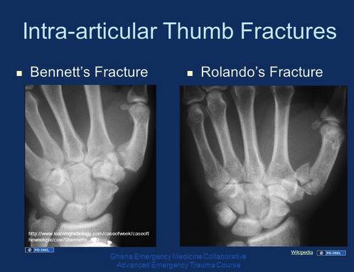
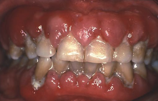
Treatment:  Strong suspicion/acutely ill:  Vancomycin +/- gentamycin.

If stable may wait until blood cultures return.

Prosthetic valves get rifampin too, increased penetration of biofilm.

Prophylaxis:  previous endocarditis, unrepaired cyanotic congential heart diease, prosthetic valve/tissue in heart.  Do this if invasive dental procedures maybe tattoos.  Use Amox.

10:30 am:  M&M; Dr. Mark Bamman

“7 Deadly Sins”....lessons learned.  AKA the Bamman confessional.

Pride: 

First chest tube-supervision offered, deferred.  Next day with new hemothorax, likely from intercostal vessel damange.  

Afib/RVR-didn’t sync for cardioversion. 

If not comfortable with procedure, get help.

“Humility is not thinking less of yourself, it is thinking of yourself less” C.S. Lewis

Envy:

“Speed envy” when self-comparingto more senior residents.  Risk=cutting corners, incomplete history, will miss things.

Gluttony:

Diet and exercise-prioritize.

Anger:

“Anger is an acid that can do more harm to the vessel in which it is stored than to anything on which it is poured.”  Mark Twain

 “It isn’t the mountain ahead to climb that wears you out, it’s the pebble in your shoe.”  Muhammad Ali

Many daily system challenges in our ED

Anger towards patients.

Must learn to let it go, in order to preserve your humanity and longevity.

Lust:

Lust for “Dr. Done”.  Elderly woman with abdominal pain, CT read said SBO, missed incarcerated inguinal hernia...look at your own CT scans, careful/repeat physical exams.  Consider why diagnoses occur (look for underlying primary problem).

Greed: 

Greedy with Time.  

Consider service for others, beyond what brings you secondary gain.

“We make a living by what we get, but we make a life by what we give.”  Winston Churchhill

Sloth:

PICU extubation:  patient extubated, then received usual push dose sedation which led to oversedation and re-intubation.  Could have been avoided with clinical reassessment before giving meds?

Re-assess your patients, especially prior to interventions, going upstairs, going home, or if nurse says change in condition.

Procastination of reading/studying-you are cheating yourself; of administrative requirements-just makes it harder to complete.

“Diligence is the mother of good luck.”  Benjamin Franklin

11:30 am:  Health Care Disparities-Social Determinants of Health; Dr. Oyinkansola Okubanjo

WHO:  Social Determinants of health:  “conditions in the environments in which people are born, live, learn, work, play, worship and age that affect a wide range of health, functioning, and quality-of-life outcomes and risks”

EG:  childhood asthma rates in children on the south side, access to public transportation, food deserts

Categories of Social Determinants of Health:

Economic Stability:  employment (access to insurance, PMD).  Employment associated with better health.  Blacks, Hispanics less likely to be in management positions.

Neighborhood, physical environment:  housing-time for EMS, smoke/CO detectors, mold, safety

Education:  language, literacy-tied into health, nutrition, employment

Food/Nutrition:  hunger in Chicago children, access to healthy options.  Food deserts correlate to areas where African Americans live.

Community and Social Context:  Diversity, Race (which is only a social construct) and discrimination, incarceration rates (normalized in certain communities)

Health Care system: insurance, provider availability, provider linguistic and cultural competency, access to health care, health literacy-pay attention to discharge instruction comprehension.

How Determinants Interact:  individual life style factors + social and community networks + socioeconomic, cultural and environmental conditions

Take home points:

-Determinants affect individual and community health directly and indirectly

-Different determinants affect different social groups

-Inequitable distribution contributes to health care disparities

-Increased knowledge of these determinants leads to providing better care for YOUR patients!

Next time:  Patient and Provider factors

12:00 pm:  Pulmonary Hypertension (PH); outside speaker Valerie Laroy, APN

Pulmonary artery hypertension-can be idiopathic or due to multiple underlying disease processes. Seen more in women, starts in small arterioles

Diagnosis with Right heart catheterization.

Pulmonary circulation:  low pressure system, low resistance, high capacitance, dynamic vascular bed. Pulmonary circulation has one fifth the pressures of systemic circulation despite the same CO as systemic circulation.

Usually several year delay from symptoms to diagnosis.  Presents with shortness of breath without hypoxia, tachycardia, fatigue, peripheral edema.  Late symptoms syncope, JVD, CP, hypotension, hepatomegaly, ascites, SOB

Patients asymptomatic until RV is affected.  How RV reacts to preload and afterload predicts outcome.

Treatment:  endothelin receptor antagonist (eg Opsumit), prostacyclin analog (eg Remodulin) and nitric oxide enhancement (PDE5 inhibitor-Viagra, Adempas).  PA pressures diagnose the disease, but goal of treatment is to remodel RV to maintain cardiac output.  Terminal treatment is therefore lung transplant, rather than heart transplant.  If you transplant a heart into patient with PH, heart will rapidly dilate/fail, death.

From Valerie Laroy (NP for PH team).  If a patient presents with PH diagnosis on PH meds, please call PH team on arrival.  They need to be on consult for the admission.   Type in “Pulmonary Hypertension” to perfect serve to identify who is on call.  If our PharmDs see the med list and identify a PH med, they will alert us.  PH meds are life saving-there are oral meds that must be continued in the ED, and pumps must stay on

Why do they present?  Fluid overload-need for diuretic assessment.  Site pain at site of infusion-possible infection.  These patients are usually baseline hypotensive.  If BP support needed, usually the preferred pressor is Neosynephine (avoid tachycardia).  Usually will go to MICU (preferred ICU), possibly 7W.  For admission, there is not a preferred admitting physician.  On the back of the patient’s IV or SQ pump (SQ pump is actually an insulin pump) will be a phone number for their specialty pharmacy-call this number for dose/rate.  NEVER SHUT OFF THE PUMP.  Inhaled meds have unique inhaler-it’s NOT our usually MDI.  Initial skin pain may not be infection-look for infectious sx beyond pain.

Testing:  Order BNP, need stat Echo

If respiratory failure, try CPAP first.  Intubation dangerous-PA pressures very high, anticipate code.   Anticipate need for pressors and inotropes.