Conference Notes 7-27-2016

Carlson/Kennedy   Oral Boards

Case 1. 67yo male with back pain and lethargy.  He also has lower extremity weakness.  ESR was elevated. Imaging showed multiple lytic lesions in the spine.   Patient had hypercalcemia secondary to multiple myeloma.  Critical actions were to diagnose multiple myeloma with hypercalcemia and treat with IV saline.   


*Hypercalcemia symptoms


*Hypercalemia management


*Multiple myeloma


Case 2.  22 month old female presents with parent saying she cannot wake the child up.  BP 52/26   HR 162,  shallow respirations.  Accuchek is 28.   History reveals that older sibling is taking phenobarbital.   CXR shows non-cardiogenic pulmonary edema.  Patient required glucose supplementation and intubation.  Hypothermia was also treated. Urine was alkalinized.  Multi-dose activated charcoal can be useful with phenobarbital ingestions.  Diagnosis was child abuse.  Mom was giving phenobarbital to keep child calm and to sleep.  Child was placed in protective custody.


*Multi-dose Activate Charcoal MnemonicsABCD for the toxins it works for.   PHAILS for the toxins that charcoal does not work for.


Case 3.  30mo male who is vomiting after taking his vitamins.  The vitamins contained ferrous fumarate.  However, calculations showed the amount the child ingested was not toxic.  If child takes less than 60mg/kg the ingestion should be non-toxic.  A serum iron level < 500 is non-toxic.   You can get vomiting with a non-toxic dose of iron.   A toxic dose typically will cause an anion gap acidosis.  This patient did not have an elevated anion gap.  Andrea made the point to get the numbers 60mg/kg and an iron level of 500 in your head regarding iron poisoning.


C & EKulstad    CV Study Guide


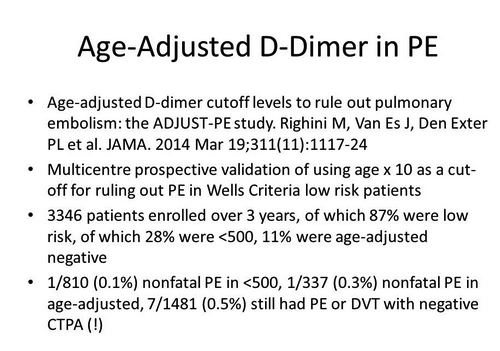
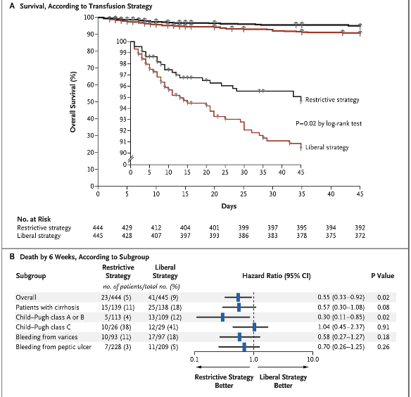
*Age adjusted d-dimer EBM


The test to evaluate for pelvic thrombosis in pregnant patients is MRI.  It may be the best study as well for identifying pelvic thrombosis in non-pregnant patients.


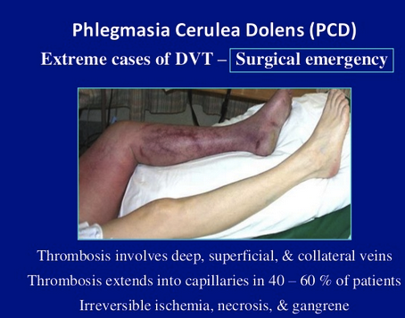
*phlegmasia cerulean dolens


*phlegmasia alba dolens


Wikipedia: The disease presumably begins with a deep vein thrombosis that progresses to total occlusion of the deep venous system. It is at this stage that it is called phlegmasia alba dolens. It is a sudden (acute) process. The leg, then, must rely on the superficial venous system for drainage. The superficial system is not

adequate to handle the large volume of blood being delivered to the leg via the arterial system. The result is edema, pain and a white appearance (alba) of the leg.

The next step in the disease progression is occlusion of the superficial venous system, thereby preventing all venous outflow from the extremity. At this stage it is called phlegmasia cerulea dolens (PAD). The leg becomes more swollen and increasingly more painful. Additionally, the edema and loss of venous outflow impedes the arterial inflow. Ischemia with progression to gangrene are potential consequence.  ePhlegmasia alba dolens is distinguished, clinically, from phlegmasia cerulea dolens (PCD) in that there is no ischemia.[1]

Emedicine/Medscape: In PAD, the thrombosis involves only major deep venous channels of the extremity, thus sparing collateral veins. The venous drainage is decreased but still present; the lack of cyanosis differentiates this entity from PCD. In PCD, the thrombosis extends to collateral veins, resulting in severe venous congestion with massive fluid sequestration and more significant edema. Without established gangrene, these phases are reversible if proper measures are taken.

Of PCD cases, 40-60% also have capillary involvement, which results in irreversible venous gangrene that involves the skin, subcutaneous tissue, or muscle.[3] Under these conditions, the hydrostatic pressure in arterial and venous capillaries exceeds the oncotic pressure, causing fluid sequestration in the interstitium. Venous pressure may increase rapidly, as much as 16- to 17-fold within 6 hours.[4


Elise comment: You now need to treat superficial thrombophlebitis with heparin or LMWH or one of the new oral anticoagulants if the thrombophlebitis is large or proximal. 

From the ACMCEM website: Thrombosis of superficial veins has long been considered benign, and deemed a separate entity from venous thromboemolism (VTE)

-- However, multiple studies illustrate a significant association with VTE (DVT and PE).

-- When patient with ST (diagnosed clinically, no ultrasound) are thoroughly evaluated, the degree and the extent of clot are underestimated 75% of the time.  Further, such patients are found to have co-existent DVT or PE 25% of the time and/or rapidly progress to DVT 10% of the time.  

---- Teaching point: get ultrasounds on all clinical superficial thrombophlebitis

-- The risk factors for ST and VTE are the same, and many argue that ST should be treated the same as VTE.  

-- In general,

anticoagulate (as you would for DVT) the patient if they have known clotting risk factors, greater than 5 cm of clot, or clot < 5 cm within the sapheno-femoral or sapheno-popliteal junction.

-- Another way to think about it is that a superficial vein thrombosis is a manifestation of a systemic clotting cascade gone awry.  To even further simplify things, seems pretty pathologic to have any blood vessel clot; ahh hello, you are clotting off blood vessels.

*Treatment algorithm for superficial thrombophlebitis


The treatment of pulmonary edema is high dose IV NTG or sublingual NTG as the mainstay of therapy.   Aggressive use of NTG allows us to avoid intubating pulmonary edema patients.

Morphine has no efficacy in the management of CHF.


*OESIL Rule for syncope is considered better than San Francisco Syncope Rule


*Rose Rule    Elise likes the Rose Rule for syncope.  She made the point that an elevated BNP is a marker for badness in syncope and can be a surrogate for CHF.


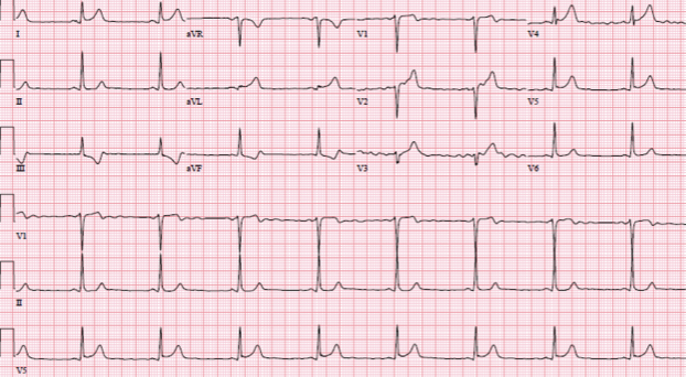
*Hypertrophic Cardiomyopathy    Patients have LVH QRS complexes with lateral inverted T waves.


Treat pericarditis with colchicine.


Hart/Regan 2015 ACLS Updates


Social media can be used to summon rescuers.

Christine, Erik and Elise spoke about an app called Pulse Point that will notify you of a nearby cardiac arrest.


*Pulse Point App


Do Chest compressions 100-120 per minute.   Compressions at a pace over 120 has a worse outcome.

Avoid leaning on chest to allow full recoil.

Vasopressin is out.  It has no advantage over epinephrine.

We discussed automated CPR machines.  Faculty present at the meeting have found them more reliable with less interruptions anecdotally, but the research does not show any benefit over human CPR.

Routine use of lidocaine is not recommended.

If ETCO2 is <10 after 20 min of downtime ROSC is unlikely

ECMO can be used in cardiac arrest with a potentially reversible cause (hypothermia, myocarditis, transplant candidate)

Any post-arrest patient not responding to verbal stimuli should be cooled to 32-36C.

Avoid and treat hypotension in a patient who had ROSC.


ACLS Workshop


McGinnis   MIDAS Reporting Update

Please place any patient safety concerns in the MIDAS system.  We really want you to make a report of any safety issues.

When placing a bed request, please check the special request drop down menu to be sure you make note of suicidal patient needing a sitter, peritoneal dialysis, LVAD, chronic trach, c-diff, andother isolation needs.  These requestswill limit the patient to certain floors that can provide that specialized care.


Conference Notes 7-20-2016

Barounis     Management of the Hypotensive Patient

Recent study on HelmetNon-Invasive Ventilation

Results  Eighty-three patients (45% women; median age, 59 years; median Acute Physiology and Chronic Health Evaluation [APACHE] II score, 26) were included in the analysis after the trial was stopped early based on predefined criteria for efficacy. The intubation rate was 61.5% (n = 24) for the face mask group and 18.2% (n = 8) for the helmet group (absolute difference, −43.3%; 95% CI, −62.4% to −24.3%; P < .001). The number of ventilator-free days was significantly higher in the helmet group (28 vs 12.5, P  < .001). At 90 days, 15 patients (34.1%) in the helmet group died compared with 22 patients (56.4%) in the face mask group (absolute difference, −22.3%; 95% CI, −43.3 to −1.4; P = .02). Adverse events included 3 interface-related skin ulcers for each group (ie, 7.6% in the face mask group had nose ulcers and 6.8% in the helmet group had neck ulcers).

Conclusions and Relevance  Among patients with ARDS, treatment with helmet NIV resulted in a significant reduction of intubation rates. There was also a statistically significant reduction in 90-day mortality with helmet NIV. Multicenter studies are needed to replicate these findings.

*Helmet NIV


*AVAPScan be used in patients who are not getting optimaltidal volumes with NIV.  It gradually increases IPAP.  Usually used in COPDer’s.

Management strategy with BIPAP: For COPDer’s move up on the IPAP.   For CHF, go up on the EPAP.   For ARDS go up on EPAP.

Dave’s first two steps when evaluating hypotensive patients are: 1. Touch the patient to evaluate their peripheral perfusion.  2. Put an ultrasound probe on the patient to evaluate their cardiac pump function.   Mitral valve movement directly correlates with LV function.  The other method he likes is calculating fractional shortening.

*Fractional shortening.  You drop an M mode line through the left ventrical and the machine will calculate the percentage change in width of the left ventrical.   Less than 50% is c/w heart failure.

Dave brought up the situation of unilateral R side CHF.  This can be caused by severe mitral valve disease causing an eccentric regurgitant jet into the left atrium forcing backflow preferentially into the right pulmonary vein.   If you see a patient with a right unilateral big infiltrate that looks like unilateral CHF, throw an ultrasound probe on the heart.  If the mitral valve looks abnormal, get a formal echo to evaluate for an eccentric reurgitant jet. 


*Unilateral Pulmonary Edema   Editorial comment: Unilateral right side pulmonary edema is a Zebra diagnosis but you could look amazingly good if you pick this one up. 

Snip20160720_5.png

 *Eccentric mitral regurgitant jets.


*Sonographic B lines go all the way to the bottom of the screen.  These indicate most commonly CHF but could also be due to other fluid in the alveoli.

In hypotensive patients with Afib and RVR don’t use diltiazem.   Give magnesium 2-4 grams.   Don’t worry to much about the magnesium level.  Patients can tolerate mag levels up to 4.9.  2nd line drug is Amiodarone 150mg bolus followed by 1mg /min.

E. Kulstad       CV Study Guide

*Indications for Transcutaneous Pacing

Compared to placebo, Heparin and LMWH do not provide morality benefit in non-STEMI acute coronary syndromes.

Compared to placebo, Heparin and LMWH demonstrate a trend toward mortality benefit in STEMI’s.


*Sgarbossa Criteria


*Heart Score Components

*HEART Score Outcomes MACE (Major Adverse Coronary Events)


Carlson       Toxicology Cases

*Acetaminophen Overdose Decision-making

*Rumack Matthews Nomogram

*Acetaminophen Toxicity Mechanism.  The amount of acetaminophen overwhelms the conjugation pathways and the excess acetaminophen instead gets metabolized to NAPQI which is toxic.

*NAC Dosing

Dextromethorphan can give you a positive toxicology screen for PCP.  It is a dissociative drug similar to ketamine.  It is commonly mixed with marijuana.  Patients can have bad trips.  Treat patients with benzo’sto manage the agitation.

Drugs that cause a positive toxicology screen for PCP: PCP, dextromethorphan, and ketamine.

Caffeine is a methyl xanthine that binds the adenosine receptor. If you are treating SVT for caffeine overdose you will need to use higher dosing of adenosine.  You should start at 12 mg of adenosine.

Caffeine will cause vomiting and hypokalemia.  Caffeine acts at the kidney to increase potassium diuresis.   Hypokalemia is variable in caffeine overdose and the level of hypokalemia should not be used to risk stratify or rule in/out the diagnosis.

Toxidrome: Anxiety/agitation, tachycardia, vomiting and hypokalemia think caffeine toxicity.

Iron toxicity causes an anion gap acidosis, hyperglycemia, and leukocytosis.  Serum iron levels >500 are dangerous.


*5 Stages of Iron poisoning.   Treat with deferoxamine.

Vitamin A is really the only dangerous vitamin overdose.  It can cause cerebral edema.

If you identify a patient with an anion gap metabolic acidosis and a respiratory alkalosis they have ASA toxicity.  Death from salicylate is a CNS death, not a pulmonary or cardiovascular death. Alkalinize serum to keep salicylate out of CNS.  Alkalinize urine to enhance elimination. Replace potassium.  Finally, hemodialysis can be life-saving.

Tinnitus is common with a salicylate level above 20. 

*Indications for dialysis in ASA toxicity

Hart    Code STEMI

Goal: Get the patient to the Cath Lab in less than 60 minutes for a STEMI.  Identify alternative critical diagnoses such as PE, Aortic dissection, perforated ulcer, GI bleed,  hyperkalemia, pericardial effusion, valve rupture.

No need for O2 if a Code STEMI patient is sating at or above 94% on room air. 

Air Versus Oxygen in ST-Elevation Myocardial Infarction (AVOID) trial compared supplemental oxygen vs no oxygen unless O2 fell below 94%.

"The AVOID study found that in patients with ST-elevation myocardial infarction who were not hypoxic, there was this suggestion that, potentially, oxygen is increasing myocardial injury, recurrent myocardial infarction, and major cardiac arrhythmia and may be associated with greater infarct size at 6 months," lead author Dr Dion Stub (St Paul's Hospital, Vancouver, BC, and the Baker IDI Heart and Diabetes Institute, Melbourne, Australia) concluded.

Methods and Results—We conducted a multicenter, prospective, randomized, controlled trial comparing oxygen (8 L/min) with no supplemental oxygen in patients with STEMI diagnosed on paramedic 12-lead electrocardiogram. Of 638 patients randomized, 441 were confirmed STEMI patients who underwent primary endpoint analysis. The primary endpoint was myocardial infarct size as assessed by cardiac enzymes, troponin (cTnI) and creatine kinase (CK). Secondary endpoints included recurrent myocardial infarction, cardiac arrhythmia and myocardial infarct size assessed by cardiac magnetic resonance (CMR) imaging at 6 months. Mean peak troponin was similar in the oxygen and no oxygen groups (57.4 mcg/L vs. 48.0 mcg/L; ratio, 1.20; 95% confidence interval [CI], 0.92 to 1.56; P=0.18). There was a significant increase in mean peak CK in the oxygen group compared to the no oxygen group (1948 U/L vs. 1543 U/L; means ratio, 1.27; 95% CI, 1.04 to 1.52; P= 0.01). There was an increase in the rate of recurrent myocardial infarction in the oxygen group compared to the no oxygen group (5.5%vs.0.9%, P=0.006) and an increase in frequency of cardiac arrhythmia (40.4% vs. 31.4%; P=0.05). At 6-months the oxygen group had an increase in myocardial infarct size on CMR (n=139; 20.3 grams vs. 13.1 grams; P=0.04).

Conclusions—Supplemental oxygen therapy in patients with STEMI but without hypoxia may increase early myocardial injury and was associated with larger myocardial infarct size assessed at six months.


 For STEMI’s make sure all patients get 4 chewable baby ASA.  If the patient received ASA from EMS or took it at home, document that.  Start IV Heparin (max dose 4000u).  Discuss anti-platelet agents with your cardiology consultant.  Some cardiologists may choose not to use an anti-platelet agent to avoid bleeding complications if the patient requires CABG.

If you have a patient with an inferior STEMI with ST elevation in lead 3>lead 2 get a right-sided EKG to identify a possible Right ventricular infarction.

*DeWinter’s Syndrome

*Dewinter Teaching Points

Lee    

Unfortunately I missed this excellent lecture. 

Nand    Observation Service and Care Management

Basically if you are admitting for a symptom, the patient should be brought in as observation.  For the most part, if you have a diagnosis for the patient then  bring them in as a full admit.  Exceptions would be TIA, syncope, and cellulitis which can be observation status.  If you have a doubt, start as an observation. Social issues should be brought in as an observation.

When patients are brought in as an observation patient, theyhave more personal financial exposure than they would as a regular admission.  If the patient is expressing concerns about the financials of an OBS admission, consult the ED Care Manager to help with this discussion.

If you have to change the admit or observation order, cancel and reorder the bed request. This action will re-generate a new level of care order.

If the patient will be staying for 2 midnights that is a patient who should be a full admit.

Don’t use the word “observation” as a verb.  Use “monitor” or “watch” instead.   For example don’t write “we will observe the patient in the ICU”.  Instead write, “we will monitor in the ICU”  Using the word “observation” as a verb in your note causes confusion for reviewers regarding whether the patient is admitted or Observation status.


Conference Notes 7-13-2016

LambertUltrasound Basics

The basis of ultrasound imaging is that every tissue in the body has a different acoustic impedance.   Sound travels at different speeds thru different tissues.   When sound waves reflect back to the probe at varying speeds, the ultrasound machine can generate an image based on these differences.


*Acoustic Impedance

Low frequency waves penetrate deeper into the tissue but provide less resolution.   High frequency waves have less penetration but better resolution.


*High Frequencyvs Low Frequency ultrasound waves

Terminology synonyms:

Longitudinal = sagittal=long axis

Transverse=axial=short axis

Coronal is basically a longitudinal view taken from the side of the body


* Sagital,  Axial, and Coronal Planes


Knobology

Mike discussed the importance of controlling the depth and gain of the image you are looking at.   You want to adjust the depth to optimize to proportion of the body structure being visualized in the image.  You want the structure of interest to take up the majority of the screen.   You need to adjust the gain to have a uniform appearing image both in the near and far fields. 


*Near field is top half of image,   far field is bottom half of image.  Gain is uniform throughout image.


Lambert      Bedside Echocardiography

Bedside echo is a game changer for identifying life-threatening illness in patients with chest pain or shortness of breath.   You can identify PE, pericardial effusion, CHF, problems with contractility/wall motion.   It is useful during cardiac arrest as well.


There are two main views of the heart when doing Bedside echo


*Parasternal Long axis


*4 Sub costal


*Pericardial effusion*


*Pulmonary embolism Note the large RV in comparison with the LV.


Mike classifies LV dysfunction as either OK or Bad.   Bad is usually obvious on bedside echo and helps you identify CHF or cardiogenic shock.   


Elise’s Journal Club Summary:

Article 1:  Flato UA, et al:  Echocardiography for prognostication during the resuscitation of intensive care unit patients with non-shockable rhythm cardiac arrest. Resuscitation  2015  Jul; 92:1-6. 

This was a Brazilian prospective, observational cohort study of 49 ICU patients with in hospital asystole or PEA cardiac arrest. Of 88 eligible patients, 39 were excluded. PEA without contractility was classified as electromechanical dissociation (EMD), and PEA with contractility as pseudo-EMD.  Two echo trained intensivists performed transthoracic echos. Rates of ROSC were 70% for pseudo-EMD, 20% EMD, and 24% for asystole.  Good ROSC percentages for all groups, but survival to hospital discharge was only seen in pseudo-EMD patients, and discharge is what counts.  Four patients survived to 180 days, all in the pseudo-EMD group, with CPC of 1, 1, 1, 2.  Echo was feasible, with maximum duration of 10 seconds, so non-disruptive to the ongoing code.  Echo also helped identify underlying etiology of arrest in selected patients, eg unexpected tamponade.  This was a very small study, and conducted in an ICU with a large number of DNR patients who were never entered into study, so different from our ED population.   

This study reinforces our usual practice of using echo to verify presence/absence of cardiac contractility and guide futility of resuscitation.  Blaivas and Fox (go ACMC!!) published a larger study in 2001 demonstrating 100% mortality in patients with asystole or PEA cardiac arrest and no cardiac contractility on bedside echo. This modality helps conserve resources (time and personnel), and may identify the underlying reason for code.  In the future, in young otherwise healthy patients with arrest, an echo demonstrating contractility may help risk stratify for ECMO.

Bottom Line:  Rather than pulse check, consider echo as the more reliable marker for viability.  Also remember end tidal CO2 to guide prognostication.  

(AHA 2015 ACLS guidelines:  “In intubated patients, failure to achieve an ETCO2 of greater than 10 mm Hg by waveform capnography after 20 minutes of CPR may be considered as one component of a multimodal approach to decide when to end resuscitative efforts but should not be used in isolation.”)


Lambert    IVC and Aorta

*Aortic Aneurysm

*IVC with hypotension. 

*The proper landmarks for observing the IVC for hypovolemia include the heart, portal vessels and IVC in the same image.

Mike feels a collapsing IVC with inspiration is the most useful sign of hypovolemia on ultrasound.  He has doubts that IVC ultrasound is the holy grail of identifying low central venous pressure.

Lambert and Team Ultrasound        U/S Lab

Conference Notes 7-6-2016

Hart/Girzadas   Oral Boards

Case 1.   15yo female suffered blast injuries in an explosion.   Patient required intubation fordyspnea, respiratory distress , and bloody sputum secondary to blast lung injury.  Patient also had a traumatic amputation of the right upper extremity that was hemorrhaging.  A tourniquet was applied to stop bleeding.  Patient was resuscitated with PRBC’s and IV LR.  Patient also had 2nd degree burns on right side of body of about 20%.   Parkland formula was started.


*Blast Injury Categories


> Mortality increases from 8% to 49% when blast occurs in an enclosed space

>  Blast Lung Injury is the most common primary blast injury causing death

>  Traumatic Amputations portend a much higher mortality from blast injury

>  Military data demonstrate that tourniquets decrease mortality from a hemorrhagingextremity from 90% to 10%.


Case 2. 5 month old child presents with bloody stool, shock, and metabolic acidosis.  Patient had an anion gap acidosis and markedly elevated LFT’s and LDH.   History revealed that grandma was giving excessive amount of ferrous sulfate supplementation.   Serum Iron level was 600.  Patient was treated with IV fluids and IV Deferoxamine.  


*CAT MUDPILES


Case 3. 35yo male presents with left hand puncture wound on the palmar surface.  A paint gun fired into his palm when he was cleaning the nozzle.  The patient has severe pain.  Treatment for high-pressure injection injury included pain control, IV antibiotics, TDAP updated, emergent surgical debridement in the OR.


*High pressure injection injuries can look innocuous on first look.


*Xrays can show the extent of subcutaneous spread of pain or grease.


Regan    M&M

Tension pneumothorax is treated initially by a needle thoracostomy followed by a chest tube.  A common complication of chest tube placement is the tube can slide up into the subcutaneous tissue and not actually enter the pleural space.   You have to make sure the tube passes thru the ribs.


*Chest tube that never passed thru ribs and is the in subcutaneous tissue. After tube placement you need to feel all the way around the tube and be sure it passes thru the ribs.  It is amazing how easily the tube can pass up thru the subcutaneous tissue and feel like it is in the pleural space.


Harwood comment:  Any time the paramedics place a needle thoracostomy in the field, the patient should get a chest tube when they arrive in the ED.


If you are managing a bloody airway, you may need to use direct laryngoscopy rather than video laryngoscopy.  Blood can obscure video laryngoscopy.

When breaking bad news to families in heartbreaking situations, don’t hesitate to get support for yourself.  Some days our job can be terribly sad and emotionally disturbing.  Reach out to our faculty, chaplain, a crisis worker or co-workers for support.


Alexander      EKG Basics


*Basic Step-wise approach to EKG interpretation


Ari suggested the Rule of 4 for initial EKG interpretation

4 Features: Clinical context (patient age, chest pain), rate, rhythm, axis

4 Waves: P, QRS, T, U

4 Intervals: PR, QRS, ST, QT


Ari then demonstrated this approach on a number of EKG’s.   If you would like further EKG training, Ari has an excellent EKG teaching blog (Christ-ECG.com) linked to the ACMC EM website (click on Enlightenment)


Regan/Hart       Intro to Codes

Code 30:  The onset time of stroke is the “last known normal.”  If a patient wakes up with stroke symptoms, the onset of symptoms is not the time of waking but rather the last time the day prior that the patient felt normal or a family member noted them to be normal. 


Check a blood sugar in anyone who you can’t have a conversation with, anyone with a seizure, and anyone with neurologic findings.

There are phone apps and paper copies of the NIH stroke scale to make it easier to perform in patients you have a concern for stroke.


Girzadas comment: You may want to use the NIH stroke scale as the new standardized neurologic exam.  If you start doing it on all your patients with neurologic complaints you can get pretty fast at performing the exam.  Some non-stroke patients may need some additional exam components such as halpike or gait testing.


In the setting of acute stroke within 4.5 hours, if you get a stat CT head and the scan shows no hemorrhage, you should consider and discuss with neurology team about moving next to getting a CTA.  CTA will evaluate for the opportunity for interventional thrombectomy up to 6 hours out from onset of stroke symptoms. CTA is used to identify large proximal clots in the MCA.  These types of clots are the ones amenable to thrombectomy.


TPA outcomes: 1 in 3 patients will have some degree of improvement.  6 in 100 will have bleeding (some studies show higher rates of bleeding).   1 in 100 will have death or serious disability secondary to bleeding.

As of 2015, all contraindications to TPA are relative.  However, you need to weigh risks carefully.  If the patient has had prior ICH or is anti-coagulated you will probably evaluate the risk of TPA as outweighing the benefit.

Code 44:  Basic preparation: Assemble your team ASAP.   Get IV access.  Start O2.  Get them on a monitor/pulse ox and get an EKG.  Check the blood sugar.  Be sure you have airway tools in the room.


Holland       Hypertensive Emergencies

Deweert      5 Slide Follow Up

Holland       Admin Update

Unfortunately I missed these 3 excellent lectures. 


Conference Notes 6-8-2016

Ede/Herron   Oral Boards

Case 1.  Middle age woman with acute weakness and diarrhea.  Electrolytes showed hyponatremia and hyperkalemia.  Further history revealed that patient was not taking her daily steroids for several days.  Diagnosis was acute adrenal crisis.  Treatment required IV hydrocortisone or dexamethasone, IV fluids and correction of electrolyte abnormalities.

Elise comment:  When dealing with an endocrine emergency always look for an inciting cause such as infection or AMI. 

*Adrenal Crisis


Case2. Firefighter trapped in a house fire suffered burns and had altered mental status and metabolic acidosis.  Treatment required Intubation for airway protection, applying Parkland Formula to manage fluids. Patient had a profound metabolic acidosis so he also required presumptive treatment for cyanide toxicity. Finally, the patient required transfer for hyperbaric O2 for elevated CO Level.   


*Parkland Formula


Elise comment:  If you get a burn victim on the boards always look for other traumatic injuries.   Look for circumferential burns on extremities that require escharotomy.  Always consider CO and Cyanide toxicity. 


Andrea comment:  A “poor man’s” rapid cyanide level in a person trapped in a house fire is a lactate.  Cyanide poisoning should give you a lactate of 10 or higher.  Hydroxycobalamine is a safe drug and there really isn’t much downside to giving it presumptively.


Case 3.   50 yo male fell from ladder and injured his wrist.  Pt has wrist

swelling and tenderness on exam.   No other injury.


*Xray is c/w perilunate dislocation.  


Girzadas comment: Lunate and perilunate dislocations can be confusing.  To me the easiest way to remember it is if the lunate is out of line with the capitate and the radius, it is a lunate dislocation.  If the lunate is in line with the radius but the capitate is out of line with the radius and the lunate it is a perilunate dislocation.  “Peri” means around, so perilunate is not a dislocation of the lunate but rather of the bone around the lunate which is the capitate.  As an aside, any time you see overlapping bones on the AP view of the wrist, you have a dislocation of some sort.  Both the lunate and perilunate dislocations require operative repair. 


Carlson   Summer Toxicology Cases


Case 1. 16 yo male ate seeds from a “loco pod”  (black seeds)  and develops anticholinergic syndrome.  The seeds were jimson weed, which is basically, plant-based Benadryl.


*Anticholinergic symptoms


Treat antichoinergic syndromes with benzos and supportive therapy.  Most cases of anticholinergic ingestions are short lived and can be observed in the ED until symptoms improve.


Case 2.  Little kids frequently ingest caustic materials.   Caustics include swimming pool products, rust removers, toilet bowel cleaners, hair treatments, detergents, bleaches, and denture cleaners, etc.   Detergent pods for dishwashers are a common caustic ingestion because kids see them as candy and try to eat them.


Never induce emesis in a patient with a caustic ingestion.   You don’t want to bring the caustic material back up the esophagus and cause further injury.

Alkali ingestions cause deeper injury by liquefaction necrosis.

Acid ingestions cause coagulation necrosis and less local injury but cause more systemic effects due to acidosis.

If the patient is drooling, has stridor, has painful swallowing, vomiting, or chest pain they have a significant caustic ingestion.

Plain films of the chest and abdomen can be useful to identify mediastinal or intra-abdominal free air. 

Endoscopy does not need to be performed emergently.  It is best done at 12-24 hours after ingestion.

For ED management of symptomatic patients, have a low threshold for starting antibiotics such as Zosyn or Unasyn.  

Steroids are really not an Emergency Physician decision.  Your GI consultant should direct steroid administration.

Andrea said the only kids she would let go home after a caustic ingestion would have to be running around the exam room, happy, eating and drinking, no tachycardia.  They have to look fantastic. Also the history has to suggest a minimal exposure.


Case 3.   Poison ivy exposure.


*Poison ivy exposure

ED treatment options:  If the exposure is recent you can advise the patient to purchase Tecnu to remove the resin.   Benadryl can help with the pruritus.    Prednisone 60mg/day for 5 days then 50mg for 2 days, then 40mg for 2 days, then 30mg for 2 days, then 20 mg for 2 days, then 10 mg for 2 days.  Steroidslessen the hypersensitivity reaction.  Finally Andrea said astringents such as domeboro solution can help dry the weeping lesions.


*Tecnu Poison Ivy treatment product


John Meyers comment:  Topical Benadryl gel is very effective for localized itching from poison ivy.  Topical steroids are also very effective for small areas of poison ivy exposure.


Case 4.  There are actually venomous snakes in Illinois.  It is rare that a patient will get a significant envenomation from a snakebite in Illinois.  Treat with local wound care, update tetanus, give antibiotics and give antivenin based on usual recommendations


*Antivenin indications.  I would also add local progression of edema/pain/skin changes at the site of envenomation


Case 5.  4 yo child ate red berries growing in the yard.  It is climbing nightshade.  Climbing nightshade causes nausea and vomiting and in general is self-limited.   Deadly nightshade is rare in the US but can cause fatal anticholinergic syndrome. 


*Climbing nightshade


*Deadly night shade


Case 6.  Sitting in the grass can result in chiggers due to a trombiculid mite.  They are very itchy but self-limited.  Treat with antihistamines and topical steroids. 


*chiggers


*trombiculid mite


Case 6.  Tick paralysis can cause weakness of bilateral feet and knees.  It is rare and all reports are in kids under age 10.  Dog tick is most common. It usually requires 5-7 days of tick attachment.  When the tick is removed the symptoms resolve rapidly. 


Case 7.  Lyme disease is cuased by borrelia burgorferi vectored by the ixodes tick .  The tick needs to be attached at least for 36 hours to cause Lyme disease.


*Erythema migrans

Patients can also have heart block and facial palsy caused by Lyme disease.


*Tick removal

Harwood comment: While pulling a tick out with forceps, you can use an 11 blade to excise the skin in which the tick is embedded.  Andrea said if you hold traction for 1-3 minutes the tick will usually release on it’s own.


Holland     

Unfortunately, I missed this excellent lecture.


Parker     TPA for CVA EBM

Dr. Parker went through all the evidence regarding TPA for acute CVA.  His conclusion was that:

If you have a patient with an acute CVA and a Rankin score of 4 or 5 (moderate to severe disability) the benefit of TPA outweighs the risks.  For patients with less severe disability the risk benefit analysis is closer to equipoise and is a tougher call. Shared decision making with the patient and their family is critical when deciding to administer TPA for acute CVA.


Okubanjo    5 Slide F/U

The management of patients with incarcerated hernias is manual reduction and out patient follow up for surgery.   Manual reduction frequently requires procedural sedation and placing patient in trandelenburg position.

Harwood comment:  To reduce a hernia, apply circumferential pressure to the hernia.  The reduction attempt may take up to 5 minutes of pressure.  After reduction, you need to observe patient for an hour or two to be sure they don’t develop peritonitis.   If you can’t reduce the hernia, consult surgery for possible emergent surgery.


Ohl    5 slide F/U

Doxylamine ingestion can cause an anticholinergic toxidrome.   The drug blocks acetylcholine at muscarinic receptors.   Patients with anticholinergic toxidromes will be grabbing at imaginary objects.  It is a peculiar aspect of altered mental status specific to anticholinergic overdoses.

Treat with IV Ativan.  This will calm the agitation and may help decrease the risk of seizures.

Physostigmine is indicated for anticholinergic toxidromes with seizures and/or severe mental status changes.

Check a CK in all these patients for possible rhabdomyolysis.

Charcoal is not indicated for anticholinergic overdoses. 

Andrea:  It is always OK to not give charcoal.


Jamieson/Walchuk    The EM-3 Final Lecture

A funny, yet sweet reminiscing of 3 years of training that went by so quickly. 


Conference Notes 5-25-2016 & 6-1-2016

Today’s Conference Notes contains 2 weeks of Learning!  Both May 25th and June 1st are included in this post.  


Meyers and Faculty        Wilderness Medicine Conference


Top things I learned without taking notes:

1.     Top causes of death in the wilderness are: 1. sudden cardiac death/ACS, 2. drowning, and 3. exposure (hypo/hyperthermia)

2.     Most common injuries in the wilderness are musculoskeletal and soft tissue injuries such as fracture, sprain, dislocation, contusions, lacerations.

3.     Prior planning can significantly decrease your risk of death, illness, injury in the wilderness. Be sure to have enough water, some food, and a rain jacket/insulating layer in case weather conditions change suddenly.

4.     We all agreed that probably the best strategy tomake a fire in the wilderness would be to pack water/wind proof matches and take some cotton balls with Vaseline smeared on them in a plastic sandwich bag.  Thewater/wind proof matches are not that expensive and the Vaseline coated cotton balls are very light and compact.  When you use the match to light the cotton balls they flame very quickly providing a great fire starter.

5.     Have a first aid kit with medications/supplies to treat anaphylaxis, clean and repair wounds, treat pain and ACS with aspirin, and have a broad spectrum antibiotic with multiple indications.  Elise recommended Levofloxacin as a once a day antibiotic with a wide spectrum of indications (pulmonary, urine, skin, bite wounds).

6.     Tourniquets are a good thing for life-threatening limb bleeding.  Recent military data demonstrates that if you apply a tourniquet proximal to life-threatening bleeding in an extremity before shock develops, the survival rate is around 95%.  If you apply the tourniquet after shock develops, the survival rate drops to around 35%.  Tourniquets can remain in place for 2 hours without much negative effect.  The tourniquet articles that John Meyers sent out are an excellent read and worth your time.

7.     Treat jelly fish stings by irrigating/rinsing with sea water.  For patients having severe pain try immersing the limb under hot water.  Some toxins are heat labile and pain may improve.  Most jelly fish stings resolve in 15-30 minutes.

8.     Moving injured people in the wilderness is energy intensive and difficult.  Splinting a patient so that they can walk or even partially mobilize them can sometimes be life-saving.

9.  Humans don't have as much fear of fast moving water as they do of cliffs but more people die of drowning or going over rapids/water falls than falling of cliffs.  Be cautious of fast moving water.

IMG_0060.JPG


Htet   STEMI Conference

Case 1.  Patient presents with chest pain, altered mental status, and hyperglycemia.  EKG shows diffuse ST elevation. 

*Example of similar EKG. 

Troponin was elevated above 8.   Second EKG shows similar diffuse ST elevation.   For the most part, the ST segments were concave upward making myopericarditis more likely.   ABG and anion gap shows DKA.  Troponin continued to trend up. Echo was performed in the ED which showed 35% EF and hypo-kinesis of the baso-inferolateral wall. Patient was managed medically for DKA.


The next day patient went to cath lab and had clean coronary arteries.   Diagnosis was myopericarditis.

Patient was transferred to ICU.   At about 6 hours in the ICU, patient suffered a PEA arrest.

A recent retrospective review of myopericarditis shows that the prognosis of myopericarditis is for the most part excellent.  3.5% of patients will have residual CHF.    A handful of patients had sudden cardiac death or tamponade.


Case 2.   Elderly patient presents with syncope. 

EKG shows   Sinus rhythm, slightly long PR interval, some inferior Q waves and poor R wave progression. 

Initial work up with labs and CT was non-specific.

3 hours later in the ER the patient had another episode of syncope.

Cardiac Catheterization did not demonstrate acute coronary occlusion but did demonstratechronic coronary disease.   Patient had episodes of V-tach in the lab and was treated with amiodarone and lidocaine. 

Treatment for Recurrent V-tach

Amiodarone

Lidocaine

Magnesium

Correct hypokalemia

Atropine

Transvenous overdrive pacing

Beta-blocker

AICD placement

Monomorphic V-tach is usually due to scarring of the ventricle.  Polymorphic V-tach is usually due to ischemia.   Cardiologist consensus was that amiodarone was the go-to drug for any form of Ventricular tachycardia.

Cardiologists felt that beta-blockers should be given for recurrent V-tach if the patient is not hypotensive, in shock, or has asthma.   I asked cardiology how it works that we are beta blocking the patient while at the same time overdrive pacing them.  They said at a cellular level it makes some sense.  You want to block ventricular beta receptors and use the pacing to shorten the refractory period.

Case3.   Patient with diffuse ST depression and ST elevation in AVR can mean proximal LAD occlusion.


*1 Proximal LAD Occlusion EKG.


Marshalla     5 slide F/U


Patient presents with sore throat and fatigue.  On exam the patient has malaise.  Initial clinical picture appeared more like a viral syndrome.   Patient requested water from the doctor.  

Lab work up shows hyperglycemia, anion gap acidosis consistent with DKA.   Patient did turn out to have positive mono test.


*Diagnostic Criteria for DKA and HHS


Treatment of DKA= VIP=Cautious Volume replacement, Insulin, Potassium replacement. 

Be cautious of the patient with malaise, weakness, and fatigue.  Keep DKA in your differential.

Einstein     5 Slide F/U

Young adult male presents with chest pain.  Exam was unremarkable.  EKG showed diffuse ST elevation consistent with pericarditis.   Troponin was elevated.  CRP was elevated.  ECHO and MRI showed acute myocarditis.   MRI of the heart can show ventricular dysfunction in myocarditis.   Diagnosis was myopericarditis.


*There is a spectrum of myo-pericarditis depending on the involvement of the pericardium and myocardium.


Kennedy        Neutropenic Fever Bundle and A-Lines


I missed this excellent lecture but with the Neutropenic Fever Bundle, nurses can now access the porta-cath to draw blood and give fluids and meds without getting a CXR prior to usage. 


Lee     Ectopic Pregnancy


The discriminatory zone for transvaginal U/S is 1500-2000 hCG units.   Viable IUP’s have an hCG increase of at least 66% over 48 hours.  15% of normal pregnancies can have a lower than expected (<66%) increase over 48 hours.  72 hour re-test may be more practical for patient convenience and getting a significant rise in hCG.   If the hCG is not rising normally over three measurements then the pregnancy is considered abnormal.    


Okubanjo  Healthcare Disparities

 Health disparities= Certain populations have different health problems due to genetic predisposition (type 2 dm, sickle cell disease, kidney stones)


Healthcare disparities= Certain populations have different healthcare access and quality due to racism, economics, or other discriminatory social factors.


Pain management has been shown repeatedly to take longer and have lower dosing in minority populations. 


Girzadas question:  How does a doctor monitor this issue in their own practice.  How do we know we are not giving disparate care?   Answer: Work to consistently treat all patients the same way.


Hayward       Heme-Onc Study Guide


*Tumor Lysis Sydrome


Petechiae and mucosal bleeding are associated with low platelets or dysfunctional platelets.  Thrombocytopenia does not usually result in deep tissue bleeding such as retroperitoneal bleeding or hemarthrosis.


*Transfusion strategies in GI Bleeding

The most recent recommended threshold for PRBC transfusion in a patient with GI bleeding is HGB of 7.  This more restrictive strategy demonstrated a better outcome than the liberal strategy of a cutoff of HGB of 9.  The faculty all felt that if the patient is actively bleeding from the GI tract and the HGB is 8 or 7.5 they are going to start a PRBC transfusion rather than wait until the HGB gets to 7.   Also patients with coronary ischemia associated with a GI bleed should be transfused at a HGB of 9.


Ted Toerne rule: In any test question or in real life, if the O2 sat is 85% and the patient is cyanotic consider strongly methemoglobinemia.    The light wavelength of methemoglobin sets the pulse oximeter to 85%.

The youngest age a sickle cell patient can receive hydroxyurea is 9 months of age.


Acute hemolytic transfusion reactions will demonstrate on lab testing: shistocytes, low haptoglobin, free HGB in the blood and urine.  Patients may develop hyperbilirubinemia over several hours.


*Cryoprecipitate components.  


*Discuss with cardiology before giving Plavix or Brilinta for acute MI patients.  If cardiac cath shows the need for CABG, use of these agents may delay or complicate surgery.


Conference Notes 5-18-2016

Urumov            Study Guide   Environmental

Harwood comment:  If a patient is shivering, they will survive hypothermia.  You just need to passively rewarm them.  If they are hypothermic and not shivering, you need to initiate active rewarming.


Treat heat cramps by removing the patient from the hot environment andgiving IV saline or oral fluids with electrolyte replacement.


Prickly heat is due to plugging of glands.


*1 Prickly Heat


Heat Stroke defined by elevated core temperature with mental status changes due to environmental and exertional heat. It should be treated by either evaporative cooling or immersion in an ice bath.


Prognosis for heat stroke is best determined by duration of hyperthermia and the patient’s comorbidities.


We had a discussion about the definition of drowning: The ILCOR recommends that the terms dry and wet drowning, active and passive drowning, near drowning, and secondary drowning no longer be used as they are confusing and not clinically relevant.

In accordance with the ILCOR guidelines, patients should be referred to as drowning victims if they have suffered a suspected respiratory injury following submersion in a liquid medium, regardless of their clinical status, which may vary from essentially asymptomatic to severely ill at time of presentation. Additional descriptors such as whether there was a precipitating event that led to drowning or whether the drowning was witnessed may be used as necessary. The primary outcome of a drowning episode is either death or survival. Adopting this clinical nomenclature will allow future studies to better characterize, study, and risk stratify drowning victims.  (Trauma Reports)


*2High Altitude Illness

Indications for antivenin: Progression of swelling. Low platelets/low fibrinogen. Hand bites.  Unstable vitals/shock.  Rhabdomyolysis.

48 hour absolute lymphocyte count is the best predictor of outcome after radiation exposure.  If the absolute lymphocyte count is <300 the prognosis is dismal.


*3Gamma rays are the most penetrating radiation and can pass through all layers of the body.  Alpha rays can be blocked by paper.  Beta rays can be blocked by aluminum.

Treatment of jelly fish stings/burningincludes using vinegar as the first choice of irrigation solution.  Ocean water is the second choice of irrigation solution.   After irrigation use a razor or credit card edge to mechanically remove the nematocysts.

Scorpion stings cause: nystagmus, tongue fasiculations, swallowing difficulty, tachycardia/hypertension, in addition to burning pain and paresthesias at the site of sting.  Treatment is pain management, wound care, tetanus prophylaxis, benzo’s, and supportive care.

Cirone/Tekwani     Oral Boards

Case 1.  Neonatal chlamydia pneumonia and conjunctivitis.   Treat with oral erythromycin.  Diagnosis is with conjunctival/nasopharyngeal culture.  You need to swab the epithelial cells in the conjunctiva.   Even in a well-appearing child, treat with oral antibiotics.

*7 Neonatal chlamydial conjunctivitis

Case 2.  Torison of appendix testes.  Rule out torsion and reassure patient’s parent about the benign nature of the disease.

Case 3.   5yo child with history of sickle cell disease. Patient has cough and fever.  CXR shows a large right side infiltrate.    Diagnosis is acute chest syndrome.  Treatment is: IV Fluids,  IV antibiotics, exchange transfusion.  This patient also required intubation for respiratory failure.

Elise comment:  In sickle cell patients, if you are considering exchange transfusion, it will depend on the HGB level.  If the HGB level is very low, the patient just needs regular PRBC transfusion.  If the HGB level is not particularly low then you do need to do an exchange transfusion.

Faculty consensus and AAP statement:  For neonatal conjunctivitis and or pneumonia, you need oral erythromycin or azithromycin for 14 days.

Girzadas   Study Guide Neurology

Faculty and Pharmacist discussion highlighting a recent change in acute ischemic stroke management: There are no longer any additional age, comorbidity restrictions for administering TPA in the 3-4.5 hours time window other than the usual contraindications  to TPA for the 0-3 hour window.


We discussed the work up of vertigo at length.  There was faculty consensus that it was difficult to do the HINTs testing in dizzy patients.  HINTs has been shown to be highly accurate when compared to ABCD2 score and MRI for identifying central causes of vertigo, but all faculty said they were not using this physical exam method due to difficulty.

*10 HINTs vs ABCD2 score

*11 Some suggested Red Flags in the setting of Dizziness/Vertigo to consider Brain Imaging.  These are red flags from my reading on this topic. This is not a validated list.   It should be noted that CT scan is much less sensitive than MRI for posterior circulation strokes.  Harwood commented that if he is going to image a patient with dizziness or vertigo he goes right to MRI.


Bamman/Ryan    Oral Boards

Case 1. Rocky Mountain Spotted Fever.  Adult male presented with rash and fever.

*4RMSF rash

Treatment is doxycycline 100mg q 12hours

Case 2. 24 yo male presents after being “Tased.”  Vital are normal.   Patient still has a Taser prong in his back.  Taser prong was removed by making an incision in the skin to allow the barb to be removed.  TDAP was updated.  Wound was irrigated.  Antibiotic ointment and bandage applied.  

Dennis made the point that most taser prongs can be removed with simple traction on the prong.

Otherwise asymptomatic patients don’t need any cardiac work up .  There was faculty consensus that if you have a young, healthy, asymptomatic patient who was “tased” and now is fine, you don’t need to do an EKG or labs.

Harwood comment: You could use a needle to cover the barb of the taser prong and remove the prong similar to the fish hook removal method.

*8 Needle over barb technique


Girzadas comment: If the taser prong resists minimal to moderate traction to remove, I think local anesthetic and making an incision to aid removal makes sense.

Case 3. 24yo female presents with depression, somnolent, and tearful. Patient is tachycardic and hypotensive.  History reveals that patient is taking amitriptyline.


*5EKG c/w TCA overdose, note tachycardia, wide QRS complex and tall wide terminal R wave in AVR.

Treatment includes: IV Bicarb bolus and drip.  IV fluids.  You can use norepinepherine if the patient is hypotensive and unresponsive to IV fluids.

Pharmacist asked when would we use intralipids for TCA overdose.  Faculty consensus wasthat they would use intralipids if no improvement with bicarbonate (still with wide complex QRS and still tachycardic).


Schmitz    5 Slide F/U

Pediatric patient presented with unilateral facial weakness, nystagmus and limited eye movements.

Diagnosis was Acute Disseminated Encephalomyelitis (ADEM).  Typically presents with fever, headache, nausea and vomiting.  Patients can have ataxia, cranial nerve palsies, altered mental status and seizures.  Treatment is steroids and IVIG.

*7 ADEM

*8 Treatment for ADEM


Denk    5 Slide F/U

Adult male presents with loss of memory.  No focal neuro findings on physical exam. CT head was normal.  Labs were all normal.

*6Transient Global Amnesia

Cause is unknown.  Leading theory is cerebral venous congestion.  Heavy lifting or valsalva prior to episode can increase venous congestion in the cerebral veins.  It is thought that possibly patients prone to TGA have incompetent head and neck venous valves.

Long term there may be possible mild cognitive impairment. No increased risk of stroke.   Harwood comment: There is an association long term with dementia.


Conference Notes 4-27-2016

Joint EM-Peds Conference   Physcian Wellness and Resilience

Misuse of Adderall is a dangerous and under-estimated risk for students and residents who use this drug for test and work performance reasons. There is a serious risk of arrhythmia in persons using this drug. The risk is especially high in persons with structural heart disease. Dr. Bunney discussed a tragic case of fatal arrhythmia in a resident using Adderall as an alertness aid to work night shifts.


Most students and residents who misuse Adderrall get the drug from friends or it is prescribed by fellow residents.  Do not use this drug unless prescribed for an accepted indication by a treating physician who is not a friend or family member and who is documenting the prescription and indication in a medical record.


Physicians make bad patients because we fear the loss of our identity as a healthy healer.  There is stigma to being ill.  We feel weak if we are sick.  We don’t like being seen as a  vulnerable person.  We also have medical skepticism.  We know the problems of the healthcare system and the errors that can occur.  We are uncomfortable putting ourselves at risk in the healthcare system.


As physicians and healthcare workers, we need to look out for each other and care for each other.


Wellness=Longevity

Sleep hygiene matters for our health and nongevity

Take care of yourselves and each other.


*Casinos have learned that giving all their workers anchor sleep (sleep at some point during the hours of 1a to 6a) improves performance and longevity. Casino’s purposely schedule night shifts to change over at 4 am so that both night and day shift workers can get some anchor sleep during that 1a-6a time period. 


As ER docs, bad things are going to happen to our patients and us.  We need to have a spiritual anchor that’s get’s us through the tough times.  We all need to find our own spiritual anchors.


Residents are at risk for burnout due to social isolation due to work demands.  Residents tend to neglect their own emotional and health needs. Residents have limited control of their schedule.  Perfectionism is a common trait of residents that can lead to burnout.  Poor relationships with colleagues can increase burnout.  Some residents may feel regret over their career choice.   Anxiety over medical errors can lead to burnout.


As educators we need to teach residents and student how we have dealt with anger, anguish, grief, fear, failure, and other strong emotions that we have faced as physicians.


Dealing with stress: Anticipate your stressors. Interpret your feelings of stress as a sign or opportunity to take positive action to mitigate that stress. Believe in yourself that you can influence events and how you react to them. Talking about feelings and emotions can be very useful for lowering stress levels.


Dealing with stress in the moment:  Take some time to remove yourself from a situation.  Meditate with deep breathing.   Rethink your strategy. 

After the event practice self-compassion and think positive thoughts about yourself or do something nice for yourself or do something you enjoy. 


Munoz/Naik   Oral Boards


Case 1.  48 yo male with severe vomiting.  Patient developed chest and abdominal pain. HR=116.  BP=100/62.   Patient has a history of pancreatitis.   Patient was drinking the night before. 

Diagnosis was Boerhaave’s syndrome.  Patient’s pain was treated with IV morphine.   IV fluid resuscitation was started.  Broad spectrum IV antibiotics were also given.  Surgery was consulted.


*CXR of Boerhaave’s syndrome.  Look for mediastinal air.  CT is more sensitive than CXR of course.

Boerhaave’s is a life-threatening disease and mortality is time dependent.  Get patients to the OR emergently.


Case 2.  74 yo male with leg pain.  Vital signs are normal.   Patient has a history of CAD, DM, and vascular disease.  He is a smoker.  Exam reveals cool lower extremity with absent distal pulses.  EKG shows Afib.

Diagnosis was ischemic limb from embolus.   Treatment is with IV heparin.  Patient also needs vascular surgery consultation.   Be sure to consider other possibilities causing an ischemic limb such as dissection, thrombosis, and trauma.


Case 3.  3 yo male with temp of 38.9.  Parents also note that patient has had fevers for 5 days.  Child is well appearing on initial exam playing with toys.  Patient also has conjunctivitis and rash.


*Kawasaki’s Disease    Medium sized vessel vasculitis. (conjunctivitis, rash, palmar/plantar erythema, red/cracked lips/tongue, and lymphadenopathy).  Consider Kawasaki’s in any pediatric patient with fever for more than 4 days.

Treatment for Kawasaki’s includes IVIG and ASA.


*Aneurysms secondary to Kawasaki’s disease


Katiyar        Toxicology


TCA’s are the most common drug overdose responsible for ICU admissions.

TCA’s have a 3 ring chemical structure.


* Tricyclic Chemical Structure


*Tricyclic Overdose EKG.  Note prominent R wave in AVR.   Also EKG shows widened QRS.


Drugs with high volume of distribution have most of the drug in the tissue rather than in the plasma.  TCA’s have a high volume of distribution Drugs with a high volume of distribution are not amenable to dialysis because the drug is predominantly in the tissue rather than the plasma and dialysis really only works on molecules in the plasma.


4 C’s of TCA overdose: Cardiovascular collapse, Coma, Convulsions, Anti-Cholinergic effect.


* TCA effects on the QRS predict the clinical severity of the overdose.  Abhi also made the point that if the patient has persistent sinus tachycardia they may be more prone to arrhythmia and OBS/monitoring should be considered.


*Anti-Cholinergic toxidrome can be caused by TCA’s.


Treatment of TCA overdose:   The antidote is Sodium Bicarb.   Give 2 amps as a bolus then run a bicarb drip (1 liter of D5W with 3 amps of Bicarb added) at 250ml/hour.

Indications for bicarb are: QRS>120msec, Arrythmia, and hypotension.


Treat TCA-induced seizures with benzos and phenobarbital, third line is propofol. 


Optimize electrolytes (K, Mg, Ca) to reduce the risk of torsades.


Avoid amiodarone(class 3 antiarrythmic) and 1a’s (procainamide) and 1c’s.


*Anti-arrythmic classification


Intralipid rescue may be useful for patients who are crashing despite the above management options.  Finally ECMO is a last ditch move.


If patients who overdosed on a TCA are asymptomatic for 6 hours they are medically clear.  If they have any cardiac or neurologic findings they should be admitted.


EDE   Lightning Oral Presentation for SAEM


Hemorrhage after thrombolysis for acute ischemic stroke.

HAT score   Hemorrhage after thrombolysis.


*HAT Score


The HAT score performed moderately well predicting ICH after thrombolysis.  In patients with high HAT scores you may want to adjust your risk assessment of intracranial hemorrhage upward when discussing TPA for stroke with patients and their families.


Jeziorkowski      M&M


Know your equipment.  Be sure you have the correct specimen collection swab or container before you collect the specimen.   Make sure it is labeled properly before you go into the patient’s room.


With Code Strokes, consult everyone as early as possible.   The time cutoffs for TPA and Invasive strategies come quickly in the ED.   You have to push to get a timely CTA if indicated.


Possible ED Imaging decision making for stroke: Bad stroke (NIH stroke score>8) or devastating deficit (aphasia) get plain CT scan first. Consult neurology and interventional neuroradiology.  If no ICH and patient within TPA window, start TPA infusion.  Get CTA looking for large proximal clot.  If CTA is positive patient may be candidate for interventional procedure.


Beware of framing bias.   Just because patients are triaged as low acuity it doesn’t always mean they have a minor problem.


Conference Notes 4-20-2016

Holland/McKean       Oral Boards

Case1.   55 yo diabetic male with right lower leg pain after getting a fish hook embedded in his right lower leg.  Patient was fishing in the Gulf of Mexico.   Xrays show SubQ Air.


*1 SubQ Air

Diagnosis was necrotizing fasciitis.  Patient was treated with surgical debridement, and broad spectrum antibiotics.  Patient required IV fluid resuscitation as well.   Vibrio vulnificus should be considered when patients have a rapidly progressive soft tissue infection after exposure to salt water related injury. 


*3 Vibrio infection


*6 Management of Vibrio Infection


Case 2.  21yo male has a syncopal event.  Vitals are basically normal except HR of 101.


*4Patient Had Lown Ganong Levine Syndrome.


*5 LGL EKG


Case 3. 31 yo male with elbow pain following scuba diving. Patient was diagnosed with decompression sickness (bends).  Treatment is hyperbaric oxygen.


Decompression Illness (the bends)  Laying the patient flat is thought to reduce the chances that an air embolus will go to the brain.

Study Guide   Pediatrics


Treat ductal dependent lesions with Prostaglandin E1.  The main side effect of PGE1 is apnea.   Consider elective intubation for patients receiving PGE1 who need to be transferred.


Most common cyanotic heart defect is tetralogy of fallot.   Key pharse is “boot shaped heart.”


*7 Management of Tet Spell.    Christine Kulstad also made the point that intranasal fentanyl may be a good choice rather than morphine. 


SVT is the most common cardiac arrhythmia in kids.   To treat, first attempt vagal maneuvers.  Ifvagal maneuvers fail, try Adenosine 0.1mg/kg (can give 0.2mg/kg for second dose).  If you give adenosine 2 doses and still have SVT, consult cardiology for either synchronized cardioversion or amiodarone.  Of course if the patient is truly unstable go ahead and cardiovert emergently.


For mild dehydration, kids need 50ml/kg of oral rehydration.

For moderate dehydration kids need 100ml/kg of oral rehydration

Faculty recommended apple juice or Gatorade diluted with pedialyte as a rehydration solution.

For moderate to severe dehydration if you give IV fuids, give 20ml/kg bolus and consider a second bolus to give a total of 40ml/kg.


*8TTP vs HUS .   TTP has low ADAMTS13 activity.   Treat TTP with plasmaphoresis.


Elise commented on malrotation with midgut volvulus: If a neonate has bilious emesis, you have to get an upper GI.   Ultrasound will miss this diagnosis.


*9 Midgut volvulus.  Apologies for blurred image but it had the best content.


*10 NEC


*11Pneumatosis intestinalis in NEC(air in the bowel wall)


Treatment of status epilepticus in kids who you don’t have an IV: valium 0.5mg/kg PR, intranasal versed 0.2 mg/kg, IM versed 0.2mg/kg


Elise comment:  In seizing kids be sure to check the glucose, sodium, and calcium.  Those are the most common metabolic causes.


Bernard    5 Slide Follow Up

Patient with AIDS and Pneumocystis Pneumonia


Estimating the CD4 count with the total lymphocyte count is imperfect.   85% sensitivity and 45% specificity for the total lymphocyte count as an estimate of the  CD4 count.


LDH is sensitive for PCP pneumonia. Often the level is over 300.

Treatment for PCP is Bactrim first line.  Pentamadine second line.


*12 Pneumocystis Pneumonia

Dean    5 Slide F/U

Pediatric patient presents with vomiting and diarrhea.   Patient is listless and has dry mucosa.  HR=125, BP 78/45.  Labs showed metabolic acidosis and elevated lactate. Patient had hyperkalemia and elevated Bun.

Patient initially responded to IV fluids in the ED. 


Dr. Dean made the point that you always need to keep up your guard for sepsis and other diagnoses even though the clinical picture looks like dehydration.


Editorial comment: consider QSOFA criteria of tachypnea, altered mental status, and hypotension when considering sepsis.


*QSOFA Criteria.  These findings of course need to be considered in the clinical context.  Diagnoses like anaphylaxis and hemorrhagic shock can have hypotension with altered mental status and are not sepsis.


Patient was later diagnosed with likely sepsis and treated with IV fluids, IV pressors, IV antibiotics.  The patient improved with treatment.


There was discussion among the faculty that this was an unusual and difficult case.  But all agreed that a broad DDX is important in ill-appearing pediatric patients.


Dr. Sullivan (visiting professor)    Medical-Legal Issues in EM


A report needs to be made to the National Practitioner Data Bank any time there is a settlement, adverse action, or payment made on behalf of a physician.  Hospitals must query the data bank when you apply for privileges. 


States with the most reports are New York, California, Florida, and Pensylvania.

There are 308,723 reports due to malpractice payments.


Average time from alleged malpractice to settlement is 4.75 years.


Most common allegation of malpractice is diagnostic error.


Residents can and do get named in lawsuits.


The statute of limitations for medical lawsuits is 2 years from the time that the patient or family learned about the issue.  This can be extended in pediatric cases.


State Medical Licensure Actions have increased significantly since 1993.

There is a broad range of actions a Medical Licensing Board can take regarding a physicians license.


Judicially tough places for docs: California, New York, Florida, Cook County IL, Pennsylvania

In Cook County, 29% of trials result in plaintiff verdicts.  Median verdict $1.1mill.  Average Verdict $3 mill.    Plaintiffs get half of the award. Plaintiff attorney’s get a 1/3 of the verdict award.

If you ever have to go to trial, don’t tick off the judge.  The judge holds the keys to the trial.

There are 4 Aspects of Medical Malpractice: Duty, Breach of duty, Causation, and Damages.

Duty is created by the physician-patient relationship.  Phone advice also creates a duty.  You may have a duty to other people who may be at risk from a patient (homicidal ideation).  If you treat a co-worker who asks for some medical treatment as a curbside.  Probably don’t do it.  You are creating a duty.  The State Medical Regulations require that a chart is created for any medical evaluation/treatment even a curbside.

Breach of Duty examples: The physician failed to uphold the standard of care.  If you don’t follow a hospital policy that is a breach of duty.

During depositions be cautious about how you describe your actions.  Your statements can be used against you to show that you did not meet the standard of care.  Bad outcomes don’t necessarily equate with breach of the standard of care.


Causation means there is a direct causal relationship between the negligent act and the injury.  There also has to be a temporal relationship between the negligent act and the injury.


Damages require that there is compensable damages or injury to the plaintiff.  Lawyers won’t usually pursue a case for less than $250,000 in damages.


Any battery, or unwanted touching of patient can result in civil and criminal liability.   Your malpractice coverage does not cover battery-related liability.

Why do patients sue physicians?   Bad feelings toward the physician.   Bad outcomes plus bad feelings toward the physician=lawsuit.

Unsatisfactory explanations.

Families don’t want it to happen to anyone else.

Patient’s feelings were ignored.

The doctor made a terrible first impression

The doctor rushed thru the visit too fast

The doctor was rude and insensitive.

80% of malpractice claims are attributed to communication problems

People won’t remember your medical knowledge but they will remember how you made them feel.


Depositions are very important.  They are intended to gather further facts, lock the deponent into a certain fact pattern, get an idea of how the deponent will act in front of a jury.   You want to present yourself as a caring, responsible doctor.  If you can do that, it makes it less likely that you will go to trial. 


Conference Notes 4-13-2016

Htet/Myers     STEMI Conference

Case 1.  65yo female with Afib & RVR.


*EKG


*CHADS2Vasc Score for Stroke risk with Afib


* ACMC ED Afib Clinical Pathway


Dr. Silverman stated that chemical cardioversion of afib with ibutalide is preferred prior to electrical cardioversion because:  1. No sedation needed, 2. If Ibutilide fails, it does makes electrical cardioversion more likely to be successful.


Elise comment: Beware of hypomagnesemia when using ibutilide.  Low mag can result in torsades when using ibutilide.


Case 2.   60 yo male presents with chest pain.   Pt had prior coronary stent placement.


*EKG shows anterior q waves.   Cath showed irregularity of LAD with no acute occlusion of stent.


Dr. Silverman comment: There is a new change to our STEMI protocol: The ED doc should ask the interventional cardiologist if they want an anti-platelet drug given in the ED.   There are some new recommendations favoringBrilinta/Effient over Plavix.  If Brilinta is given however, cardiac surgery is recommended to be delayed for 5 days.   To keep it simple in the ED just give heparin and asa and ask the interventionalist whether they want Plavix, Brilinta, Effient, or no additional anti-platelet drug given .    The P2Y12 anti-platelets are not time-sensitive and can be given in the cath lab.

Dr. Avula comment: No benefit to IV beta blockers in the ED. 

Elise comment:  Just to be clear we are

not routinely giving beta blockers in the ED.  All the cardiologists agreed.

Risk of restenosis of a stent increases with 3 factors: DM, multiple stents, and small vessel lumen(<3mm).


Case3.   40 yo male with chest pain.


* EKG

Cath showed 100% LAD occlusion.

All cardiologists agreed this was a tough EKG to call a STEMI.  Everyone felt bedside echo was useful in this case to identify focal wall abnormality.  Also it was important to see that the EKG was evolving over minutes to an hour.  Pericarditis does not evolve over minutes to an hour.   Josh Eastvold/Jason Thomasello comment:  Benign early repol does not have focal ST depression and the QTc will be less than 380.   With STEMI’s, the  QTc is usually >380.   Early repol almost always has prominent R waves in V2-4.  Pericarditis never has ST elevation in V1.


Follow up email from Elise:   Dr. Silverman asked that we not give P2Y12 platelet inhibitors (Plavix, Brilinta, Effient) in the ED routinely for STEMI.  There is not a time sensitivity to giving these agents in the ED as opposed to the cath lab, and Brilinta/Effient have received a higher level of recommendation in the latest iteration of AHA recommendations, so some cardiologists will prefer a different agent than Plavix.  He will be discussing this with the interventionalists with the anticipation that these medications will routinely be given in the cath lab rather than in the ED.  For now, it's reasonable to ask the interventionalist if they want Plavix or not, and please document if given.

Also, a reminder that the new Atrial Fibrillation pathway is active and on the Advocate website. It includes the option for Flecainide for chemical cardioversion of stable patients with Afib for < 24 hours of duration and no structural heart disease.  (Flecainide + Structural heart disease = higher risk of bad dysrhythmias).  Another alternative discussed this morning although not on pathway is Ibutilide. If using this agent be sure Mag and K are normal (Ibutilide + hypomag = Torsades).

Navarette      M&M

60yo female with DM, HTN, CHF and smoking history.  Patient presents with SOB and increased O2 requirement at home.

Exam showed 97% O2 sat on 4l.  Patient has some lower extremity edema and scattered wheezing with diminished air movement bilaterally.

CXR shows infiltrate.

Initial treatment was nebs,steroids and antibiotics.  

Patient had increased respiratory distress so team moved to intubate.

After intubation, BP drops to 60 and heart rate drops to 60 as well.

Re-evaluation of patient determined that  auto-Peeping and air trapping in lung was the cause of patient’s rapid deterioration.


*Flow diagram of auto-Peep.


Strategies to counter Auto-Peep.

Increase the expiratory time by using an I:E ratio of 1:5

Decrease the respiratory rate

Use a tidal volume of 6ml/Kg

Sedate and Paralyze the patient

Use a large ET tube

Suction frequently

Use bronchodilators and steroids

Elevate the head of the bed


Chastain     Study GuideEndocrinology


* Management of Thyroid Storm


* Management of Myxedema Coma.   You also need to identify/treat the underlying cause such as infection or MI.   Be careful giving thyroxine. IV thyroxine can cause an MI. Use small doses and give slowly. 


* AKA can have normal or  only slightly elevated serum glucose levels. 


*Adrenal Crisis Identification


* Adrenal Crisis Management

DKA management: no insulin bolus, no bicarb unless ph<6.9, no fluid bolus in kids unless they are in shock.  Supplement potassium early on as long as patient is making urine.   Activate the DKA protocol as soon as possible.


*Rule of 50 Glucose replacement for kids.   Use D10 if child is <30 days old.  D25 for kids 30 days to 2 years.   D50 for kids 3 years and up.


Eastvold/Thomasello        Lessons from the Community ED

Make sure you sedate patients adequately if you are going to use a neuromuscular blocker.

No need to rate control Afib with Cardizem if the rapid rate is due to fever or sepsis.  Treat the sepsis and the rate will come down.

Don’t under-resuscitate the septic patient with a history of CHF or ESRD. Try to get as close to the 30ml/kg recommendation as possible. They can handle more fluid than you think. 

When using insulin/glucose for hyperkalemia consider giving 2 amps of glucose and checking blood sugar on an hourly basis.  There are many cases of hypoglycemia from this giving 10 units of insulin and 1 amp of glucose.

Kayexelate has no value for acute management of hyperkalemia.   It takes hours to work  and sorbitol by itself is similarly effective.   Kayexelate has significant risks (intestinal necrosis, bowel perforation and concretions)  Faculty in the room said they would still give it if nephrology advised it. 

Strategies toimprove Press-Ganey scores:  Look the patient in the eye.  Shake the patient’s and family member’s hand. Sit down. Listen. Try to make 2-3 contacts with patient during their ED stay.  The power of saying yes.   (Avoid saying No directly to patient. Try to lower their expectations without using the word no.  Say, “sure I will definitely try to do X but if we can’t, this will be our alternative plan.” )

Girzadas comment:  Try to make a conscious note of the patient’s eye color.  It is a mental strategy to spend enough time looking the patient in the eye. 

Ways to calm a patient: Sit down, use calmest voice, and clarify any confusion.  Don’t say "calm down", it doesn’t work.  Feed the patient.   Food is very effective in calming patients.   If patient or family is upset, do a more thorough or protective work up.  Ask the patient, “Did I do something to upset you?”    Ask the patient, “What are you most worried about?”

Christine comment: If patients don’t like you, a more cautious work up may counter your internal bias against that person. 

Nate West comment: Use the phrase, “We did extensive blood work today to evaluate your problem” (He learned this from Christian DenOuden)

If patients are very ill and you expect them to die, don’t sugar coat the prognosis to the family.  Giving false hope may lead to blame down the line.  Tell them,  “the next 48 hours could be very rocky and you may want to call family to the hospital.  Your family member is that sick.

Be alert for pyelonephritis with obstructing kidney stone.  These patients get very sick very fast.   Consider imaging the kidneys with bedside ultrasound in all urosepsis patients.  Patients with pyelonephritis and an obstructing stone need emergent ureteral stent or urostomy tube placement.

Kelly comment: I have recently changed my practice to do a bedside renal ultrasound on all patients with pyelonephritis or urosepsis.  I am looking for signs of hydronephrosis.

Eastvold comment: In any male with a UTI, you need to rule out 3 things: Ureteral stone, urinary retention, and prostatitis.

Be very concerned about pelvic fractures:  Wrap the pelvis as soon as possible.  Get blood/plasma started early.  Transfer the patients to a Trauma Center if you are not at a Level 1 facility.  If you are at the Trauma Center, look for free fluid in the belly with ultrasound.  If free intra peritoneal fluid is present go to OR, if absent go to IR.

When intubating sick kids, just use ketamine.  Don’t paralyze them.   The acidotic pediatric patient can deteriorate so quickly that neuromuscular blocking increases your risk greatly.

Patients with trauma or sepsis who have transient hypotension with etomidate or pain meds are under-resuscitated.     They need fluids/blood products and possibly pressors.     Josh has observed that Tylenol in febrile septic patients can result in hypotension as their fever resolves.   He won’t give Tylenol to febrile, septic patients until he has 2 liters of fluid on board.

PCARN guidelines do not apply to non-accidental trauma.


Conference Notes 4-6-2016

Kennedy/Walchuk       Oral Boards

Case 1. 71 yo diabetic male with fever and altered mental status.  Patient had seizures pre hospital and in ED.  Patient was given Ativan and propofol to halt seizures. Patient was intubated.  Dr. Kennedy ordered an EEG to determine if patient was still seizing while intubated/neuromuscular blocked.   Physical exam showed otitis media.  CT head showed extensive mastoiditis.  INR was supra-therapuetic so LP was contraindicated.  Patient required management of sepsis with IV antibiotics and IV fluids.  ENT consultation was also indicated.

Diagnosis was otitis media with severe mastoiditis resulting in sepsis, seizures, and encephalopathy .

Case 2.  65yo male who crashed his motorcycle and presented with neck pain and upper extremity weakness.  Mechanism of injury suggested hyperextension injury of the neck.

Diagnosis was Central Cord Syndrome.  Immobilize the neck, careful neuro exam, consult neurosurgery.  No steroids.  Decompressive surgery within 8 hours is optimal.


*2Central Cord Injury


Snip20160406_3.png

*3 Central Cord Injury


Case 3. 35 yo female with erythematous rash after taking Bactrim.  


*erythema multiforme

Patient had no blisters or mucosal lesions.

Diagnosis is erythema multiforme.   Stop the offending agent.  Get a thorough rash history including medications, travel, and sexual history.  Perform physical exam looking for blisters and mucosal lesions, which would indicate EM major/SJS.  Treat with antihistamines and topical sterooids for itching.  Oral steroids are controversial.  Consider testing for mycoplasma, HSV, TB.  However, testing is not usually indicated unless history suggests one of these diagnoses.


*4 Causes of Erythema Multiforme

Comments:

Elise: For my rash exam I document there are no mucosal lesions and no blisters.  I also note whether the rash blanches.

Trushar: Make the statement “I will put the patient in spinal precautions”


Lambert       Soft Tissue Ultrasound


Snip20160406_5.png

*5Cellulitis


*6 Abscess


*7 Necrotizing Fasciitis.  Look for StAF=Soft tissue thickening, Air, and Fluid.   Air shows up on ultrasound with a hyperechoic band with downward streak artifacts/shadowing.  Ultrasound sensitivity for necrotizing fasciitis is mid 80% range. Specificity though is upper 90’s%.


Mike showed multiple examples of using ultrasound to diagnose clavicle fractures, shoulder dislocations, and AC joint separations.


The supraspinatus is the most common muscle/tendon injured (97%) in a rotator cuff injury.


*8 Supraspinatus Injury .  You position the probe anterior/superior on the right shoulder.  Position the patients arm with their hand on their buttock like a “hand in a the back pocket position”  Aim the probe in the direction of the patient’s ipsilateral ear.


*9 Hip Effusion   Position the probe anteriorly with the hip slightly externally rotated.


US is very good to identify quadriceps tendon, patellar tendon, and Achilles tendon ruptures.


*10 Patellar tendon rupture


Lambert           US Guided Nerve Blocks

 Mike discussed multiple nerve block techniques.


Team Ultrasound                     Soft Tissue/MSK Ultrasound Lab


Conference Notes 3-30-16

8:00 am   Pediatric Appendicitis Update:  Drs. Raghavan and Slidell

Pathophysiology:

Luminal obstructionà Increased pressure with continued mucus/fluid secretion à growth of bacteria, recruitment of WBCs/purulent fluid à higher pressures à venous outflow obstruction leads to wall ischemia à bacterial invasion of the appendiceal wall and subsequent extravasation of bacteria à “perforation”

5X higher rate of perforated appendicitis if 48 hour time of symptoms c/w less than 24 hours of symptoms

Complex/Complicated appendicitis:

Phlegmon, abscess, perforation or gangrene.  +/- appendicolith.

Pediatric Appendicitis Score (as opposed to Alvarado more used in adults)

8 components, total possible score of 10.  Score does not include time; much less likely to be appendicitis if prolonged/intermittent pain.

Imaging Choices:  US (fast/cheap/safe but operator and patient dependent), CT (accurate, makes other diagnoses, but radiation) MRI (accurate, but cost, time, availability).  Each with pros/cons

Normal US:  appendix less than 6 mm, compressible, no free fluid, normal hypoechoic muscular layer and echogenic mucosa, no peri-appendiceal inflammatory changes

Appendicitis on US:  larger than 6 mm, non-compressible, hypervascular, appendicolith, associated findings of periappendiceal fat, free fluid, abscess, and point tenderness over appendix

Transverse and Lateral thickened noncompressible appendix on US= appendicitis

MRI gaining traction, accurate.  Unlike in US, MRI can be called “negative” even if can’t see appendix, as long as no secondary signs of appendicitis on MRI

So far at ACMC:  100 cases in new protocol, with 30 positive cases by MRI, 29/30 true positives, 1 false negative

In general, more of a push for antibiotics/IV hydration, both while waiting for OR, as well as the potential for antibiotics as definitive care.  Antibiotics are especially important in complicated appendicitis.

ACMC Pathway:

Start with Pediatric Appendicitis Score:  PAS

PAS less/equal to 2 unlikely appendicitis

PAS greater/equal to 3 possible consider imaging (MRI if daytime weekday)

PAS >7 probable appendicitis, consider imaging, consult surgery and discuss antibiotics

As soon as diagnosis of uncomplicated appendicitis (no phlegmon, perf, abscess), then start Cefoxitin in the ED).

If complicated appendicitis then start Ceftriaxone and Flagyl

Basically, as soon as diagnosis of appendicitis is made, please start antibiotics, NPO, 1.5 x maintenance IVF

The future???  There will likely be another arrow in the pathway for uncomplicated appendicitis, with medical management using IV antibiotics only, as is reflected in evolving adult literature.  Anticipated one-year success rate with IV antibiotics instead of surgery of 80%.


9:00 amM/M Dr. John Meyers

Case I:  17 yo female, MVC trauma patient 3 days prior seen at OSH, unremarkable initial eval, returned to ED with back/neck pain and vomiting.  In ED with hypotension/tachycardia, developed fever, renal failure, ultimately with gram negative sepsis (EColi) due to UTI.  Ultimately did well, renal function returned after treatment of sepsis.  Received IVF, pressors, antibiotics, admission PICU.

Bias due to trauma history, one set of normal VS in ED (disregarded other multiple sets of abnormal VS).  Patient given ketorolac (Toradol) in ED, which in retrospect not a good choice with the renal failure, hypotension.  Was a good opportunity to switch from System I (intuitive) to System II (deliberate, reflective) thinking when initial evaluation and reaction the presenting symptoms don’t make sense.  If stepped back, may have considered the differential of shock and reached diagnosis of sepsis more quickly. 


Case II:  Septic patient from NH. 

Early central line placement, inadvertent arterial placement in femoral artery.  Picked up by MICU nurse who read entire report of CT (mentioned “arterial line” in body of report).  Teaching point-pay attention to your gut and any concerns about possible arterial placement.  Confirm placement!  Can use blood-column monometer (http://emcrit.org/central-lines/), or bubble test for IJ/subclavian (https://www.youtube.com/watch?v=XBNQw0BFJLI), or just US the wire to verify that wire is in the vein (both transverse and longitudinal views of the wire).  Look at your imaging studies, and read the whole report!

Case III:  Busy signout, in hurry to get to conference after overnight shift.  Pending BMP on a patient that was signed out as “doctor done, nothing to do”, missed K of 7. Teaching point:  signout is a dangerous time.  Although signing out tasks for others to complete has negative juju, all outstanding labs and testing must be accounted for with a physician taking responsibility.  Before hitting  “doctor done”, look at all the data again.  Remember, a new set of eyes can be very helpful-both for having coordinated signout of data, and to re-consider complicated/sick/undifferentiated patients.


10:00 am Fast Track Pitfalls-beware the snakes in the grass!  Dr. Steve Anneken

Worry about these common/minor presenting complaints that are often missed on initial ED eval, that actually require urgent specialist followup, where outcomes may result in serious morbidiy for patient and medico legal exposure for the doc.  Use dynamic stress testing with exam.  Special xray views can improve sensitivity of diagnosis, CT when in doubt, and always look at your own images!  If in doubt immobilize and refer, and carefully document your concern and plan of care/referral plan.

Top 12 MSK “snakes”

1.  Game keeper’s or skier’s thumb-don’t worry about stress testing in ED, just splint and send to ortho!

2.  Infectious Flexor Tenosynovitis-remember Kanavel’s cardinal signs.  IV abx, early OR.  Evil dorsal cousin....Human fight bite.

3.  Recurrent branch of Median nerve “million dollar nerve” laceration.  Think about it with laceration to palm at the base of the thumb/thenar eminence.  Need to test opponens (opposition) strength.  If concern, contact Hand.  Needs repair within 2 weeks.

4.  Snuff box tenderness, FOOSH, negative Xray = occult scaphoid fracture.  Try axial load scaphoid pressure, and get scaphoid view xray, splint and send to ortho if unsure. Scapholunate dissociation another “miss” in that area.  Use the “clenched fist view” xray to diagnose.

5.  Elbow effusion (large anterior/”sail sign” or any posterior fat pad) without fracture on xray = occult fracture.  75% of fracture, long arm splint and ortho followup.  Kids typically have occult supracondylar fx, adults usually have occult radial head fx.

6.  Shoulder injury with lateral impact mechanism: posterior sternoclavicular dislocation.  Rare, but can be clinically subtle, usually need CT to make diagnosis and look for associated injuries.  Huge risk for mediastinal injury.  Needs OR for reduction.

7.  Quadriceps tendon rupture-sudden violent contraction of quadriceps with a slightly flexed knee-doesn’t require much force.  Many not have a lot of pain if complete and may not have obvious deformity (swelling fills in the defect).  Can’t lift heel off the cart!  (extensor mechanism injury; other 2 possibilties are patella fracture and patellar tendon rupture). Xray with patella baja (low riding) in quads tendon rupture.  Knee immobilizer, urgent ortho referral for OR, best result if OR in 72 hours.  Could also use US to help with diagnosis.  Tibial plateau fracture also a potential low impact fracture, esp. in elderly and the obese.  Obese also with higher rate of occult knee dislocation!  Patella fracture most common knee fracture, usually from fall onto flexed knee; consider sunrise and oblique xray or CT.

8.  “Twisted ankle” with widened mortise/medial tenderness and tenderness over syndesmosis (squeeze tib and fib together about 6 inches proximal to ankle = squeeze test) concern for Maisonneuve fracture.  Need to get tib/fib xray to look for associated proximal fibula fracture!  Usually need operation. 

9.  “Twisted ankle” with lateral tenderness...think of peroneal tendon dislocation.  More common in past with low ski boots.  Other mechanism when walking down stairs, feels “snap.”  Posteriorly located peroneal tendon ruptures, will have tendernessposterior to the posterior mallelous rather than anterior to malleolus as typical for simple sprain.  Can do a physical exam stress test of the tendon.

10.  “Snowboarder’s fracture”, when lands after jump, fracture of the lateral process of talus.  Looks like a little chip, missed as an ankle sprain, tenderness is in the same spot as sprain.  Look carefully at mortise view.  May need OR.

11.  Jones vs. Avulsion (Dancer’s) fracture of based of fifth metacarpal .  Distinction if fracture goes into cuboid space (Avulsion) vs intointer-tarsal space (Jones). Danger of Jones = non-union, needs immobilization, non-weight bearing, and possible OR.  Avulsion fracture can walk in a cast shoe.  Also look for anterior process of calcaneus avulsion fracture.

12.  Lisfrance:  Can do weight bearing stress view xrays to help with diagnosis.  5% will also have compartment syndrome.


11:00 am Safety Lecture Dr. Nathan West:  Morphine, Dilaudid, Fentanyl Oh My!  Opioid safety.

--Remember different potencies of opioids, and mg vs mic dosing for morphine/dilaudid (hydromorphone) vs fentanyl. 

--Morphine:Dilaudid 7:1 potency ratio.  Assess your patients within minutes of medication dosing to determine effect/need for more meds. 

--Duration of action 3 hours for morphine/dilaudid, one hour for fentanyl. 

--Higher risk patients for adverse effects:  extremely of age, obesity with risk of sleep apnea, opioid naïve, concomitant use of other sedation drugs, preexisting cardiopulmonary disease/major organ failure, thoracic trauma/incision/disease that may impair breathing.

--safety story of delayed apnea after ketamine and dilaudid administration

--Joint Commission recs:  identify tolerance, find hidden fentanyl patches, opioid pumps, consider starting non-narcotic, goal of tolerable pain, if opioid naïve, start low and go slow.  Take extra care when dosing patients who are being discharged. Avoid using opioids to meet an arbitrary pain rating.

11:30 am Dr. John Meyers Wilderness Medicine Elective/Avalanche Awareness course

Great stories, great pictures, thanks!


Noon:  5 slide FollowupMatt DeStefani

64 yo female, healthy, usually completely independent, now confused, found at home, generalized weakness.  H/o one month of abdominal pain, h/o kidney stones.

Exam:  unkempt, slow to respond, obese, dry mucous membranes, diffuse abdominal tenderness no guarding.

Workup:  Hypercalcemia, normal TSH, CT abdomen/pelvis with gyn tumor, metastatic disease.

Hypercalcemia:  90% due to malignancy or hyperPTH.

ECG short QT, J waves, arrthymia

Treatment IVF, correct K, Mg, bisphosphonates, calcitonin, hemodialysis.  NO loop diuretics-will worsen dehydration/electrolyte disturbances

Mnemonic:

·        Stones (renal or biliary)

·        Bones (bone pain)

·        Groans (abdominal pain, nausea and vomiting, constipation)

·        Thrones (polyuria) resulting in dehydration

·        Psychiatric overtones (depression, anxiety, cognitive dysfunction, insomnia, coma)


Patient received 3 liters NS, 150 cc/hr, IV zoledronate, Calcium normalized by HD#3, poorly differentiated adenoCA, started chemotherapty, d/c HD #20


Conference Notes 3-2-2016

We had our first Wellness Retreat. 

I did not take notes at this retreat but a few key take home points:

1.  Andrea spoke about burn out in EM physicians.   Across the country, 70% of EM physicians have burn-out as measured by the validated Maslach assessment tool! Burn out includes 3 components: Loss of enthusiasm,  cynicism/depersonalization, and low sense of personal accomplishment.   We have a great career/calling but we have to be very aware of the toll our work can take on us. 

2.  Andrej introduced the Maslach burn-out survey.  All present at the retreat took the survey and were able to see their own level of burn-out.

3. We practiced yoga,focused breathing, and meditation with yogi Danny B.   These practices are all useful tools to maintain our mental health and compassion.  Danny taught us his Triad: Daily physical movement/breathing practice/meditation.  He recommended a practice of 5 minutes of each component daily to keep ourselves mentally healthy.  

Yogi Danny B   comment: 

Leo Tolstoy on practice and slow growth..........

“The greatest changes in the world are made slowly and gradually, not with eruptions

and revolutions. The same things happen in one’s spiritual life.”

“To be good at any activity requires practice: no matter how hard you try, you cannot do

naturally what you have not done repeatedly.”

“A person uses the wisdom of those who lived before him. The education of mankind

reminds me of the creation of the ancient pyramids, in that everyone who lives puts

another stone in the foundation.”

Hugs and high fives!!

ASANA (5min)

In general, move your body for 5min in a mindful way. Below is an example of what you

could do.

-establish your breath seated

-move into table top and find some movement of the spine, neck, hips

-down dog

-ragdoll

-standing-intention, breath

SUN SALUTE A

-raise your arms

-gentle back bend

-forward fold

-half way lift

-forward fold

-rise up to stand up with arms raised

-a breath of rest (arms at sides or at heart)

(do 3 times if you have the time)

SUN SALUTE B

-down dog

-lunge

-stand up to high crescent lunge

-open up to warrior 2

-extended side angle (tick tock your torso forward keeping side bodies long)

-reverse warrior (tick tock your torso backwards keeping side bodies long)

-table top or plank

-lower to belly or half way down

-cobra or upward facing dog (lift your torso up with your legs down, toes untucked)

-simple twist; standing, seated or on back

-bridge or wheel pose (a back bend)

-svasana for at least 30sec (laying flat on back, arms at sides, be the witness to the

energy you’ve created.)

-----------------------------------------------------------------------------------------------

PRANAYAMA (5min)

Two essentials for pranayama: a stable (achala) spine and a still (sthira) but alert mind.

Find a tall, comfortable seat and an alert spine.

Shoulders comfortably pulled back to feel the chest expanded.

Don’t over exert.

Soften your skin.

Close your eyes. Gaze downward.

Set alarm for a 5min alert. DO NOT look at the timer!! Trust it is working.

The most simple breath is to simply BREATH. Take your breath off of auto-pilot, use

your ears and control the sound of the breath in and out so it sounds the same. Soft,

smooth, eased. Tension creates dis-ease. If the breath becomes “work”, stop. Take a

few normal breaths and start over. We don’t want to fight with the breath. We create

more harm than good.

More challenging “beginner” breath technique:

Samavrtti Pranayama (4 part equal breath)

Ideal ratio is equal. 1:1:1:1

If INHALE is 4 counts, HOLD breath at top for 4 counts, EXHALE for 4 counts, HOLD

breath out at bottom for 4 counts.

Don't stress yourself with holding breath out after exhale if this creates tension. It is

challenging. All of the air is out of you. Don’t panic. Find ease. This may take time to

build to. No problem. Your starting breath could be a 3 part breath, ratio 1:1:1. Example:

4 count inhale, 4 count hold at top, 4 count exhale, repeat.

-----------------------------------------------------------------------------------------------

MEDITATION (5min)

Set alarm for a 5min alert. DO NOT look at the timer!! Trust it is working. With all your

might, stay seated with eyes closed and find as much ease as you can. Be the sky (all

of the thoughts), not the cloud (a thought). Try not to follow a single thread of thought,

be a witness to all that is happening. Don’t participate, just witness the subconscious tell

its story. The mind will race. This is normal!! Chatter will happen. Normal! Let the story

happen. Acknowledge it as being part of the process, but try to place it in your

periphery. This takes time and practice. There is NO such thing as "this isn't working".

It's working. Be there. Be present. Allow what comes to come and go. No judgement.

End your practice by acknowledging your efforts, loving yourself and smiling!

-----------------------------------------------------------------------------------------------

"We either make ourselves miserable or we make ourselves strong. The amount

of work is the same." - Carlos Castenada

Yogi DannyB

Website:

www.yogidannyb.com

Email:

yogidannyb@gmail.com

Facebook: Yogi Danny B

Instagram: @yogidannyb

Twitter: @yogidannyb

-----------------------------------------------------------------------------------------------


4. Kelly discussed key info regarding healthy eating.  She focused on the benefits of the Mediterranean Diet.  A short hand phrase to remember about eating is "Eat food, mostly plants, not too much." (Pollard)  Don't eat food-like substances (basically don't eat processed food that your great-grandmother would not recognize).   Don't drink your calories.  Don't eat food with more than 5 ingredients listed on the label.

5. Christine lead the group in a high intensity aerobic training session.  She used the website Fitness Blender that offers free aerobics videos.  The video-led exercise was challenging and many people had sore muscles over the next few days.

6. Natalie Htet led the residents thru a team building exercise.  Human interaction, strong working relationships and valued friendships are important aspects of long term health and resilience.

7. We shared a lunch consisting of Mediterranean diet components.  During lunch Andrej discussed the results and implications of the Maslach Burn-out survey.  Faculty and residents shared personal experiences with wellness practice.


IMG_0585.JPG

Conference Notes 1-20-2016

Lee/Felder      Oral Boards

1.  33yo female had a seizure.   Patient is post-partum.  Vitals: HR=118, BP=95/60 otherwise normal.  Patient gave history of peri-partum hemorrhage followed by difficulty with breast feeding over the last 2-3 weeks.  Labs show sodium of 113 and potassium of 6.2.   Patient also had signs of UTI.   Concern was raised foradrenal insufficiency and stress dose steroids were given.  Diagnosis is Sheehan’s syndrome.


*Sheehan’s syndrome

Be alert for associated hypothyroidism due to lack of TSH.   Treatment for this case included Normal Saline if patient is no longer seizing.  Mark Pharm D comment: If patient is still seizing give hypertonic saline 30ml/hour. Goal is to raise the serum sodium level by 12 in 24 hours. 

Christine comment:  I like this idea from Emcrit http://emcrit.org/podcasts/hyponatremia/

Basically for the seizing hyponatremic patient, give 100 ml of hypertonic over 10-60 min, then re-evaulate. Should be ok for 50 kg or larger adult. And bonus for me, easy to remember.

Give hydrocortisone 100mg Q8 hours.


2.  10mo male noted by parent to have had a seizure in the crib.  Seizure lasted less than 5 minutes.  No Fever.  No one else is ill in the home.  No PMH.  On exam, the child is playful.  On further history, the babysitter noted that child had fallen out of car seat earlier on the day of ED visit.   A head CT and skeletal survey were ordered.  Imaging shows a skull fracture.   The skeletal survey shows a metaphyseal chip fracture and old rib fractures.


*Metaphyseal chip fracture


Diagnosis was seizure secondary to non-accidental head injury.  DCFS was notified and child was admitted to ICU.  This case illustrates the need to always be alert to the possibility of non-accidental trauma.


3. 68yo female transferred from the NH with ongoing seizure.    HR= 120,   BP=130/80,  Pulse OX=98%,   Dexi=25.  It was learned through discussion with NH that patient accidentally received long-acting insulin instead of lovenox.   Patient was given IV dextrose followed by D10NS drip to prevent further hypoglycemia.    

Be aware of adverse drug events in patients coming from a nursing home.

Check blood sugar in every patient who seizes and every patient who cannot speak normally with you. 

Elise comment: If a patient who is seizing does not respond to initial Ativan, you have to start thinking about more uncommon causes of seizures such as hyponatremia, INH toxicity, hypoglycemia, hypocalcemia. 

PharmD comment: When treating severe hyponatremia you can go up by 12 meq over 24 hours.

Harwood comment:  For Sheehan’s you need to test for and treat adrenal insufficiency and hypothyroidism.  For the boards, verbalize your assessment of the problem and your plan of management so the examiner can accurately evaluate your medical knowledge.   In my experience, the NH will never admit to a drug error.  If you suspect accidental insulin administration,  you can order an insulin level.  A high level will indicate exogenous insulin in the patient.


Den Ouden           The Force Awakens/Thoughts on Being an Emergency Physician

1.  We have an impossible job.    You have to accept the fact that you can’t meet all the demands placed on you.

2.  Patient satisfaction is a trickytask.  A UC Davis study showed a 26% higher mortality rate in the most satisfied patients.


The Cost of SatisfactionA National Study of Patient Satisfaction, Health Care Utilization, Expenditures, and Mortality FREE

Joshua J. Fenton, MD, MPH; Anthony F. Jerant, MD; Klea D. Bertakis, MD, MPH; Peter Franks, MD

Arch Intern Med. 2012;172(5):405-411. doi:10.1001/archinternmed.2011.1662.

Text Size:

Background Patient satisfaction is a widely used health care quality metric. However, the relationship between patient satisfaction and health care utilization, expenditures, and outcomes remains ill defined.

Methods We conducted a prospective cohort study of adult respondents (N = 51 946) to the 2000 through 2007 national Medical Expenditure Panel Survey, including 2 years of panel data for each patient and mortality follow-up data through December 31, 2006, for the 2000 through 2005 subsample (n = 36 428). Year 1 patient satisfaction was assessed using 5 items from the Consumer Assessment of Health Plans Survey. We estimated the adjusted associations between year 1 patient satisfaction and year 2 health care utilization (any emergency department visits and any inpatient admissions), year 2 health care expenditures (total and for prescription drugs), and mortality during a mean follow-up duration of 3.9 years.

Results Adjusting for sociodemographics, insurance status, availability of a usual source of care, chronic disease burden, health status, and year 1 utilization and expenditures, respondents in the highest patient satisfaction quartile (relative to the lowest patient satisfaction quartile) had lower odds of any emergency department visit (adjusted odds ratio [aOR], 0.92; 95% CI, 0.84-1.00), higher odds of any inpatient admission (aOR, 1.12; 95% CI, 1.02-1.23), 8.8% (95% CI, 1.6%-16.6%) greater total expenditures, 9.1% (95% CI, 2.3%-16.4%) greater prescription drug expenditures, and higher mortality (adjusted hazard ratio, 1.26; 95% CI, 1.05-1.53).

Conclusion In a nationally representative sample, higher patient satisfaction was associated with less emergency department use but with greater inpatient use, higher overall health care and prescription drug expenditures, and increased mortality.


3. What scares EM docs?  Lawsuits, Peds emergencies, big misses, difficult airways.

4. Make a human contact with patient and family. Shake their hand. Make eye contact.  Find out their fears.   A good emergency physician is like being a waiter.  You have to guide patients thru the system and keep them updated.

5. Repeat your plan of care multiple times to the patient.  Make a verbal contract with the patient.

6. Tell patients you want to make them feel better. Tell patients you will rule out anything emergent.  

7.  You have to do some “doctoring” that is reassuring the patient, review the “extensive” testing you did in the ED, express a caring nature to patients.

8. Everyone has bouncebacks.

9. Tools to screen for aortic dissection: HX of migratory pain and uncontrolled hypertension, bilateral blood pressure, CXR, d-dimer (imperfect test, you can’t document that you used it to screen for dissection), CTA.  CTPE will find dissections as well.


*Adam Bonder’s Reference: AHA Algorithm for working up Aortic Dissection


*Harwood’s Reference for using d-dimer for screening for aortic dissection.  This applies to low risk patients in the above algorithm


D-Dimer as a Test for Aortic Dissection: Relevant or Not?

Daniel J. Pallin, MD, MPH reviewing Asha SE and Miers JW Ann Emerg Med 2015 Mar 24.

A meta-analysis finds that a low D-dimer level excludes aortic dissection but only in low-risk patients, who might not be tested anyway.

Previous studies have shown that d-dimer levels tend to be high in the presence of aortic dissection and low in its absence. But is d-dimer testing sufficiently sensitive to contribute to clinical decision making? To find out, investigators conducted a meta-analysis to assess the negative predictive value of a normal d-dimer result among patients evaluated for aortic dissection. They analyzed the results according to pretest risk determined by an American Heart Association risk score. The score designates a patient as low risk if there are no high risk findings, which range from Marfan syndrome to new aortic insufficiency murmur, but, importantly, include chest, back, or abdominal pain of abrupt onset, severe intensity, and ripping or tearing quality. The pre-test probability (prevalence) of aortic dissection in low-risk patients is 6%.

In pooled analysis of data from four studies including 1557 patients, a d-dimer level <0.5 µg/mL had a sensitivity of 98% and a negative likelihood ratio of 0.05 to rule out aortic dissection. For non–low-risk patients, the authors concluded that d-dimer testing was not sufficiently sensitive to rule out aortic dissection and should not be used.


10.  Simple epinepherine drip for anaphylactic shock: Put 1 mg of cardiac epinepherine in 1 liter of NS.  Give 1 drop per second thru an antecubital IV.  You can titrate up as needed. You can do this with any pressor, 1 mg in 1 liter of saline gives you a 1mcg/ml solution.

11. Win your patients over. Make human contact with the patient and their families. Set the expectations of the ED visit.


Htet         Jeopardy


Boutoniere and swan neck deformities are seen with rheumatoid arthritis.


*boutoniere and swan neck deformities, mallet finger is also pictured.  Mallet finger is due to trauma.


*monteggia vs galeazzi fracture


*Kanavel’s cardinal signs


The incidence of appendicitis during pregnancy is the same as in non-pregnant patients.  The incidence of perforation during pregnancy is increased however.

Harwood comment: The location of the appendix does not change during pregnancy.  It is an urban myth that the appendix rotates up to the RUQ during pregnancy.


Rocky Mountain Spotted Fever is treated with doxycycline in adults and kids.  Even though we traditionally don’t use doxy for kids, RMSF has high enough mortality that doxy is recommended even for kids.  There was debate about the best antibiotic choice for a pregnant patient with RMSF.


CDC Reference:

The use of doxycycline to treat suspected RMSF in children is standard practice recommended by both CDC and the AAP Committee on Infectious Diseases. Use of antibiotics other than doxycycline increases the risk of patient death. Unlike older tetracyclines, the recommended dose and duration of medication needed to treat RMSF has not been shown to cause staining of permanent teeth, even when five courses are given before the age of eight. Healthcare providers should use doxycycline as the first-line treatment for suspected Rocky Mountain spotted fever in patients of all ages.

Other Treatments

In cases of life threatening allergies to doxycycline and in some pregnant patients for whom the clinical course of RMSF appears mild, chloramphenicol may be considered as an alternative antibiotic. Oral forumulations of chloramphenicol are not available in the United States, and use of this drug carries the potential for other adverse risks, such as aplastic anemia and Grey baby syndrome. Furthermore, the risk for fatal outcome is elevated in patients who are treated with chloramphenicol compared to those treated with doxycycline. Other antibiotics, including broad spectrum antibiotics are not effective against R. rickettsii, and the use of sulfa drugs may worsen infection.

Harwood comment: I would give a pregnant patient with RMSF an initial dose of IV erythromycin and consult ID and the CDC.   There is no great answer in this very difficult situation.


*Centor Criteria.   There was debate among faculty whether to perform a rapid strep test on the patients with 4 criteria or just treat them.


*Commotio cordis occurs when the pediatric chest is struck with a blunt object at just the wrong time during the QRS cycle to cause V-Fib.  It has to do with the potassium channel currents.


Button battery ingestions are high stakes situations.   If the button battery is in the esophagus it needs to be removed emergently.  If it is in any other area of the GI tract, it probably can be watched to see if it passes.   On xray you can identify if the object is a battery by looking for a circumferential crimp (double ring or Halo sign). 


*Button battery on xray vs. coin on xray.   Note the button battery has a double ring or Halo sign.


*Elise reference on the management of button battery ingestion


Kadar       Critical CareM&M


Consider using a lower dose of sedation and a higher dose of paralytic when intubating the unstable patient.  For example 10mg of etomidate and 1.5 mg/kg of succinylcholine.   Harwood comment: I would never go past 2mg/kg of succinylcholine.  Mark our PharmD said high doses (>2mg/kg) of succinylcholine can cause bradyarrythmias.   Very high doses of rocuronium can cause tachyarrythmias.

Harwood comment: Consider using ketamine and topical lidocaine for an awake intubation/sedated intubation in the unstable patient.   It is preferred to intubate in the ED versus other areas of the hospital.   We have more resources at hand in the ED.


*Delayed Sequence Intubation


*Awake intubation.   Rachel made the point that you could safely use both nebulized lidocaine and atomized lidocaine to anesthetize the throat.    If you don’t have atomized lidocaine, you can use nebulized lidocaine and viscous lidocaine.


Snip20160120_13.png

*3-3-2 rule


Cirone          Physical Restraints in the ED


Restraint must be considered a last resort in the setting of the patient being at risk of harming themselves or others.


It is wrong to use physical restraints to prevent a patient from leaving the ED, punish them, or maintain an orderly environment.  


Within 1 hour of initiating any physical restraint, the physician must evaluate the patient with a face-to-face clinical exam and document it on the correct form or in the medical record using an appropriate template.


Physical restraint has risk of aspiration, injury, and death for the patient.   It also puts medical staff at risk of injury. 


Conference Notes 1-13-2016

Katiyar      Toxicology Rapid Review 2016

The toxic level of acetaminophen is 150mg/dl. It is important to know the units mg/dl because different labs use different units.  Just think MD for mg/dl

Elevated liver enzymes usually first develop 18 hours after acetaminophen overdose.

The typical metabolic picture of aspirin overdose is metabolic acidosis and respiratory alkalosis.

Urinary alkalinization is effective for aspirin and phenobarbital overdoses.  Urinary alkalinization traps the drug in the urine and it gets excreted.

Dolobid (an old NSAID) can give a false positive drug screen for aspirin.


*mnemonics for organophosphate poisoning signs


The treatment of seizures due to INH overdose is pyridoxine. Pyridoxine improves the production of GABA.  INH causes an anion gap acidosis.


*anion gap acidosis


Valproic acid overdose can cause CNS depression, miosis, prolonged QT, electrolyte abnormalities, and elevated ammonia level.  Treat with airway control, give L-carnitine, consider hemodialysis.   Indications for dialysis are respiratory depression,  metabolic acidosis and high valproic acid levels.


When looking at ethanol and toxic alcohols,  the more carbons that are present in the molecular structure the more inebriating the alcohol.  Isopropyl has 3 carbons so it makes you more inebriated than ethanol which has 2 carbons.   The less carbons present in the molecule, the more toxic the substance is.  Methanol has one carbon so it is more toxic than ethanol.   Ethylene glycol has two carbons but the glycol moiety makes it renal toxic.


*anion gap calculation


*osmolalgap calculationIt is ok to round 2.8 to 3 and 4.6 to 5 when taking the test to make the calculation easier.


*ciguatera poisoning


Jimson weed has anticholinergic effects.


Iron poisoning:  The most common iron preparations such as ferrous sulfate have 20% elemental iron.  If the patient ingests 20mg/kg of elemental iron they have risk of toxicity.   So for test-taking the number to remember for iron toxicity is 20.  20% elemental iron is most common in pills, and 20mg/kg is a dangerous ingestion.   A concerning serum iron level is above 350mcg/dl.     If the child is not vomiting by 6 hours after ingestion, significant toxicity is unlikely.    Treat iron toxicity with whole bowel irrigation, supportive care, IV fluids, deferoximine.  If liver toxicity is identified transfer to a transplant center.   Deferoximine changes urine to vin rose wine color.


Girzadas     PD Update

1.     Please speak with the MAR whenever admitting a patient to inpatient medicine or OBS.

2.     When collecting a specimen of any kind, bring a patient label in with you to the room before you obtain the specimen.  Check the label against the patient’s wristband. Initial/time/date the label.

3.     Please complete the ACGME survey ASAP.

4.     Please sign up for you 6 month eval

5.     If interested in a global health experience in Tanzania this coming fall, please speak with me.

6.     Wellness issues:  Be alert to your own and your fellow residents’ mental and physical health.

7.     Avoid excessive caffeine use.

8.     Adderal has significant arrythmogenic risk and should never be used off-label for alertness when working nights or taking tests.  It should only be used under the supervision of a physician experienced with it’s use for FDA indicated diagnoses.


Lambert           Ultrasound Guided Procedures


When doing an ultrasound guided central line be sure you can fully compress the vessel prior to puncturing the skin with the needle.  If you are able to compress the vessel, that rules out a clot in the vessel you are planning to place the line.


Elise Hart comment:  To make sure your probe is lined up properly (right to left) with the screen, put some gel on the probe and touch the right side with your finger and then touch the left side of the probe with your finger and make sure that you are seeing the corresponding image on the right and left side of the screen as you touch the probe.


When doing an IJ place the left side of the probe into the angle of the SCM and the clavicle.  Keep the probe perpendicular to the surface of the neck.  Angle the needle at about 30-45 degrees from the neck.


Mike prefers the long axis view of the vessel when doing an ultrasound-guided peripheral line.  Mike will identify the vessel and the course it is taking in the arm. He then has the nurse cannulate the vein. He feels that if he can identify the course of the vessel, the nurse can cannulate the vessel no problem with a longer needle.  The reason the nurse can’t cannulate the vessel without ultrasound is usually that they don’t know which direction the vessel is going.  The longitudinal view of a vessel is the best way to identify the direction the vessel is taking proximally. 


Snip20160113_6.png

*Cellulitis with cobblestoning on ultrasound


*Abscess on ultrasound


Harwood comment:  If a patient says the abscess is draining some already, still take a look with the ultrasound.  You will be surprised that there is still significant abscess collection that requires I&D despite some drainage of fluid.


Mike discussed the technique of placing a femoral nerve block.   He identifies the femoral nerve lateral to the femoral artery with ultrasound and slowly injects bupivacaine without epi around the nerve.


Mike discussed axillary nerve block technique.


When doing pericardiocentesis find the area of greatest fluid and direct the needle right into the fluid from the closest point of entry.    If you can identify a significant volume of anterior pericardial fluid there is no lung interposed between skin and pericardial space.  No need to use the subxyphoid approach if you are using ultrasound and can identify  significant fluid.

*Ultrasound-guided pericardiocentesis

When doing thoracentesis, you can use ultrasound to identify the location of the fluid.  Also, using a linear probe you can identify the rib level that the diaphragm moves up to with expiration and make sure you are placing the needle above the maximal upward movement of the diaphragm.


Lambert/Chan/Frazer/Burns and other Team Ultrasound Members

Vascular Access Lab


Conference Notes 1-6-2016

Barounis        Hypoxemic  Respiratory Failure

Peak pressure is calculated on resistance and flow.

Plateau pressure is calculated on tidal volume and compliance.  There is no flow in the measurement of plateau pressure.   You get a plateau pressure when the ventilations are paused.

If peak and plateau pressures are both high then you have a compliance problem.

Compliance is affected by water, pus, air, or blood in the lung, or fat compressing the lung.

If the peak pressure is high and the plateau pressure is normal then there is a resistance/flow problem.  Think mucous plug or kinked ET tube or bronchospasm or right mainstem intubation.

Lungs are very sensitive to high plateau pressures.   As noted above, plateau pressure is affected by tidal volume.    High Tidal volumes have been found to cause ARDS.

Obesity can affect lung compliance.  It is important to put the patient in reverse trandelenburg (feet down, head up) to displace the stomach off the diaphragm.

Dave made the point: Use Low Tidal Volumes in All Patients

For most male patients a TV of 500 is a reasonable starting point.  In most women a TV of 400 is a reasonable starting point.  You have to give a slightly higher respiratory rate around 16 when using these lower tidal volumes.   Start with 5 of PEEP but you can go up to 10 or 12 if patient is still hypoxic.   Of course, asthmatic patients will need lower ventilation rates.

Don’t give a lot of fluids to patients with ARDS.  They will third space fluid into their lungs.   Dave wants to keep his ARDS patients as dry as possible.  In fact intensivists are using a lot of Lasix in ARDS patients to keep them as dry as possible.

Inhaled nitrous oxide can be useful to improve VQ mismatching.  Nitrous oxide improves the measurements of lung function but it has not been shown to improve mortality.

For refractory hypoxemia, prone positioning works to improve survival.

Finally ECMO can be life saving in a select group of severe ARDS patients.


Hart /Chan   Oral Boards

Case 1.  59 yo female with abdominal pain. Vitals normal except BP of 102/52. Labs are normal except an elevated lactate.  Repeat exam shows persistent pain and diffuse abdominal tenderness.  Upright Chext X-ray shows free air.


*Free air on Chest X-ray

Upright Chest X-ray is 80% sensitive for free air.   Give IV antibiotics, pain medication, fluids, and get patient to the OR.

Elise comment: Check an EKG on elder patients with abdominal pain.  AMI can present with abdominal pain.

Harwood comment:  Upper GI perforations will present early with severe pain.   Lower GI perforations from a perforated diverticulum will present in a delayed fashion with less severe pain and significant amount of free air.  The CT findings will seem inconsistent with the patient’s clinical presentation.


Case 2. 21 mo male refusing to walk.  Vitals and Dexi are normal.  Xrays show a Toddler’s Fracture

*Toddler’s fracture.  These are not associated with child abuse.


Case 3.  69 yo male with an episode of near syncope. Vitals are normal. Dexi is normal.   EKG shows markedly peaked T-waves.


*Hyperkalemic EKG note the Tall Narrow T waves. P waves are still present and QRS is still narrow.


*Hyperkalemic EKG changes


Patient had potassium of 6.7.  He had signs of renal failure.


*Treatment of Hyperkalemia


Elise comment: In a male with new renal failure, use ultrasound or place a foley to identify acute urinary retention.


Katiyar        Billing and Coding


Doctors are now almost universally evaluated by the RVU system.   If your group generates more RVU’s, you have more money to hire physicians and make capital investments such as purchasing an ultrasound machine.  If you are a low RVU generator you will be at risk to be fired.


There are two ways RVU’s are factored into physician compensation.  1. Pure RVU reimbursement.    2. As a factor in a bonus system above the guaranteed base salary.


*RVU’s per EM Code


Remember “Fortuntenate”   4-2-10-8.    4HPI factors,  2History Items,  10 ROS items,  8 physical exam items are required for a level 5 EM code.

Use the EM caveat for patients who cannot communicate effectively with you.  For kids under age 6 most faculty felt it is reasonable to use the caveat for age.   Our coder felt that age was problematic sometimes as an EM caveat.  She said to get as much history from the parent as possible. Language barrier is not a factor that can be used to invoke the EM Caveat.  You have a responsibility to get an appropriate translator.   Our coder suggested that using clinical acuity as the EM Caveat is probably the best factor you can use.


Risk management issues:

1.     Document “no FB” in all lacerations.   Harwood comment: I document that I asked the patient if they feel any foreign body or if they are concerned there is a foreign body. I document that I looked for a foreign body.  I document that the patient declined an x-ray.

2.     Document “tendon intact” for all lacerations

3.     Document pregnancy status in all female abdominal pain patients

4.     Document re-evaluations and status of patients

5.     Document time and content of conversations with PMD and Consultants


If you want the PICU attending to see a patient in the PED, put in a consult order for the PICU attending.

If you are sending images to another physician regarding patient care, be sure you have patient and family consent.  Also use Perfect Serve to send the image.  It is HIPPA compliant and time stamped.


Look at your all your xrays.  The radiologist can miss stuff because they don’t know the clinical picture.


Nejak     ED Crowding


ED Crowding is when the number of patients in the ED put such a strain on resources that ED care for patients is hampered or limited.


One thing we can do to improve patient throughput is to order antibiotics as soon as we know we are going to give them. 


Elective surgeries at the beginning of the week have been identified as an important factor in ED crowding.  Some are advocating 24/7 work culture for all areas of the hospital not just the ER and ICU’s.  The OR’s could go 24/7 to smooth out the weeks’ workflow.


Consequences of Crowding: Increased LOS, increased LWBS, increased ambulance diversion, increased medical malpractice claims (by a factor of 5 if the patient waits more than 30 minutes to be seen)


Fixing crowding requires an enormous effort on all departments in the hospital.  However, fixing our crowding problem will result in better patient outcomes.


Walchuk     Study Guide Pediatrics


*Bacterial Pathogens by age


*Physiologic vs Pathologic Jaundice


Strep pneumo is the most common bacterial cause of otitis media in children.


Strep throat is very uncommon in kids <3years of age.


*Potts Puffy Tumor


A single CT has a 1:2000 risk in young children for causing a fatal cancer sometime later in life.   A single CT has a 1:5000 lifetime risk of causing a fatal cancer in older children.


*Neonatal acne is most common around week #3.


*Erythema toxicum    Erythema toxicum neonatorum[1] (also known as erythema toxicum,,[1] urticaria neonatorum and toxic erythema of the newborn[1]) is a common rash in neonates.[2]:139[3] It appears in up to half of newborns carried to term, usually between day 2–5 after birth; it does not occur outside the neonatal period.

Erythema toxicum is characterized by blotchy red spots on the skin[4] with overlying white or yellow papules or pustules.[5] These lesions may be few or numerous. The eruption typically resolves within first two weeks of life, and frequently individual lesions will appear and disappear within minutes or hours. It is a benign condition thought to cause no discomfort to the baby.   (Wikipedia)


*Kawasaki’s Disease

0.3mg/kg po of decadron is equally effective as 0.6mg/kg of decadron for croup.  So use the lower dose.

Alexander/Ohl/Einstein      Discharge Pilot Project

We are trying to improve ED throughput. Optimize discharge process. Improve patient understanding and satisfaction.

Main interventions: Click Discharge Home but not Dr. Done.  Print up DC instructions, work note, prescriptions.   MD and RN go together to discharge patient.    After discharge click Dr. Done in the computer.

There was good discussion about this proposed pilot plan. It will begin on Monday 1-11-2016


Conference Notes 12-23-2015


Happy Holidays!  


Lovell       Study Guide     Peds 3


Congenital adrenal hyperplasia:  These kids need dextrose, saline, and hydrocortisone.

Dextrose dosing:  %Dextrosex ml/kg should always equal 50

Adult D50: 50% dextrose X 1ml/kg=50

Child D25:  25% dextrose X 2ml/kg

Neonate/Infant: D10: 10% dextrose X 5ml/k=50


CAH occurs when the adrenal glands do not produce enough cortisol and aldosterone hormones, and instead produce too much of the male-like hormones, androgens.

The overproduction of male-like hormones can affect a baby before it is born. Girls with CAH may have an enlarged clitoris at birth, and may develop masculine features as they grow, such as deepening of the voice, facial hair, and failure to menstruate or abnormal periods at puberty. Girls with severe CAH may be mistaken for boys at birth. Boys with CAH are born with normal genitals, but may soon become muscular, develop pubic hair, an enlarged penis and a deepening of the voice sometimes as early as two to three years of age. The testicles of boys with untreated CAH cannot function well and may not make sperm normally.

Children’s growth also may be affected. Their long bones have growth plates at the ends. These plates allow for growth and eventually “close” when normal adult height is reached. High levels of androgens may cause rapid early growth. However, if these high levels of male-like hormones continue, the growth plates may “close-up” too early resulting in a very short adult.

In its most severe form, called salt-wasting CAH, a life-threatening adrenal crisis can occur if the disorder is not treated quickly. An adrenal crisis can cause dehydration, shock, and death within 14 days of birth. Other forms include Simple Virilizing CAH and milder forms.   (Texas Department of Health Reference)


*Torus Fracture


*Kerion needs 8 weeks of oral griseofulvin.  Don’t I&D this!


*Red Flags for Syncope in Kids.  We also discussed the minimal work up in the ED for syncope is listening for a murmur, check a glucose, and get an EKG.


*Eczema Herpeticum.  This is a complication of eczema.  If you see vesicles where the patient normally has eczema then treat with anti-viral and admit.  These kids usually look somewhat ill.

*Crash and Burn mnemonic for Kawasaki’s(Thanks to John Meyers)

The leading cause of death in sickle cell disease is infection.  Sickle Cell patients have functional asplenia.  Consult with hematology for kids with fever and sickle cell disease.  Get a blood culture and give ceftriaxone.  Some kids with sickle cell disease and fever can go home on a case by case basis if well appearing and OK’d by Hematology.

For sickle cell pain crises don’t give a lot of IVF.  Over-aggressive IV fluids can increase the risk of acute chest syndrome.  If kids can drink then let them just drink.  Elise was OK with maintenance fluids but felt that boluses were not indicated.  In a similar fashion don’t give O2 unless the patient is hypoxic.


Snip20151223_6.png

*MEmnemonic for Salter Harris Fractures

Christine made the point that on a recent EMRap podcast, there is a growing sense that Salter 1 injuries generally do very well and don’t necessarily need a post mold.  Elise felt that it depended on the level of pain and mechanism of injury.  Harwood made the point that Salter 5 fractures are a serious problem for kids.  You don’t want to miss these injuries.  Salter 5 injuries will look on xray like the growth plate is compressed or absent in comparison to the other side.  Kids with Salter 5 fractures will have arrested limb growth and will have assymetric limb lengths making it difficult to walk or use their arms

For hemophiliacs get Factor 8 into them as soon as possible.  Give the Factor before diagnostic studies.  For head injury give 50u/kg to attain 100% activity.   For hemarthrosis give 25u/kg to attain 50% activity.


*Toddler’s Fracture is a spiral fracture of the distal tibia in a toddler.  It is not a fracture of abuse. 


Bonder     M&M

I will not include details of the case to maintain confidentiality.  I will only give the teaching points.

Steroids have never shown any functional improvement in patients with spinal cord injury.  Since 2013 steroids are no longer recommended for spinal cord injury.

Surgery is still indicated for spinal cord injury if there is a possibility of decompression o the cord or stabilization of spine.

It is important to re-evaluate all patients in the ED.  Assess them carefully if they have any new complaints.   Document your re-evaluations in the chart.

Be cautious of cognitive biases that alter your thinking about the patient.  Our normal human responses to patients’ behavior can sometimes cause us to not evaluate them optimally.

McDowell    Thrombolytics in Sub-massive PE

Case: 30yo male with PE.  Patient has enlarged RV on echo.  BP is 110 systolic.  HR is 112.  BNP and Troponin are both elevated.  EKG has signs of right heart strain.

*Definition of Sub-massive PE.  Massive PE has hypotension, shock, or arrest.

*Daniels Score for EKG findings of right heart strain in the setting of PE.

Ekos Catheter is a catheter that goes into the pulmonary artery.  It gives off sound waves that weaken fibrin strands of clot and the catheter also gives intra-arterial thrombolytic in a smaller dose than intravenous thrombolytic.

Back to the case, we discussed the management of the initial patient.  Elise, Erik, and Harwood made the point that there is no consensus on how to manage this patient.  The benefit of thrombolytic treatment is that it prevents post-PE pulmonary hypertension.  This seems to be more important in younger patients who are more active, need towork and have longer lives ahead of them.  However whether you choose heparin, LMWH, TPA, or Ekos catheter  it is on a case by case basis.  Erik felt that for sub-massive PE he would lean toward TPA if the bleeding risk was low.  He would definitely give TPA for massive PE regardless of the bleed risk.  


*Management of PE


Alexander          Pediatric TraumaSafety Lecture


*Children are not just small adults.  It’s worth reading this slide.

We discussed a proposed Pediatric Trauma Protocol with defined roles for EM/PICU/Surgery responders at the Trauma Resuscitation.

There seemed to be consensus that there should be the same number of people responding to Pediatric Trauma Codes every time.  That number of responders should be capable of a maximal response for severe trauma cases.  The team captain can dismiss responders rapidly if the patient is not severely traumatized.   Also there was consensus that assigned roles and assigned locations around the bed were good ideas.


*Trauma Team Assignments


Iannitelli     M&M

I will not include details of the case to maintain confidentiality.  I will only give the teaching points.

Comfort measure for a terminally ill patient who is DNR:  2mg of morphine Q 1 hr PRN dyspnea/tachypnea/respiratory rate >24.

If a patient has a public guardian, call the public guardian to find out about the patient’s medical problems and their DNR wishes. 

Make a copy of the DNR/POLST form and affix it to the patient’s bed.

If you are admitting a patient for Hospice, call the physician who will be writing the admit orders to be sure there will be no confusion about DNR status.

West comment: If you have an elder family member who has specific DNR wishes, have the DNR form on every entrance to the home and on the wall above their bed.

Samir Patel comments:  I always give the family a clear picture of what the patient’s life will be expected to be like for the next six months.  The most common response of families to a patient who will not be independent is to “just make the patient comfortable and do heroic interventions”.

A Healthcare Power of Attorney can overturn a DNR form.

Munoz      Safety Lecture   Medication Errors

Medication errors are the most common type of safety events.

In the ED we have time pressure, multiple patients, patients are strangers, we use high-risk mediations and deal with high risk populations (elderly, pediatrics, pregnancy, comorbidities), and we have multiple distractions and interruptions.   We work in the almost perfect ecosystem for error. 


*Phases of Medication Dispensing


*Errors in Medication Dispensing

Most medication errors occur in the prescribing stage: wrong medication, wrong dosing, lack of knowledge about medication.

Avoid trailing 0’s when writing drug dosing.  It can result in over-dosage.


Conference Notes 12-16-2015

Burt/Paquette      Oral Boards

Case 1.    Patient had carbon monoxide poisoning with an ischemic appearing EKG.  Patient has criteria for hyperbaric treatment.


* Criteria for Hyperbaric Therapy for CO poisoning (#4 from bottom)


Case 2.  68yo male developed V-fib in the setting of  hypokalemia and pneumonia. 


*V fib

Patient was defibrillated to sinus rhythm but remained unresponsive.  Consequently patient needed to receive therapeutic hypothermia.


Elise made the point that the appropriate order of response to V-fib is Ciculation, Airway, Breathing. Circulation first with CPR and defibrillation.  After you address circulation then move to airway with an LMA if needed and then breathing.


Case 3.  24 year old female with foot pain.  Patient twisted her foot while dancing.

 Xray shows pseudo-jones fracture


*Jones vs Pseudo Jones fracture


*Jones vs Pseudo Jones fracture


Shannon Staley MD   ACMC Pediatric EM Faculty       Why Kids are not Small Adults

 Case 1.  22mo with head injury. No loss of consciousness but more fussy since injury.   Child has a frontal hematoma.   Should you scan or not?   It is estimated that around 5000 cases of cancer  may result from the 4 milliion CT scans done on kids per year.   The Choosing Wisely Campaign promotes doing less head CT’s in children for head injury.    Shannon advised using the PCARN guidelines to help make decisions on head-injured children.


Snip20151216_6.png

*PCARNHead Injury Rule


Snip20151216_7.png

*PCARN Head Injury Rule    A reasonable observation period is 6 hours from time of injury.


*TEN-4 Bruising Rule.   If you identify any of these types of bruises in an injured child, you need to investigate the injury a bit further to figure out if there was non-accidental trauma.


Case 2.  10 month old male with cough, congestion, wheezing in the winter time.  Diagnosis is bronchiolitis.   The go-to treatment for bronchiolitis is suctioning.   If there is overt wheezing , you can try  4 puffs with an albuterol mdi with a spacer and mask.  If the child improves,  home use is worth a try.  

In hypoxic patients, hi flow O2 is a good option. 

Which kids end up in the PICU?   Children under age 2 with history of low birth weight kids (<5 lbs)  and current RR>70.  Low birth weight kids are more prone to delayed lung development.   For full term kids admit all under 3 months of age for risk of apnea. 

Don’t get CXR on kids with bronchiolitis unless you suspect pneumonia for some reason.  

Give supplemental O2 for Pulse Ox <90%

No need to send PCR testing for RSV.  Testing does not change management.


Case 3. 7.5-week infant with fever to 101.2.     Child looks ok.   WBC is 11.  UA shows signs of UTI.   6.5% of UTI’s at this age will have bacteremia.  2.8% of  febrile neonates with uti will have serious neurologic complications such as meningitis or require intubation.    We had an animated debate about how much of a work up these kids need to have.  Many faculty wanted to do a limited septic work up in such a child with no LP.   However, everyone agreed that 2.8% rate of serious neurologic complications is concerning and makes you think twice that maybe doing an LP is more indicated than we thought.  


Case 4.    Shannon discussed complications of septic joints.  The main point was that septic joint patients can get severely septic and possible die or loose a limb more rapidly than you would expect.  If you suspect septic joint and there will be a delay to joint aspiration for several hours, she recommends giving IV antibiotics to cover MRSA and MSSA. 


Regan/Kennedy/Holland/Cartalano/Omi          Thoracic Trauma


 200,000 Americans die every year from trauma.   50,000 of those are from thoracic trauma.   1/3 of deaths due to thoracic trauma occur prior to arrival to hospital.   Another 1/3 of these deaths occur in the first 1-3 hours after arrival to hospital.  The main causes of early death are aortic injury, cardiac injury/pericardial tamponade, and airway obstruction/aspiration.


Fractures of ribs 1-3 suggest hi energy injury.  Lower rib fractures suggest lung and diaphragm injuries.   Diaphragm can move as high as the 4th intercostal space on expiration.


*Cardiac Box


The most common area of aortic injury is at the take off from heart.  This location of injury is always fatal.   The most common area of survivable aortic injury is just distal to the left subclavian artery because the aorta is tethered there. 


Unfortunately, I was called out for a large portion of this excellent lecture, so the notes are missing a lot of info.  

Harwood Reference:     Blunt Cardiac Injury, Screening for
Published 2012
Citation: J Trauma. 73(5):S301-S306, November 2012

Level 1
An admission electrocardiogram (ECG) should be performed on all
patients in whom BCI is suspected (no change).

Level 2
If the admission ECG reveals a new abnormality (arrhythmia, ST changes,
ischemia, heart block, and unexplained ST changes), the patient should
be admitted for continuous ECG monitoring. For patients with
preexisting abnormalities, comparison should be made to a previous ECG
to determine need for monitoring (updated).
In patients with a normal ECG result and normal troponin I level, BCI
is ruled out. The optimal timing of these measurements, however, has
yet to be determined. Conversely, patients with normal ECG results but
elevated troponin I level should be admitted to a monitored setting
(new).
For patients with hemodynamic instability or persistent new arrhythmia,
an echocardiogram should be obtained. If an optimal transthoracic
echocardiogram cannot be performed, the patient should have a
transesophageal echocardiogram (updated).
The presence of a sternal fracture alone does not predict the presence
of BCI and thus should not prompt monitoring in the setting of normal
ECG result and troponin I level (moved from Level 3).
Creatinine phosphokinase with isoenzyme analysis should not be
performed because it is not useful in predicting which patients have or
will have complications related to BCI (modified and moved from Level
3).
Nuclear medicine studies add little when compared with echocardiography
and should not be routinely performed (no change).

Level 3
Elderly patients with known cardiac disease, unstable patients, and
those with an abnormal admission ECG result can safely undergo surgery
provided that they are appropriately monitored. Consideration should be
given to placement of a pulmonary artery catheter in such cases (no
change).
Troponin I should be measured routinely for patients with suspected
BCI; if elevated, patients should be admitted to a monitored setting
and troponin I should be followed up serially, although the optimal
timing is unknown (new).
Cardiac computed tomography (CT) or magnetic resonance imaging (MRI)
can be used to help differentiate acute myocardial infarction (AMI)
 from BCI in trauma patients with abnormal ECG result, cardiac enzymes,
and/or abnormal echo to determine need for cardiac catheterization
and/or anticoagulation (new).


Pulmonary contusions are treated based on age and severity of contusion.  Dr. Cartalano advised ICU observation, aggressive pain control,  judicious IV fluids, bipap and intubation if needed.  The pulmonary contusion will be at it’s worst on day 3.  If the patient is in distress on day 1 they likely will need intubation to survive day 3. 


Flail chest requires 3 or more consecutive ribs with segmental fractures.  It is problematic mostly for the underlying pulmonary contusion.  Treat the pulmonary contusion as noted above.  There is debate among Traumatologists about the utility of internal fixation of the fractured ribs causing flail chest.


If intubating for thoracic trauma use a lung protective strategy of low tidal volume of 6-8 ml/kg,  and PEEP titrated to maintain oxygenation.


*Lung Protective Strategy


Snip20151216_11.png

*Needle decompression of tension pneumothorax.  Trauma surgeons also said that if you can’t find a 3-inch catheter, just put in a chest tube if you suspect a tension pneumothorax.


Definition of massive hemothorax is 1500ml of blood into the chest tube initially or 200ml of blood output per hour for 4 hours.  Patients meeting this criteria should go to the OR.


We had a robust discussion on ED Thoracotomy.  Trauma faculty felt that if you are an emergency physician in an ED with no thoracic surgery back up, don’t ever do a thoracotomy.  Even if you can fix the problem with thoracotomy, you need some one to definitively treat the surgical problem.  If these patients get transferred with open chests they won’t survive.  If you have Trauma or Thoracic surgery back-up where you are working then consider following an algorithm such as the one below.


*Decision to Perform ED Thoracotomy


Tekwani/Watts     Research in Residency


Reasons to do research: Confidence builder, opportunities to travel and present your work, networking, career builder.


5 keys to a successful resident project: Start early, brainstorm multiple clinical questions that are interesting to you,  evaluate your ideas using FINER criteria (see below),   find a friend to collaborate on projects with,  create and follow a timeline. Pick an appropriate journal for publication.


Feasible

Interesting to you and the EM community

Novel: Based on literature search

Ethical

Relevant: Does it pass the “so what ?“ test


“Easy” IRB studies are “hands off” studies: chart reviews, education research, meta- analysis, etc.


“Difficult” IRB studies involve potential harm or cost to patients, RCT’s, studies on critically ill patients.


IRB approval not required: QI studies, Case reports, Image submission.


Conference Notes 12-9-2015

Sherman   Joint Pediatric and EM Conference           ENT Emergencies       


Case 1. We discussed a case of a 1.5-year-old child who had stridor after eating some food.    Inspiratory stridor indicates an upper airway obstruction.  With inspiratory stridor, CXR is not indicated.  Dr. Sherman recommended also not placing an IV in the ER in such a child.  He recommended keeping the child calm and getting the child to the OR ASAP.   The management of upper airway foreign body is best performed by ENT and Anesthesia in the OR.


*The pediatric airway is more anterior and superior than the adult airway.   The epiglottis is more floppy.   The occiput is larger.   The vocal cords are higher in a child than an adult.  Based on these differences a straight blade is usually thought to be a better choice than a curved blade to intubate in children.  Dr. Sherman felt whatever blade you are most comfortable with is optimal.  There is also the option of the pediatric glidescope.


If you have to intubate in the ER use a sedative (Ketamine) to keep respirations spontaneous.  Avoid positive pressure ventilation if possible as it may move a FB to a more problematic location.


Case 2.  We discussed periorbital/orbital cellulitis. 


*Orbital Cellulitis


*Chandler Classification of Orbital Infections.  Classes 2-5 require ENT/Ophthomology Consultation


*Pictures of Chandler Classes 1-4.


Case 3.   We discussed neutropenic patients with mucormycosis.   The inferior nasal turbinate is the most common site of findings indicating mucormycosis.   CT is not sensitive or specific for invasive fungal sinus disease.


Case 4.   We discussed a teenage patient who had persistent headache.   There was no improvement of a course of 2-3 weeks despite oral antibiotics.  CT showed mass in the ethmoid sinus and sphenoid sinus.  Patient had chronic allergic fungal sinusitis.


*Chronic allergic fungal sinusitis.  This is usually due to aspergillis.  Patients usually improve with oral steroids and surgery.  They do well in general.


Case 5.    Potts Puffy Tumor is more common in patients that have had prior frontal skull surgery.   We saw six cases at ACMC this year.   One ID specialist feels the increased incidence is due to vaccines selecting out more invasive bacteria in the nose.


*Potts Puffy Tumor


*Potts Puffy Tumor CT


Case 6.  Nasopharyngeal Angio Fibroma is a disease of boys.  They can’t breath thru their nose and have epistaxis.


*Juvenile Nasalpharyngeal Angio Fibroma


Juvenile Nasalpharyngeal Angio FibromaCT


Jamieson/Marynowski        Oral Boards

Case 1.  Adult with drooling and stridor and difficulty breathing.  Diagnosis was adult epiglottitis.   Patient could not be intubated and required cricothyrotomy.   Patient also required IV antibiotics.    Fiberoptic nasal intubation is the preferred approach for intubation in the patient with epiglottitis.


Case 2.   52 yo male hit in the face with a falling tree limb. Patient has left eye pain.  Exam was consistent with retrobulbar hematoma with orbital compartment syndrome  (Elevated intra-occular pressure, loss of vision, and non-reactive pupil) requiring lateral canthotomy.


*Retrobulbar hematoma


*Retrobulbar Hematoma with Orbital Compartment Syndrome


*Lateral canthotomy.  If IOP is above 40 following cutting the superior tendon, the next step is to cut the inferior tendon. 


Steve immobilized the patient’s C-spine appropriately.   He consulted ophthomology and obtained serial IOP measurements.  

Adjunctive therapy for retrobulbar hematoma with orbital compartment syndrome is osmotic therapy with mannitol and carbonic anhydrase inhibitor in addition to lateral canthotomy.

Case 3.  43 yo male injured his right upper extremity when he fell from step stool at work.   Patient has forearm deformity and a small laceration of the distal forearm.   Xrays show a Galeazzi Fracture/DLX.   With laceration you have to consider open fracture and give antibiotics.  Galeazzi fractures need surgical reduction and internal fixation.


*Galeazzi FX/DLX


 Town Hall Meeting

We discussed a number of issues affecting our residents.


ENTWorkshop

The workshop featured multiple stations covering common ENT topics such as epistaxis treatment, FB removal, and peritonsilar abscess management, and much more.


Conference Notes 12-2-2015

Much Thanks to Erin Frazer for Providing Lunch today for the Residents!!   If any attending or graduate would like to purchase a lunch for the residents on any conference Wednesday please contact Rose or the Chiefs. 


Meyers/Htet           STEMI Conference


Case 1. We discussed the management of out of patients who have V-Fib Arrest out of hospital.   EKG on arrival to the hospital shows STEMI.  CurrentLevel 1 Recommendations are  to proceed with urgent Cath Lab activation and therapeutic hypothermia for these patients.  


Now what do you do if the EKG is non-specific?  It has been shown that 31% of patients who are post –arrest and have a non-specific EKG have a culprit coronary occlusion on coronary angiography.   A non-randomized, retrospective analysis of post-arrest patients who go to the cath lab shows they have better survival with good neurologic outcome than patients who did not go to cath lab.  Cardiology comment: This is retrospective data and likely represents selection bias.


*Algorithm for post-arrest patients.   We focused on the risk stratification factors in the central portion of the algorithm.  Most faculty present felt these were reasonable markers of poor prognosis.   Harwood felt that patients with ESRD can sometimes be resuscitated by lowering the potassium level.  He also felt that drug overdose victims could sometimes be saved with ECMO.


Paul Silverman comments:  Even though 31% of patients have a culprit lesion there is no data to demonstrate that coronary angioplasty improves mortality in post-arrest patients.  We are developing a cardiogenic shock protocol for patients who may benefit from a support device in clinical situations such as a drug overdose.   A recent study showed that balloon pumps don’t improve mortality.   We still use them but there is no proven mortality benefit.

The problem with cathingpost-arrest patients is that we can open the artery and stent the artery but these patients don’t recover their brain function and they still die.

If a patient has an arrest, wakes up and has chest pain and a nSTEMI on EKG they may not benefit from emergent cath.   All cardiologists agreed that if a patient had recurrent V-Tach or V-Fib following arrest and they had neurologic function, they would take the patient to the cath lab.   Please talk to the interventional cardiologist and discuss these cases prior to activating a CODE STEMI.  Cooling is very important for all unconscious patients after arrest.


Erik Kulsad comment:  I agree with Dr. Silverman.  It is very ill-advised to base therapy on non-randomized retrospective data.  It is quite common that when these topics are re-examined with a prospective randomized trial the exact opposite results are found.


Case 2.   Patient with chest pain and evolving EKG with signs of posterior MI. Posterior EKG shows STEMI.   Most commonly (85%) posterior MI is due to an RCA lesion.  15% of posterior MI’s are due to a left circumflex occlusion.   Patient had V-Tach and needed to be cardioverted.


*Posterior MI


Be sure to repeat EKG’s in the ED when chest pain is continuing or worsening.  ST changes are dynamic and STEMI can develop over time.   If the patient is going to the cath lab be sure to pre-emptively place the Zoll pads on the patient to be prepared for V-Tach or V-Fib.


Paul Silverman Comment:  In most cases, I would take the patient to the cath lab based solely on the anterior EKG showing posterior STEMI.  If the posterior EKG shows ST elevation that is added confirmatory data but a suspicious anterior EKG is enough to cath the patient.  The only caveat would be maybe to give some nitrates and see if the patient’s pain and ST changes improve. If they do, this could be anterior ischemia/angina rather than posterior MI.


Case 3.   We discussed the difficult decision of taking patients with multiple co-morbidities to the cath lab.  The Cardiology Faculty felt that poor renal function, anti-coagulation, DM, age, cancer all portend a poor outcome including possible renal failure due to contrast administration for the patient.   Cardiology felt it would be totally fine to speak with the interventionalist on call and discuss these complicated cases prior to activating the CODE STEMI.  


Girzadas            M&M

Take home points:

•       Respect Asthma (Common, Deadly, Deceiving).  Patients can present with  typical wheezing and dyspnea, altered mental status, or only cough.

•       Be cautious with non-selective beta blockers (labetalol, propranolol, sotalol, carvedilol, and topical timolol)  These drugs have B-2 blocking effects and can cause fatal bronchospasm in asthmatics.  Topical ophthalmic beta blockers have caused fatal asthma attacks.     Even selective beta-blockers can have an adverse effect on FEV1 in asthmatics.


*Effect of Beta Blocker in Asthma


•       Optimize your communication with patients and their families.  Be sure to discuss your plan for the patient’s care with their family. 

•       Think about your clinical thinking. Beware of  “What You See Is All There Is”  (WYSIATI).  Our system 1 thinking works to make a coherent story out of limited and poor information.  Use your critical thinking (System 2) to calibrate youSystem 1.   To read more about this concept a great book is “Thinking Fast and Thinking Slow”

•       LMA is a bridge device that should be used in a failed intubation scenario to obtain a temporary airway.  You can then intubate thru the LMA or perform cricothyrotomy while you are bagging the patient through the LMA.

•       If you are going to perform cricothyrotomy, don’t over-delay the start of the procedure.  It is a common pitfall to start the procedure too late.


Navarrete         Triage


Reasons for triage:  Prioritize incoming patients, helps with appropriate bed assignment, and provides demographic data.


*ESI Triage system.   The more severe the presentation, the lower the number. 


*ESI Triage System Resources and non-Resources.  The resources in this chart are used to differentiate levels 3-5.


Theresa used the ESI algorithm to work through multiple triage case studies.


Cirone        HIV


HIV is a single stranded RNA that is enveloped.  The virus has a spherical shape.


In 1982 the virus was named HIV.


Mangabies and Chimpanzee’s are the animal reservoirs of the virus.


HIV1 makes up 95% ofHIV cases world-wide.   HIV2 is more prominent in West Africa.  HIV2 accounts for 5% of HIV cases and is more indolent than HIV1.


 AIDS= HIV infection plus CD4 count of 200 or less, CD4T <15%, or an AIDs defining illness.


*AIDS defining illnesses


*CD4 and Disease


The expected period of seroconversion after sexual assault is about 8 weeks.  If a patient has a negative HIV test 8 weeks after sexual assault, they are negative.


The most common presenting symptom of acute HIV exposure is sore throat (mono-type clinical picture)


Michael then discussed his research that he will be presenting at the national CDC Academic Assembly.  He found that ED patients are receptive to HIV testing.  HIV testing does not interfere with ED flow.  They were able to identify patients with HIV infections in the acute phase and also patients with AIDS.


Felder     OB/Gyne


Treatment for chlamydia infection during pregnancy is Azithromycin.


Fetus at 2-8 weeks:  Organogenesis, radiation is teratogenic

Fetus at 8-15 weeks:  Radiation can affect neurologic development


Snip20151202_9.png

*Radiation exposure from various tests during pregnancy.


*HELLP Syndrome.   Consider this diagnosis in every pregnant patient with RUQ pain after 20 weeks gestation.


*Treatment of ecclampsia


Get a pelvic ultrasound in patients with abdominal pain or vaginal bleeding and a beta-hcg below 1500.   The ultrasound may show an ovarian mass, free pelvic or intra-abdominal fluid.  These ultrasound finding can help diagnose ectopic pregnancy.  If the ultrasound is unremarkable with no IUP then you will need to do serial b-hcg’s and advise the patient about the possibility of ectopic pregnancy.


*Management of Amniotic Fluid Embolism


*Kleihauer Betke Test


* Kleihauer Betke Test Interpretation