Conference Notes 5-31-2017

McKean/Schmitz

Case 1. Digoxin poisoning from botanical cardiac glycoside. Child was bradycardic and EKG showed bradycardia and heart block.  

2nd Degree heart block.  2:1 block. Can't determine whether it is Mobitz I or II based on this ECG.

2nd Degree heart block.  2:1 block. Can't determine whether it is Mobitz I or II based on this ECG.

Andrea comment: For boards, he most common plant that will cause botanical glycoside toxicity is Oleander.

Case 2. Black widow envenomation.  75% of bites are on extremities and are asymptomatic.  For patients with significant pain and vital sign abnormalities, antivenom can be a critical treatment modality.   Andrea comment: No hospitals have black widow antivenom on site.  You will need to call poison control. Poison control will get antivenom from the zoo.  Calcium is no benefit for black widow spider envenomation. Benzo's however, can symptomatically improve the muscle spasm.

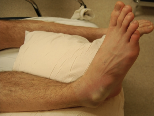
Case 3. Achilles tendon rupture can be diagnosed with Thompson's Test. (96% sensitive, 93% specific).

Picture A shows normal plantar flexion with squeezing the calf=negative Thompson's Test.  Picture B shows no plantar flexion with squeezing the calf=positive Thompson's Test.   Edtor's note: I can never remember which is positive and …

Picture A shows normal plantar flexion with squeezing the calf=negative Thompson's Test.  Picture B shows no plantar flexion with squeezing the calf=positive Thompson's Test.   Edtor's note: I can never remember which is positive and which is negative.  So in the chart I write Thompson's test showed intact Achilles tendon function. Or, Thompson's test indicated Achilles tendon rupture.  For the Thompson's test to be valid you need to have the knee flexed.  So either lay the patient on their stomach and flex their knee or have them kneel with one knee on a chair or cart.

Lee   M&M

No case details, just some take home lessons.

Hypotension in association with aortic dissection indicates either the patient has an ascending aortic dissection with pericardial tamponade or the patient has a surgical indication for a type B dissection. Either way consult Cardiothoracic surgery.

Cardiothoracic surgery is the service to consult for suspected aortic dissections.

Do a bedside echo on patients on whom you have concern for aortic dissection and all critically ill patients.  Elise comment: Do serial bedside echo's to determine if pericardial fluid is accumulating.  This can help identify a dissection of the ascending aorta.   Elise Hart comment: TEE has high sensitivity for identification of ascending aortic dissection.

Dr. Lee then lead a fascinating discussion about the clinical situation of very high levels of ethanol intoxication and concern for toxic alcohol ingestion.  Very high levels of ethanol can cause death by respiratory depression, and at levels around 600, cardiovascular instability.   There are two online osmolar gap calculators when you do a google search, 1. University of Iowa and 2. MD Calc.   The University of Iowa calculator uses a different calculation than MD Calc.  It uses a conversion factor of 1.2 to account for some invitro studies that showed very high levels of alcohol affect the osmolar gap.  Andrea said that this new calculation used by the University of Iowa Calculator has not been fully validated.  She feels the MD Calc osmolar gap calculator is safer for patients.

Traditional Osmolar gap calculation used by MD Calc calculator.

Traditional Osmolar gap calculation used by MD Calc calculator.

To account for ETOH, this formula uses 1.2 x ETOH/4.6University of Iowa Calculator.   Andrea felt this calculator is not ready for prime time.   Harwood is going touse this calculator. He feels it cuts down on the number of patie…

To account for ETOH, this formula uses 1.2 x ETOH/4.6

University of Iowa Calculator.   Andrea felt this calculator is not ready for prime time.   Harwood is going touse this calculator. He feels it cuts down on the number of patients who need to have fomepizole and dialysis.

Serum alcohol level only measures ethanol.  Toxic alcohol ingestion has no impact on the blood alcohol level.

Chinwala     5 Slide Follow Up

Diagnostic algorithm forAnaphylaxis

Diagnostic algorithm forAnaphylaxis

To quote Dr. Chinwala, "EPI is King" when treating anaphylaxis. 

To quote Dr. Chinwala, "EPI is King" when treating anaphylaxis. 

Hart/Regan     Ortho Updates

Segond Fracture is highly associated with ACL tear and meniscal injuries.


This is one of the most common ankle fractures in snowboarders.  It was a rare high-energy type injury prior to the sport of snowboarding.

This is one of the most common ankle fractures in snowboarders.  It was a rare high-energy type injury prior to the sport of snowboarding.

Quadricep tendon ruptures are more common than patellar tendon ruptures.

Donapudi        5 Slide Follow Up

Treatment priorities for cholangitis:  Agressive fluid resuscitation (think sepsis fluids),  broad spectrum antibiotics, ERCP, and long term the patient will need cholecystectomy.  In patients who are not good surgical candidates acutely, IR can place a percutaneus drain to get source control of an infected gall bladder.   It is prudent to consult GI and General Surgery on all cholangitis patients.

Predictors of need for ERCP:  High bilirubin, thrombocytopenia, low albumin, and tachycardia.

Friend        5 Slide Follow Up

Vitamin K deficiency affects factors 2,5,7 ,9, 10, C, and S.   Vitamin K deficiency acts like a warfarin overdose.  It is rare in healthy adults. It can be caused by antibiotics, fat malabsorbtion, high doses of vitamins A and E.    Factor 7 is the factor affected most early and severely due to vitamin K deficiency or warfarin overdose.

Erbach    5 Slide Follow Up

Brain AVM's present with bleeding or seizures. 

AVM's are a high pressure malformation.  When they rupture they have big bleeds.   Cavernous Malformations are relatively low pressure malformations and present with seizures and smaller bleeds.

AVM's are a high pressure malformation.  When they rupture they have big bleeds.   Cavernous Malformations are relatively low pressure malformations and present with seizures and smaller bleeds.

Olfactory hallucinations are associated with temporal lobe seizures.

Denk       Safety Lecture   How to determine if a patient has fluid responsive septic shock

Fluid responsiveness is defined by increase in stroke volume of 10-15% with a 500ml fluid bolus.

There are new gadgets that can determine fluid responsiveness:  The EV-1000 can calculate stroke volume variation based on non-invasive finger probe readings.   The problem with this device is that patients have to be in sinus rhythm, not tachycardic, and have to be intubated to get accurate readings.

The other device is the Cheetah NiCom. 

There is no Tech fix or fool proof way to measure fluid responsiveness in septic patients. We need to continually re-evaluate our septic patients.  Give fluids mindfully and assess the patients' response to fluids.

Elise comment: From our Process Trial experience, most septic shock patients whether or not they have heart disease, CHF or ESRD can tolerate 30ml/kg of fluids. You need to frequently re-evaluate these patients.  But in general give 30ml/kg for septic shock. 


Conference Notes 5-24-2017

Putman/Ohl    Oral Boards

Case 1.  50 yo male with fatigue and low grade fever.  Patient has a petechial rash/skin bruising. Labs show anemia and low platelets. Peripheral smear shows shistocytes. Bun/Creatinine was elevated.   Patient has mental status change as well. Diagnosis is TTP.

Classic Pentad for TTP.  However, the Triad of anemia, thrombocytopenia, and neurologic symptoms without fever and renal dysfunction is more common than the Pentad.

Classic Pentad for TTP.  However, the Triad of anemia, thrombocytopenia, and neurologic symptoms without fever and renal dysfunction is more common than the Pentad.

Treatment of TTP includes Plasma Exchange (PEX), andIV steroids.   TTP is due to ADAMTS13not cleaving VWF and platelets. Because of this ADAMTS13 dysfunction, the patient gets long VWF and marked platelet aggregation.   When considering TTP, check the level of ADAMTS13 activity.   Do not transfuse platelets unless there is life-threatening bleeding.  More platelets can cause larger platelet aggregates.  Get plasma exchange done as rapidly as possible.

Elise and Harwood comment: Avoid doing an LP and central line in non-compressible sites when the platelet count drops below 50,000.  


Case 2.  36 yo male presents with dental pain, fever, and brawny edema of the submandibular area.

*Ludwig's Angina is the diagnosis.  Treat with broad spectrum antibiotics and consult for ENT surgical evaluation.   Be prepared for difficult intubation.

Case 3.   35 yo female bit by her cat.  Treatment includes prophylactic antibiotics to cover pasturella multocida.  Augmentin is the usual choice for patients who are not PCN allergic. For PCN allergic patients, bactrim + Clindamycin or doxycycline + clindamycin or cipro + clindamycin are options.   Update tetanus status. Cat bites are low risk for rabies.  Check an xray for fracture or foreign body.

*Tip of the Cap:  Bristol Schmitz was nominated for an MVP Award*

Lovell/Williamson      Spiritual Wellness & Mindfulness

Resilience is foundational to wellness.   

One simple thing you can do to develop wellness is at the end of each day think of 3 good things that you experienced that day.

Spiritual Wellness includes: Nurturing your deliberate actions.  Having the ability to reset your attitude or thinking.  Finding purpose and meaning in your work.  Employing mindfulness, meditation, and or religion to reach spiritual wellness.

Mindfulness is a tool to improve our spiritual wellness.   Mindfulness is paying attention to the moment with purpose, and not passing judgment on the moment or the thoughts we have at that moment. This can include meditation or conscious breathing or just presence in the moment.  This is the opposite of multi-tasking. It is focused mono-tasking.  

Victor Frankl made the point that mindfulness helps us learn about the space between a stimulus we experience and our response to that stimulus. We can control that space and positively impact our behavior.

You can start to meditate by doing it 10 minutes a day for 10 days in a row.  "Head Space" is a website developed by a buddhist monk that can help you meditate.   Research has shown that Meditation develops your anterior cingulate cortex.

Tekwani       Study Guide  

WASH regimen for anal fissures: Warm water (sitz baths), Analgesia, Stool softeners, and High fiber.

With exposure to hepatitis B infection patient will develop anti-HBS and anti-HBC.   With vaccination, the patient will only have anti-HBS and not anti-HBC.  Editorial comment: For test purposes maybe remember C stands for Close Contact with source person. Kinda weak mnemonic but the best I could come up with.

Amebic abscess is usually single and has a rim of edematous tissue.  Amebic abscess is treated with metronidazole.  Patients with amebic liver abscess may have an associated right side pleural effusion.

Amebic abscess is usually single and has a rim of edematous tissue.  Amebic abscess is treated with metronidazole.  Patients with amebic liver abscess may have an associated right side pleural effusion.

Strangulated hernias will have abdominal and or systemic signs due to impaired blood supply to the affected bowel.  Patients will have fever, tachycardia, and peritonitis. Don't reduce suspected strangulated hernias in the ED.  You may perforate the bowel. Consult surgery and start antibiotics.   Hernias with no fever, no peritonitis, and no systemic symptoms can have an attempt at reduction in the ED. 

Cecal volvulus seen in young patients,  usually has dilated bowel in left upper quadrant on test questions.  Also look for dilated small bowel loops associated with cecal volvulus.   Sigmoid volvulus seen in elder patients, usual…

Cecal volvulus seen in young patients,  usually has dilated bowel in left upper quadrant on test questions.  Also look for dilated small bowel loops associated with cecal volvulus.   Sigmoid volvulus seen in elder patients, usually has dilated bowel in right upper quadrant and has dilated large bowel most prominantly. 

Dawson        Pediatric Airway Emergencies

Failure to manage the airway is the leading cause of preventable deaths in children.

The Pediatric airway is narrowest at the subglottic cricoid ring. The pediatric larynx is more superior in the neck compared to adults. The infant tongue is larger than the adult tongue.

Dr. Dawson rarely uses an uncuffed ET tube. As a general rule cuffed ET tubes are preferred for most pediatric patients.

Dr. Dawson rarely uses an uncuffed ET tube. As a general rule cuffed ET tubes are preferred for most pediatric patients.

Lemierre's syndrome (or Lemierre's disease, also known as postanginal shock including sepsis and human necrobacillosis) refers to infectious thrombophlebitis of the internal jugular vein.[1] It most often develops as a complication of a bacterial sore throat infection in young, otherwise healthy adults. The thrombophlebitis is a serious condition and may lead to further systemic complications such as bacteria in the blood or septic emboli.

Lemierre's syndrome occurs most often when a bacterial (e.g., Fusobacterium necrophorum) throat infection progresses to the formation of a peritonsillar abscess. Deep in the abscess, anaerobic bacteria can flourish. When the abscess wall ruptures internally, the drainage carrying bacteria seeps through the soft tissue and infects the nearby structures. Spread of infection to the nearby internal jugular vein provides a gateway for the spread of bacteria through the bloodstream. The inflammation surrounding the vein and compression of the vein may lead to blood clot formation. Pieces of the potentially infected clot can break off and travel through the right heart into the lungs as emboli, blocking branches of the pulmonary artery that carry blood with little oxygen from the right side of the heart to the lungs.

Sepsis following a throat infection was described by Schottmuller in 1918.[2] However, it was André Lemierre, in 1936, who published a series of 20 cases where throat infections were followed by identified anaerobic sepsis, of whom 18 patients died.  Wikipedia reference.


Conference Notes 5-17-2017

Anderson/Traylor    Oral Boards

Case 1. 56 yo male with shortness of breath. HR 126, BP 88/59,  RR35.  On exam, patient has a diastolic murmur and rales bilaterally.   EKG shows LVH.  Echo shows signs of aortic root dilation and aortic regurgitation.  Diagnosis is aortic dissection with aortic valve failure.  CXR shows pulmonary edema.

Aortic regurgitation can occur due to aortic root dilatation or direct involvement of the aortic valve

Aortic regurgitation can occur due to aortic root dilatation or direct involvement of the aortic valve


Case 2. 36yo male with low back pain and bilat lower extremity pain.  Patient is tachycardic. He states he has an aching in his thighs.  Patient gives a history of ETOH and cocaine use the night prior. Urine showed large blood with minimal RBC's.  CK was 24,000.

Cocaine-induced rhabdomyolysis with secondary acute renal failure

By Karthikram Raghuram, MD, Department of Radiology, University of Alabama at Birmingham and Birmingham VA Medical Center, Birmingham, AL

The incidence of rhabdomyolysis in patients who use cocaine varies from 5% to 30% in published reports. It is unclear why cocaine causes rhabdomyolysis. Hypotheses include cocaine-induced vasospasm with resultant muscle ischemia, excessive energy demands placed on the sarcolemma, and direct toxic effects on myocytes. Seizures, agitation, trauma, and hyperpyrexia may also play a role. In general, the severity of the rhabdomyolysis parallels the severity of the cocaine intoxication; patients with very high CK levels tend to have the most severe complications from this disease. Intravenous cocaine use may be associated with a higher incidence of rhabdomyolysis-induced acute renal failure (ARF) compared with smoking cocaine.1

Patients with rhabdomyolysis classically present with complaints of muscle weakness, swelling, and pain. The myalgias may be focal or diffuse, depending on the underlying cause of the disease. The patient may also note dark- or tea-colored urine. However, a high clinical suspicion for rhabdomyolysis must be maintained in patients at risk because up to 50% of those with serologically proven rhabdomyolysis do not report myalgias or muscle weakness.1


Case 3. 45 yo male with bilat lower extremity numbness.  Vitals are normal. Numbness is localized to lateral thighs.  Patient is obese.   Neurologic exam shows diminished sensation on the lateral aspect of thighs bilat.  Remainder of motor and sensory exam is normal. Diagnosis is meralgia paresthetica.

Geraghty    Endovascular Treatment of Stroke

The typical stroke patient loses 2 million neurons per minute

Only 8% of stroke patients are eligible for TPA.  Also, TPA does not work well for larger clots.

Patients with life changing or potentially fatal strokes are candidates for endovascular management.   Minimum NIH stroke scale of 6 for considering endovascular therapy.

Stent thrombectomy after TPA resulted in earlier neurologic recovery and improved functional outcome at 3 months compared to TPA alone for patients with proximal clots.   NNT=3. 

Who gets endovascular therapy?   No bleed on plain CT.  CTA shows clot in proximal anterior circulation. Patiet received TPA within 4.5 hours and can get endovascular procedure within 6-12 hours.  New study just released today shows that even patients who wake up from sleep with stroke symptoms can benefit from endovascular therapy without receiving TPA.

If a patient wakes up with stroke symptoms and has an NIH score >6 send them for both plain CT and CTA.  If they have a large vessel occlusion they may be eligible for endovascular therapy without TPA.  Dr. Garaghty will consider patients for endovascular therapy based on imaging and the overall clinical picture not just strictly on time endpoints.  

We had a general discussion of how this new data will affect the ED approach to stroke.  We will need to start considering endovascular therapy for a much larger subset of patients than those that present with stroke symptoms within 4.5 hours. Because evolving evidence is suggesting that patients with proximal clots may benefit from endovascular therapy even if they are not TPA eligible based on time of onset.

Berkelhammer/Carlson     Acute Liver Failure

The INR is the best indicator of liver function/dysfunction.

Acetaminophen overdose is the most common cause of acute liver failure.  Acetaminophen causes centrilobular necrosis due to the concentration of cellular mechanisms to to detoxify acetaminophen in the centrilobular areas. 

Mechanism of acetaminophen detoxification in the centrilobular region of the liver. NAPQI is the toxic metabolite of acetaminophen. NAC/glutathione detoxifies NAPQI.

Mechanism of acetaminophen detoxification in the centrilobular region of the liver. NAPQI is the toxic metabolite of acetaminophen. NAC/glutathione detoxifies NAPQI.

The RM nomogram can only be used for acute single ingestions of acetaminophen.  You don't need to adjust the nomogram for patient factors such as chronic or acute ETOH use, P450 inducing medications, or malnourishment.  There is enough saf…

The RM nomogram can only be used for acute single ingestions of acetaminophen.  You don't need to adjust the nomogram for patient factors such as chronic or acute ETOH use, P450 inducing medications, or malnourishment.  There is enough safety built into the nomogram to cover all patients.

NAC prevents severe liver injury if given within 8 hours of ingestion.  There are benefits though even if NAC is given later. 

If you use the 21 hour IV protocol you need to verify there is no detectable acetaminophen in the blood and the LFT's are normalizing prior to stopping therapy. If not, then you need to treat beyond 21 hours.  There have been some reported case…

If you use the 21 hour IV protocol you need to verify there is no detectable acetaminophen in the blood and the LFT's are normalizing prior to stopping therapy. If not, then you need to treat beyond 21 hours.  There have been some reported cases of liver failure when the patient was treated with IV NAC for 21 hours only and the acetaminophen level was not 0.

Dr. Berkelhammer only will allow a total of 2 grams of acetaminophen per day in patients with alchoholism or cirrhosis. In patients with cirrhosis he also avoids NSAID's to reduce the risk of GI bleeding and renal failure.   If patient needs more pain control he favors prescribing norco with higher doses of hydrocodone (norco 7.5 or norco 10)

Twanow   5 Slide Follow Up

Erythrodermic Psoriasis

Erythrodermic Psoriasis can look like TEN.  Treatment of Erythrodermic Psoriasis includes IV fluids, systemic steroids, and local wound care.  Care best provided in a burn unit.

Erythrodermic Psoriasis can look like TEN.  Treatment of Erythrodermic Psoriasis includes IV fluids, systemic steroids, and local wound care.  Care best provided in a burn unit.


Denk      Trauma Airway

To obtain an airway in this patient, you can cut the vertical wires with trauma shears or wire cutters to open the mouth enough to get a Video or Direct Laryngoscope in the mouth.  If you don't have time to do that you can either attempt nasotr…

To obtain an airway in this patient, you can cut the vertical wires with trauma shears or wire cutters to open the mouth enough to get a Video or Direct Laryngoscope in the mouth.  If you don't have time to do that you can either attempt nasotracheal intubation or perform a cricothyrotomy.

Dr. Denk discussed 2 other difficult trauma airway situations.  She discussed a recent EmCrit Podcast "Having a Vomit SALAD"  which discusses using the yankaur suction to lead the laryngoscope blade into the supraglottic space to suction out blood or vomitus.  You can then move the suction catheter to the left side of the mouth and keep the tip in the upper portion of the esophogus.  This way the suction catheter continues to clear blood or vomitus from the airway while you are trying to intubate.

Einstein        Extremity Trauma

Every extremity injury requires an evaluation of vascular status, nerve function, soft tissue injury, and bony injury.

When evaluating for vascular injury in an extremity, examine for hard signs, soft signs, and get ABI's.

When evaluating for vascular injury in an extremity, examine for hard signs, soft signs, and get ABI's.

Conference Notes 5-10-2017

Munoz   M&M

Pay attention to vital signs in kids.  Pediatric patients can look OK despite significantly abnormal vital signs.  Vital sign abnormalities, especially tachycardia above age-appropriate values may be the only early clue to serious illness.

Early IV antibiotics and IV fluids are as critical in kids as in adults for treating sepsis.

Be cautious of pediatric patients who have a return visit to the ED for the same illness. They may warrant a more detailed evaluation.   The average emergency physician during their career will send home 44 patients who will die within 7 days.

Risk factors for death in discharged patients: abnormal vitals especially tachycardia, failure to recognize worsening of chronic illness, atypical presentations of disease, patients with psychiatric illness or substance abuse. If a patient has one or more of these risk factors, consider further evaluation or a period of observation prior to discharge.

Another interesting 2017 study discussing deaths in discharged medicare patients.  Higher admission rates are associated with less deaths.  ED discharge diagnoses such as altered mental status, dyspnea, and malaise, were the highest risk f…

Another interesting 2017 study discussing deaths in discharged medicare patients.  Higher admission rates are associated with less deaths.  ED discharge diagnoses such as altered mental status, dyspnea, and malaise, were the highest risk for death after discharge.

BMJ 2017; 356 doi: https://doi.org/10.1136/bmj.j239 (Published 01 February 2017) Cite this as: BMJ 2017;356:j239

Elise comment:  Once you make the determination that a patient is ill and needs time-critical interventions,  you need to physically look at the clock and set definite time constraints on your team to get things done.  If you don't hit those time marks you need to stop all other activity and re-focus the entire team on the patient you are concerned about.

Patients who are boarding in the ED for prolonged periods of time can have significant changes in their condition.  Try to at least lay eyes on these long term sign out patients at least once during your shift. 

Submassive PE is defined by RV dysfunction or elevated troponin

Massive PE is defined by hypotension <90 mm HG lasting more than 15 minutes or requiring inotropic support, shock, profound bradycardia, or cardiac arrest

*Management of submassive and massive PE

Katiyar     Toxicology

The intrinsic and extrinsic coagulation pathways converge at Factor 10

 Excerpt from DeSancho TM, Pastores SM The Liver and Coagulation:   The liver is the primary site of synthesis of most of the clotting factors and the proteins involved in the fibrinolytic system. These include all the vitamin K-dependent coagulation proteins (factors II, VII, IX, X, protein C, protein S and protein Z), as well as factor V, XIII, fibrinogen, antithrombin α2-PI and plasminogen. The notable exceptions are von Willebrand factor (VWF),tPA, thrombomodulin, TPFI and uPA. The VWF, tPA, throm-bomodulin and TFPI are synthesized in endothelial cells, while uPA is expressed by endothelial cells, macrophages, renal epi-thelial cells and some tumour cells [4].

Vitamin K, a fat-soluble vitamin, is required to achieve proper levels of procoagulant factors (II, VII, IX and X) and anticoagulant factors (proteins C, S and Z). These factors require vitamin K as a cofactor for post-ribosomal modification to render them physiologically active.

Warfarin blocks Vitamin K reductase and affects the factors 2,7,9,10, and proteins C,S, Z

Warfarin side effects include blue toe syndrome, skin necrosis, hair loss, and urticaria in addition to bleeding problems.

*Warfarin Blue Toe Syndrome is a rare complication of warfarin.  If is thought to be due to cholesterol emboli released from bleeding within an arterial plaque.  If you stop the warfarin, the toes will remain blue but the pain will improve.  After stopping warfarin, changethe patient to a 10a inhibitor for anticoagulation. Consider other sources of thromboembolism such as an aortic aneurysm.


*Foods that have a high level of vitamin K.  Abhi discussed a case in which an elderly patient was not eating any vegetables because she lost her dentures and had an increasing INR because her dietary vitamin K intake had significantly decreased.

If a child ingests rat poison (super-warfarin) you don't need baseline labs on initial presentation.  Kids need coagulation labs 24-48 hours after ingestion. If at that time INR is prolonged or child has bruising/bleeding you would initiate vitamin K therapy.  Life threatening hemorrhage in this situation may require FEIBA for management.


*Factor 10a and 11a Inhibitors

Reversal of 10a and 11a inhibitors is accomplished by administering FEIBA.

Altman/Katiyar/Williamson        Medico-Legal Small Group Workshop

Chan/DeWeert     Oral Boards

Case 1.  67 yo female with abdominal pain.  Patient has history of Atrial fibrillation. She is not on warfarin or 10a inhibitor.  On exam pt has diffuse abdominal tenderness. Stool is heme positive. Lactate is 4.6.  Diagnosis is mesenteric ischemia due to an embolism to the SMA confirmed.   Diagnosis made by CTA showing occluded SMA.  Treatment is IV fluids, IV morphine, IV heparin, and IV antibiotics.  Emergent consult to Vascular Surgery and IR.

Which consultant provides definitive treatment (Vascular surgery, GI, or IR) will have to be determined on a case by case basis.  The clinical situation will determine which consultant can bring the most applicable skill set to the patient's care.  Some of the residents discussed cases where the GI consultant scoped the patient prior to surgery to determine the extent of necrosis. Depending on the type and extent of the vessel occlusion and how much the bowel is affected will determine whether Vascular Surgery or IR treats the patient.


Case 2. 33yo male with right ankle and foot pain after a fall.  Xrays show a calcaneus fracture.  Examine the spine and entire lower extremity for associated injuries in a patients with a calcaneus fracture.  Initial management is splinting, elevation, non-weight bearing with crutches or walker, pain management and orthopedic consultation.  Many patients with calcaneal fractures can be discharge home with outpatient follow up.  However, there are risks of compartment syndrome of the foot, fracture blisters, and associated injuries of the lower extremities, spine, pelvis and abdomen in patients with calcaneal fractures. .

Case 3.  54yo male with epigastric pain.  He is tachycardic.  Finger stick blood sugar is >600.  Labs are consistent with DKA (anion gap, metabolic acidosis, ketonuria). Patient has an elevated troponin but no STEMI on EKG.  Diagnosis is DKA with NSTEMI.  Treat with IV fluids, IV insulin, potassium, po asa, and IV heparin.

Dave Barounis taught me to focus on the anion gap and urinary ketones.&nbsp; If both are present in the setting of hyperglycemia, you have DKA.

Dave Barounis taught me to focus on the anion gap and urinary ketones.  If both are present in the setting of hyperglycemia, you have DKA.

Okubanjo      Safety Lecture

I missed this excellent lecture

Chiefs    Ortho Cases

I missed this excellent lecture

Conference Notes 4-19-2017

Thanks to Elise Hart for her help with the Conference Notes for the Ophthomology Lecture by Drs. Farooq and Shah this week. 

Okubanjo/Ryan     Oral Boards

Case 1.   19yo male passes out at a movie theater. No seizure activity identified.  EKG shows a Brugada pattern.

Brugada Pattern in Leads V1-2

Brugada Pattern in Leads V1-2

Patient required admission to cardiolgy service for AICD placement.

Case 2. 41 yo male with right leg pain following a bad ankle injury.  Vitals are normal.  Xrays show maisoneuve fracture.

Maisoneuve fracture pattern has a medial maleolar fracture or tear of deltoid ligament with associated proximal fibular fracture and tear of the syndesmosis between the tibia and fibula.&nbsp;&nbsp; On exam, palpate both the ankle and the proximal f…

Maisoneuve fracture pattern has a medial maleolar fracture or tear of deltoid ligament with associated proximal fibular fracture and tear of the syndesmosis between the tibia and fibula.   On exam, palpate both the ankle and the proximal fibula to identify this fracture pattern.  Treatment is splinting followed by ORIF.


Case 3.  45 yo male with weakness and vomiting. Patient has tachycardia with thready pulses. Patient has history of daily ETOH use and recently has had vomiting and cannot keep down any food/fluids.  Diagnosis is AKA. 

Lovell      Town Hall Meeting

Traylor     Stroke Outcomes in Patients over 80yo Receiving TPA at ACMC

Logan gave his upcoming ICEP presentation.  I did not want give away the info prior to his presentation.  You will have to attend ICEP to get the outcome info. 

Okubanjo      Healthcare Disparities

Oyin gave her upcoming CORD presentation describing the Healthcare Disparities Curriculum she created.

Einstein     Wilderness Medicine

Noah gave his upcoming CORD presentation describing the Wilderness Medicine Curriculum he developed.  

Pastore     5 Slide F/U

There are 2 conditions in LVAD patients that require immediate notification of LVAD team.

#1 Pump failure which will be indicated by screaming LVAD alarms

#2 Pump thrombosis indicated by dark or brown urine.  Patients will have an elevated LDH and signs of hemolysis on CBC.  Patients may have new heart failure symptoms.  The incidence of pump thrombosis has increased recently due to a trend toward lower anti-coagulation INR targets for LVAD patients. 

Treatment of pump thrombosis is heparin drip and IV bicarb drip. Patient may go to OR for LVAD exchange or ECMO.  TPA can also be used. Get the LVAD team involved in the patient's care as soon as possible.

Ashley recommended asking all LVAD patients what their urine looks like to screen for pump thrombosis/hemolysis.  Also get an LDH on all LVAD patients to screen for pump thrombosis/hemolysis.  Compare the LDH to prior levels. 

Tran       5 Slide F/U

Treat delerium tremens with IV Ativan and IV phenobarbital.

Etoh withdrawal seizures don't typically have prolonged post-ictal periods.

The later a patient starts having withdrawal symptoms following cessation of ETOH, the worse the prognosis/severity of withdrawal.

Phenobarbital works at the GABA receptor and also lowers glutamate in the CNS.

Dexmedetomidine is a newer sedation medication that can be very effective in ETOH withdrawal. The downside is that it is very expensive.

Kennedy comment: When giving patients big time bnezo's and phenobarb keep them on an end-tidal CO2 monitor to be alert for potential apnea. 

Hart/Regan    Ortho Updates

Rotator Cuff Tears can be diagnosed with the following exams:

Treat with sling and Ortho Management.  Some patients will require surgery

Farooq/Shah   Visiting OphthalmologyConsultants from U of C     Eye Emergencies

Ruptured globes - mechanism important. look for abnormality to pupil as one clue


Usually no rush to get FBs out unless contributuing to increased IOP (very rare) or if wood/vegetable matter (very inflammatory) - o/w FBs unlikely to get infected.


lateral canthotomy/cantholysis:

1) inject lido WITH epi to help w/ hemostasis/can also clamp down with hemostat

2) cut skin with 15 blade first (scissors often too dull, though )

3) Get scissor between globe and cathus, keeping dull side of scissor against globe. Cut posterior (lateral canthotomy) first (note this part doesn't do much for IOP), then inferior limb (cantholysis)


Pearls: 

- If you can pry open lid and their EOMs are OK they're unlikely to need lateral canthotomy

- You can strum with scissors to see if you feel the cord of the tendon to know if you got it

- Don't worry too much about messing up the lid/skin - oculoplastics can always fix that later

- If pressure <22 they're probably fine

- On call Ophtho should come in if this is being done - OK to start without them, but their job is to ensure it was done adequately

- If being done for compressive optic neuropathy should also give steroids (recommended dose: 500 mg? solumedrol).  Exception: Traumatic Optic neuropathy (posterior orbit sphenoid fx w/ effect on vision but not EOMs and not a lot of external swelling --> this involves direct trauma to optic nerve, akin to lack of efficacy of in spinal cord injury)

If there is a severe eyelid laceration exposing the globe, place antibiotic ophthalmic ointment as soon as possible to protect the globe.  Talk to plastics or ophthomology to emergently approximate the lacerated lid to get coverage over the globe.   If you as an emergency physician need to repair a gaping eyelid laceration that exposes the globe use 5.0 or 6.0 vicryl or 5.0 or 6.0 fast absorbing plain gut.  Keep the sutures in the tarsal plane external to the mucosal lining as much as possible.

Injuries to the medial canthus area that damage the canaliculi system need to have a plan to repair the canulica within 3 days.  Beyond 3 days there is significant scarring and it limits ophtho's ability to repair the problem.

Orbital floor fractures are almost never an emergency in adults unless there is an associated serious eye injury.   Pediatric orbital floor fractures need to be addressed on an emergent basis because pediatric fractures under age 16 can entrap and strangulate the inferior rectus muscle causing life long diplopia.  If a pediatric patient has inability to elevate the eye in the injured orbit they need emergent surgery.

Chemical Burns Check the ph in 4 quadrants of the eye.  Check visual acuity, pupils, and eye pressure.  Alkali burns cause more damage than acid burns because alkali causes liquifaction necrosis.  Acute conjunctival irritation is actually a good sign, better than a whitish appearing cornea.   Copious irrigation of the eye is the key management to chemical burns. Irrigate until the ph is 7 in all 4 quadrants.  It may take 10-16L of NS to get ph down to 7.  Chemical burns can cause severely elevated intraoccular pressure.   Adjunctive therapy for chemical burns includes topical steroid, topical antibiotic, and cycloplegic drop.  Other therapies include vitamin C, doxycycline, and intraoccular pressure lowering drops.

Central Retinal Artery Occlusion is characterized by painless unilateral vision loss.  Patient should be admitted or stroke work up.  Neurology should be consulted.  There is no proven therapy for this disease.  Occular massage may be helpful.  Long term visual prognosis is poor.  Rare diagnosis in children.  Can be associated with malignacies or patent foramen ovale in children. 

Globe rupture signs: 360 degree conjunctival hemorrhage, 8 ball hyphema, flat anterior chamber, irregular pupil.  Get a CT scan to evaluate the globe.  For adults, Avelox is the optimal antibiotic for globe rupture because of it's abilty to attain high levels in the globe.  Levaquin is the second choice.   In kids, discuss antibiotics with ophthalmology.  Some pediatric specialists may advocate for a single dose of avelox in kids as well.

Acute Glaucoma treatment

Acute glaucoma management

Don't send topical ophthalmic anesthetics home with patients with eye pain.  It can cause complete vision loss in the affected eye.    For pain relief for corneal abrasions use topical antibiotic ointment and an oral analgesic.

Conference Notes 4-12-2017

Hart/Regan      STEMI Conference

Case 1.   Patient had environmental hyperthermia associated with lateral STEMI changes.  Decision between Emergency Physicians and Cardiology agreed that rapid cooling was indicated rather than emergent cardiac catheterization.   EKG changes resolved with rapid cooling.   Patient had markedly elevated CK due to Rhabdomyolysis.   Rhabdo was treated with IV fluids and eventually dialysis.  

There is a paper describing EKG changes in patients participating in the Haaj in Mecca who had hyperthermia.  Common EKG abnormalities seen were sinus tachycardia, QT prolongation, non-specific ST changes, and focal ST changes.  There are other case reports of STEMI appearing EKG changes in patients with hyperthermia.  Typically patients had resolution of EKG changes with rapid cooling.  Many had clean caths.   The EKG findings of hyperthermia are thought to be due to physiologic stress, dehydration, electrolyte abnormalities.

Harwood comment:  The treatment is to cool these patients not take them to the cath lab.  The Cardiology faculty present agreed.  They said treat the patient, not the EKG.  Cooling is the main therapy in this situation.

Case 2.  Elderly patient with prior CAD, stents, HTN presents with syncope.  Patient's initial EKG showed clear-cut STEMI changes.  Patient had history of placement of coronary stents for a prior STEMI about a week prior to this presentation. Cardiac Cath at this second presentation showed thrombosis at proximal portion of recently placed stent.

There is no clear-cut time frame for STEMI changes to resolve following MI.  If ST elevation is identified beyond 1 week post-STEMI that is probably an acute abnormality or an aneurysm or pericarditis.

Stent thrombosis is uncommon with about a1-2% frequency.  Stent thrombosis sually presents with STEMI or death.  Patients not taking anti-platelet therapy are at higher risk for stent thrombosis.  Bare metal stents most commonly occlude in the first 48 hours.  Drug eluting stents most commonly re-occlude within 30 days.  STEMI's secondary to stent thrombosis have worse outcomes than STEMI's caused by plaque rupture.  Cardiac Cath is very time-sensitive in these patients.

Cardiology comment: Most of the time stent thrombosis is due to either medication non-compliance or mechanical/placement issues related to the stent.

Cardiology comment: It is very important in patients with prior CAD and/or stents to compare their ED EKG with a prior EKG.  The patient may have aneurysmal changes or LVH changes that can look like acute STEMI.  If you have an old EKG showing similar changes it can help clarify the situation.

Case 3. Elderly patient presents with dyspnea. EKG showed a paced rhythm with anterior concordant ST depression.


Sgarbossa criteria for identifying STEMI in patients with LBBB and Paced rhythm.&nbsp; The main thing to look for is concordant ST depression or elevation.

Sgarbossa criteria for identifying STEMI in patients with LBBB and Paced rhythm.  The main thing to look for is concordant ST depression or elevation.


Smith Modification of Sgarbossa criteria: If you identify concordant ST elevation or depression, that is significant.  If you have discordant ST change more than 25% of the main deflection of the QRS, that is also considered significant.   This last criteria replaces the 5mm discordant criteria from Sgarbossa.

Cardiology comment:  The decision to take patients to the cath lab with paced rhythms is difficult.  The decision has to be made on a case by case basis using Smith modified Sgarbossa criteria and looking at the overall clinical picture.

KennedyM&M

I only noted the take home points to avoid divulging the specific aspects of these cases.

When pacing, you have to set the asynchronous rate above the patient's native rate or you can get a pacer spike landing on a QRS complex.  This can cause V-tach, torsades, or V-fib.  If you are technically adept with the pacer device, placing the pacer in VVI mode also avoids this R on T phenomenon.

Harwood and Girzadas comment: There is a value to keeping things simple when placing a transvenous pacer.  The "Emergency" button on the pacer give you asynchronous pacing at the rate of 80 which does the job in the vast majority of patients who have a very low native rate.  Harwood made the point that once the patient is more stable you can then adjust the pacer box to the VVI mode. 

Carlson comment: Check the pacer battery while you are prepping to place the line.  It is not uncommon that the pacer battery may need replacement. You don't want to find out the battery is not working right when you need the device to pace.

Check vital signs prior to discharging patients. Document any re-assesments thatyou perform on patients.

Consider getting an EKG on patients that are persistently tachycardic with no good clear diagnosis.  Be on the lookoutfor myocarditis.

Tekwani comment: If you get a gut feeling about a patient in signout, go to the bedside and carefully re-evaluate the patient.  Don't ignore that gut feeling.

Lovell comment: Be cautious about chalking up symptoms and signs to anxiety.  Most of the time the diagnosis is actually anxiety but rarely it can be an occult pathology.

Sedation and positive pressure intubation can markedly decrease cardiac output and increase the chance of cardiac arrest.  Be sure to resuscitate the patient prior to intubation. Have pressors ready to go or even started prior to starting intubation.

Williamson comment: Take some time prior to every acute resuscitation to speak with the family and appraise them of the gravity of the situation.  It should be in your pre-resuscitation check list.

Menon/EinsteinOral Boards

Case 1.  Botulism secondary to IV drug use.  Treat botulism with anti-toxin.  You can obtain anti-toxin from CDC.  The Illinois poison control also has access to anti-toxin

*Botulism

Carlson comment: Heroin is the #1 cause of wound botulism.

Case 2.  Patient with recurring fever following trip to Africa.  Malaria was suspected based on CBC showing anemia and schistocytes.  Thick and thin smears confirmed malaria. 

Elise comment: You need to know this diagnosis for the boards.

*Thick and Thin smears for Malaria

Case 3.  Flexor tenosynovitis Treat pain, update tetanus, give IV antibiotics and consult hand surgery. Admit patient for further management and surgery.

*Knavel's 4 signs of Tenosynovitis

Destefani         Safety with Pediatric IV fluid Administration in DKA

IV fluid administration in pediatric patients with DKA needs to be done with caution to avoid cerebral edema. However, research illuminating how exactly to give fluids in Peds DKAis lacking.  Cerebral edema in DKA is rare and relatively unpredictable.

Do not bolus NS in DKA patients unless the patient there are signs of shock. 

PECARN group is currently doing a study looking at various IV fluid strategies in Pediatric DKA patients.

Any change in mental status in a pediatric DKA patient should raise your suspicion for cerebral edema.  Also be alert for headache, incontinence, and vomiting.

*Diagnostic, Major and Minor Criteria for Cerebral Edema

Small studies show that central lines in pediatric patients with DKA may be more at risk for DVT.  If you can't get peripheral IV access go to an IO line first.  Favored locations for pediatric IO lines are proximal tibia, distal tibia, and distal femur. Avoid placing IO lines above the femur in skeletally developing kids. 

Harwood comment:  Most DKA patients are not in shock and don't require rapid fluid boluses or large fluid resuscitation.  Give them fluid and insulin per protocol only and they will gradually improve.

Garrett-Hauser     Ethics     Reporting Medical Errors

We discussed a case scenario regarding end of life decisions.

The act of withdrawing care is not furthering a patient's death.  It is returning the patient to their original disease state.   However when you withdraw care, you do need to fill out a "Goldenrod Form" justifying the withdrawl of care.  Two attendings need to co-sign the Goldenrod Form.

A patient had unintended imaging.  Patient was informed of imaging and radiation exposure. A discussion with billing office was had to avoid the patient receiving a bill for an unintended test.

If a patient has an adverse outcome from a medical error, the appropriate approach is to discuss the error, the cause, and the expected outcome with the patient and family.   Families want to know how this problem will be prevented going forward.    It is good to have Chaplain support with these conversations.

All these cases highlighted the importance of error disclosure AND the challenges we all have with informing patients and families about errors. 


Conference Notes 4-5-2017

Hart & Regan     Wound Management

Strategies to lessen the pain of injection of local anesthetic: small gauge needle, slow injection, inject through wound margin (not intact skin), buffer with bicarb, warm to body temp, and pre-treat with topical anesthetic such as LET.   Harwood comment: Use an Insulin syringe initially. It has a 31 gauge needle. Then use the longer 27guage needle to anesthetize more deeply.

Pasturella strains from cat bites are felt to be more virulent than pasturella strains from dog bites.  Additionally, cats have smaller, sharper teeth that can puncture more deeply further increasing the risk of infection.

Care of Amputated digit: Wrap the digit in saline soaked gauze, place the wrapped digit in a plastic bag, place the bagged/wrapped digit on ice.   Finally, keep the digit with the patient on the patient cart so the digit does not get lost.

Fight bite wounds are high risk for infection.  Examine all fight bite wounds through full ROM of the MCP joint to check for tendon injury.  Give Augmentin or Unasyn as initial antibiotic if patient is not penicillin allergic. Consult with Hand Surgery.  If there is evidence of extensor tendon injury, joint penetration, infection, or fracture, the patient should be considered for admission for washout.

To release skin entrapment in a zipper, you need to get sturdy wire cutters or a bolt cutter to cut the median bar of the zipper.  Dennis Ryan comment: This can be a tough procedure to get the median bar cut.

For lacerations around the eye, emergency physicians should probably not be repairing lacerations around the medial canthus, involving the lid margin, or if you see exposed fat.  If you identify any of these, you needto consult ophthomology.

When can packing after I and D be removed?  When purulence has stopped draining.

Wound VAC's work by decreasing edema, improving vacular and lymphatic flow, decreasing bacterial density and increasing granulation tissue.

Ohl/Yappo   CPC Presentations

I did not want to divulge the cases because they will be presented at CORD this month.

Lorenz    5 Slide Follow UP

Patient presents with dizziness.  EKG shows polymorphic ventricular tachycardia (Torsades)

The patient was alert and talking only because he had an LVAD.   Patient was initially given IV magnesium without effect.   Patient was next defibrillated into sinus rhythm.   In hospital patient was treated with amiodarone and AICD.

Data shows that 2 year survival with an LVAD is about 23%.  LVAD's can give patients a longer life. On the other hand, patientsmay experience challenges and complications from an LVAD.   This risk/benefit balance was discussed.

Harwood comment: If you see a patient with torsades who does not have an LVAD you need to defibrillate and also start magnesium and an anti-arrhythmic.  These patients will continue to revert to torsades after defibrillation until you get therapuetic levels of an anti-arrhythmic.

Wing    5 Slide Follow Up

Myasthenic crisis can be precipitated by infection, pregnancy, and medications (aminoglycosides, flouroquinolones, beta blockers, and magnesium).

Evaluate the patient with a measurement of Vital Capacity and NIF.  Normal VC should be around 3L (bad is around 1.5L).  Normal NIF should be around -60(bad is around -20).   If the NIF is -20 neuro may use plasma exchange.   Harwood would intubate if NIF is worse than -20.

Treatment is prednisone, IVIG, and plasma exchange.   Intubation for respiratory support.

Faculty comments: Respiratory therapy can measure both VC and NIF in the ED.

Snip20170405_8.png

Nejak    STEMI Equivalents

Dan's recommended steps to evaluating an EKG: Get an old EKG to compare.  Look at AVL for isolated depression.  Check for loss of precordial T wave balance.  Look for LMCA ischemia, Check for Wellen's and DeWinters signs. Check for reciprocal changes.  Check for hyper-acute T waves.


Loss of precordial T wave balance


LMCA Occlusion shows diffuse ST depression and ST elevation in AVR and V1


DeWinter's ST depression in V2-5 with rocket shaped T waves and Subtle AVR ST elevation.  This is an anterior STEMI equivilent and indicates LAD occlusion.


Wellen's is a sign of critical LAD stenosis.  These patients should have Cardiology evaluation.

DeWeert, Stanek,  Traylor      Abdominal and Pelvis Trauma

There was a ton of information in this excellent lecture that I could not encapsulate into these notes.  I put in a few key points made in the lecture.

Every 10 minute delay to give blood in trauma patients has an increased odds ratio of 1.27 for mortality.

Give TXA within 3 hours of initial trauma.  It is safe and number needed to treat is somewhere between 7-67 to decrease mortality.  Beyond 3 hours after injury, TXA increases mortality for unclear reasons.

Image shows the effect of a pelvic binder to close down the potential space in the pelvis.   Have a low threshold to place a pelvic binder early in the course of managing a multiply injured trauma patient.  There was consensus that the old strategy of "rocking the pelvis" to identify pelvic fracture on physical exam risks increasing pelvic bleeding.  In the multiply injured trauma patient, bind the pelvis early on in the resuscitation, don't rock the pelvis,  and get a pelvic x-ray to identify fracture.

Seat belt sign is associated with increased risk of intra-abdominal injury and Chance Fracture of the lumbar spine.

Algorithm implementing FAST exam in blunt abdominal trauma


Algorithm for Blunt GU Trauma

Algorithm for Blunt GU Trauma

There was a ton of information in this lecture that I could not encapsulate into these notes. 

Conference Notes 3-22-2017S

Stanek/Walchuk      Oral Boards

Case 1.   Patient with history of schizophrenia on multiple antipsychotic medications (Latuda, Lithium, Cogentin) presents with altered mental status. Patient has history of ataxia.  Patient has clonus on neurologic exam.  Lithium level was elevated.  Management included contacting poison control and initiating dialysis.  Any lithium level over 3.5 is considered severe.  Any change in mental status or significant neuro symptoms indicate dialysis.

Main point of discussion was to also consider serotonin syndrome and neuroleptic malignant syndrome in the polydrug psychiatric patient.

Case 2. 86yo female presents with 4 days of dyspnea. Patient has history of rheumatic heart disease and a mitral valve problem.   Lung exam reveals crackles. CXR shows cardiomegaly and congestive changes.  BNP is markedly elevated.  Patient's echo shows incompetent mitral valve.  Patient is in congestive heart failure due to severe mitral valve regurgitation.  Patient has valve rupture.  Treatment is afterload reduction with nipride followed by CV surgery consult.

Case 3.  7yo child suffered bite on finger from rat.  Treatment is copious irrigation of wound. Give antibiotics. No suturing or dermabonding. Tetanus prophylaxis is not indicated for rat bites.  People are at risk of Rabies from bites by Bats, Foxes, Raccoons, skunks, cattle, and horses.  No rabies risk from rats, squirrels, hamsters, guinea pigs.  The only lagamorph that can transmit rabies is groundhogs.  However, you can get rat bite fever which has a 13% mortality.   You can prevent rat bite fever with penicllin or augmentin at the initial treatment of wound.  Treat with penicillin, unasyn,or ceftriaxone.

Carlson     Oral Board Day Debrief

1. Unstable Afib requires cardioversion.  Cardioversion requires procedural sedation and a pre-sedation assessment. You also need to get informed consent, and do a Time Out.  Choose a sedative with minimal hemodynamic effects.

Cardioversion has a 90% success rate for unstable A-fib.  100J biphasic is the most commonly cited dosage for normal weight patient.  For obese patients start at 200J.  Anterior/Poster electrode pad placement seems to have a higher success rate.

Components of Pre-Sedation Assessment: Mallampati score, ASA Class, Prior Complications, Allergies, PO intake.

2. Consider abuse in the setting of an infant with altered mental status.  Check a blood sugar. Do a thorough physical exam to look for injuries.  Take custody of child.  Report the case to DCFS.

Infants are prone to head injury for shaken baby syndrome due to large heavy head compared to overall body size and large amount of water in head.   On exam look for bulging fontanelles and retinal hemorrhages, and inappropriate bruising. CT will show subdural or subarachnoid hemorrhage. Get a skeletal survey to look for prior fractures.

3. Patients with sickle cell disease are at increased risk for cholecystitis. They are also at increased risk for meningitis and infection from any encapsulated bacteria due to funtional asplenia.

4. Treat salicylate toxicity with alkalinization of serum/urine (goal: urine ph of 7-7.5) and hemodialysis. You have to replace potassium to effectively alkalinize the patient.  Method of alkalinization is initial 2 amps of bicarb bolus followed by bicarb drip of 3 amps in D5W and run at 4 hour rate.   This is an easy diagnosis to miss to elderly patients with chronic toxicity. Many OTC products have salicylate as a component. Goodies is an example.  Look for metabolic acidosis and respiratory alkalosis on the ABG.

5.  For neonates unstable with suspected congenital heart disease, give prostaglandin infusion. These kids will have mottling or cyanosis. Be prepared for apnea as a side effect of prostaglandin.  Prostaglandins can also have the side effect of fever and hypotension which can mimic sepsis.   If you have to transfer a neonate who is receiving prostaglandin, intubate prior to transfer. Lovell and Harwood felt that a septic work up in an unstable neonate may be potentially risky if you attempt to perform an LP.  Andrea felt you should still give antibiotics even if you have concerns about doing the LP.  

6. Be alert for testicular torsion in kids with lower abdominal pain.  Pediatric patients frequently will not say their testicle hurts.  Consult GU, Attempt detorsion. Get an ultrasound and get them to surgery.  Two peaks of incidence: First year of life and in adolescence.  If you can detorse or get to surgery in less than 6 hours there is a 90% salvage rate. 

7. Treat CO poisoning with 100% FIO2 with NRB and transfer for HBO. 

8.  Consider pericardial tamponade in patients with dyspnea and or hypotension/tachycardia. Electrical alternans is a sign of pericardial effusion. 

Electrical Alternans

Electrical Alternans


Echo is key to the diagnosis of pericardial tamponade.  Treat with pericardiocentesis and consultation for pericardial window. 

                                              Pecha Kucha

Alexander           Lipids to the Rescue!

Lipid emulsion has a protective effect from local anesthetic toxicity.  The best studied toxicity is from bupivicaine.

Lipid emulsion can be used also for toxicity due to tricyclics (amitriptylene, buproprion), beta-blockers and calcium channel blockers.

Hart    Vocal Cord Dysfunction

Predominantly seen in Caucasian females.  Can be confused with asthma, upper airway problems and panic attacks. Probably is due to combiniation of physiologic and psychiatric etiolgies. Standard dyspnea work up will be negative.  Flexible laryngoscopy will show paradoxical closure of the cords with inspiration.  The cords will have an opening at the base.

 Can treat with dissociative doses (0.5mg/kg) of ketamine.

Einstein   TEE for Cardiac Arrest

TEE gives you a better view of heart. Less frequent pulse checks.  It stays out of your way during resuscitation.  More sensitive test for cardiac contractility.  EM case reports and studies show anecdotal unexpected saves and improvement of CPR quality and diagnoses of dissections.  The probes are expensive.

Erbach   Cyanotic Congenital Heart Disease in the ED

If a child with a shunt has no murmur that indicates a big problem. Contact Peds CV surgery right away.

Use O2 to get patient back to their usual baseline, not higher than that.

Patients with a single ventricle have a higher risk of stroke.

Consider endocarditis in cardiac kids with fever.  Get blood cultures.

When consulting CV and Cards, know the patient's last procedure and when it occurred.

Donepudi       U/S in the Acute Management of Elevated ICP

Normal ICP is under 20mm Hg

Monroe-Kellie Doctrine

Monroe-Kellie Doctrine

You can use a high frequency probe to measure to optic nerve sheath diameter.  Use alot of ultrasound gel on a closed eyelid.  You can use tegaderm to keep the eyelid closed.  Measure 3 mm from globe.  Any diameter greater than 6 mm is abnormal.

Kennedy    Status Asthmaticus

Treat with aggressive bronchodilators, 10-20mg nebs.  Give 2grams of IV magnesium over 20 minutes.  IV steroids. Bipap using IPAP as the key to relieving work of breathing . Terbutaline 0.25mg SubQ. Epinepherine titrated as a drip.  Ketamine can be used for sedation for bipap or induction for intubation.  Intubation requires careful management to avoid air trapping and barotrauma.   Work to keep a limited plateau pressure.  Use a high I to E ratio.  Use smaller tidal volumes and lower respiratory rates.   Heliox can be used to improved air flow.  Heliox can't be used in hypoxic patients due to low FIO2 with Heliox.  Last ditch strategies are general anesthesia and ECMO/ECCOR (simplified ECMO).

Carlson    Toxicology

QT prolongation due to TCA can be treated with IV Bicarb and if that is ineffective, IV Lidocaine drip can be effective.

Physostigmine can be used in pure anticholinergic toxidromes that have coma, severe agitation, or intractable seizures.  Don't use empirically in patients with undifferentiated coma/agitation or polysubstance overdose.  Contraindications are TCA overdose, QRS wide, AV block, bronchospasm, bowel or bladder obstruction.   Andrea feels physostigmine is most indicated in pure anticholinergic overdose in pediatric patients. 

There was some difference of opinion between Harwood and Girzadas about the use of physostigmine.  Harwood felt he would use it in kids and teenagers with pure anticholinergic overdose and coma or severe agitation. It could potentially avoid intubation or complication of agitation. Girzadas was concerned about side effects of physostigmine particularly risk of bronchospasm and would do supportive care only (benzos for agitation, airway protection for coma) and avoid physostigmine. 

The common board question about an anticholinergic plant is Jimson weed.  Deadly nightshade, climbing nightshade, and Mandrake are other botanical anticholinergics. 

Treatment of TCA overdose includes sodium bicarb to a serum ph of 7.5.  Serum alkalinization is the goal not urinary alkalinization.  You are not trying to trap ions in urine. You are trying to keep the TCA out of the CNS.   Give bicarb for wide QRS, acidosis, hypotension, ventricular arrythmia.

TCA EKG from Life in the Fast Lane

Ohl    Safety Lecture

Sean discussed the content and organization of our ENT equipment.

Quick note on pancreatitis management: We need to treat pancreatitis with IV dilaudid for pain and somewhere around 350ml/hour of LR for the first 12 hours. The message from our GI consultants is to be more aggressive with our IV hydration for pancreatitis patients.


Conference Notes 3-8-2017

Wellness Retreat at Lake Katherine

We had our 2nd annual Wellness Retreat this last Wednesday.  Multiple aspects of wellness were covered including Financial Wellness (Special thanks to Nick Kettaneh), Yoga, Meditation/Spirituality, Healthy Eating and Team Building.  And..... Therapy Dogs!  

Snip20170310_5.png

Conference Notes 3-1-2017

Lovell     Procedural SedationJeopardy

Key Principles of Pain Management

1. Assess Severity 2. Use appropriate doses of analgesics 3. Titrate your pain meds 4. Monitor the patient's status.

Local anesthetics can be divided into amides and esters.  Amides are longer acting.  Amides have lower incidence of allergic reactions than esters.   Amides all have two i's in the name (lidocaine, bupivicaine).   Esters only have one i in the name (procaine).   If a patient has an allergy to local anesthetics it is usually due to the preservative.  Cardiac lidocane has no preservative so is usually considered safe in regard to allergies.  Subcutaneous diphenhydramine can also be considered as a substitute for local anesthetic in the setting of severe local anesthetic allergy.

*ASA classification of patients for sedation and anesthesia

The risk of using ketamine in kids with an active uri is laryngospasm.  If this develops you can bag the patient through this complication most of the time.  Ketamine is OK in head injury.   You can use ketamine in children down to 3 months of age for sedation.  For patients younger than age 3 months ketamine is not approved and there may be a higher risk of laryngospasm.

3 drugs for treatment of regional complex pain syndrome: corticosteroids, calcitonin, and bisphosphonates.  These patients present with burning pain, edema, warmth to skin, local sweating, and allodynia (pain to light touch).   To treat, get the patient's affected limb re-mobilized (take off splint or cast) and start oral prednisone.  They need follow up with PMR.  If you don't identify and treat these patients early they can develop severe chronic pain. 

Healthcare Disparity:  A large study showed that African American pediatric patients had a 60% chance of getting pain medications for abdominal pain compared to white pediatric patients. In other words African American children were 40% less likely to get analgesia for abdominal pain compared to white patients.

Ways to reduce the pain of local anesthetics: 1. inject thru the open margins of the wound. 2. buffer with sodium bicarb 3. Inject slowly4. Warm the anesthetic5. Use a small (25 or 27 guage) needle 5.  Reassure or distract the patient

Non-ASA NSAID's are noted by the FDA to cause heart attacks and CHF.  Elise avoids giving NSAID's in elderly patients, patients with heart disease, HTN, or renal disease.

Benzocaine and prilocaine are the two local anesthetics that can cause methemoglobinemia.

Max dose of lidocaine is 4mg/kg without epinephrine or 7mg/kg with epinephrine.  For bupivicaine, the max is 3mg/kg plain or 5 mg/kg with epinephrine.   Girzadas comment: Draw up your local anesthetic before you enter the child's room so you never enter the room with a potentially toxic dose of anesthetic and the child does not see the needle.

Toxicity from local anesthetics initially causes dizziness, facial or extremity paresthesias, ringing in the ears.  Patients may progress to seizures and then cardiovascular collapse (V-tach).  Bupivicaine has the highest risk of cardiovascular toxicity.  Treatment for local anesthetic toxicity is lipid emulsion therapy.

* Levels of procedural sedation

Recent large study in NEJM shows that if emergency physicians are high intensity opioid prescribers (prescribed opioids to 24% of their patients) the patients receiving opioids have a 30% higher relative risk of having long term dependence on opioids. Overall absolute risk of about 2% of becoming addicted.  Elise comment: the pendulum has swung away from opioid use so be cautious of prescribing opioids to patients. Discuss with patients that there is a 2% risk of becoming addicted.  Abhi comment: Warn patients about nausea and constipation to better inform them and encourage them to limit their use of narcotics. 

Scoring system to determine who can be safely discharged after procedural sedation

Scoring system to determine who can be safely discharged after procedural sedation

KatiyarEM Billing and Coding

Optimizing your RVU's requires optimizing your documentation.  Medical decison-making documentation is critical to your charting.    Also make sure you write a procedure note for any procedures.  Another key documentation item is to document your plan of care for a fracture or sprain.  Also document a re-exam after a splint was placed to show neuro vascualar status is intact and the splint is not too tight.  Ortho cases in general have high RVU's.  Appropriate fracture and joint reductions have very high RVU values.

If you incise an abscess, probe, break down loculations and pack, that is considered a complex abscess.

Any wound checks following abscess drainage can be billed as a level 2 or 3.  If you have to re-pack or give antibiotics, it becomes a level 3 chart.

Measure the length of the lacerations you repair.  There is a ruler printed on the paper wrapping of a tongue blade that you can use to measure the wound.

If you make management changes with oxygen for a low pulse ox, document your thought process regarding the pulse ox and oxygen therapy.  This is important for the medical decision making and for the RVU documentation.

The diagnosis you place on the chart is critical for determining the ED's case mix index.  If you document acute STEMI rather than just Chest Pain it better characterizes the acuity your ED is seeing.  Try to be as specific as you can in the ED.  If the patient has an nSTEMI, document that diagnosis rather than Chest Pain.  If the patient has DKA document that rather than hyperglycemia.  If the patient has Pyelonephritis, document that instead of uti. 

When you are describing patient behavior use objective terms as much as possible.  For example, instead of writing the patient is "beligerent", write the patient was screaming obscenities at staff, violently rocking the cart and throwing punches at staff or other specific actions.

Review all chart documentation including what the nurses and the techs and EMS personel wrote.  I any court case, the lawyers will go thru all charting with a fine tooth comb.

Marshalla      Patient Safety LectureMassive Transfusion Protocol

We now have refrigerated blood in the ED for any patient in hemorrhagic shock.  In that refrigerator there are 2 units of O pos blood for all males and for females over age 49.  There are 2 units of O neg blood for females under age 49.

ED Attending physicians and Trauma Attending physicians can order blood from the ED blood supply for all causes of hemorrhagic shock.  Only nurses can physically access the blood from the ED refrigerator.

Einstein       ED EKG's

Noah discussed strategies to improve the information flow of EKG's in the ED.

Sedation Workshop

We broke into small groups and discussed different sedation scenarios. 


Conference Notes 2-8-2017

McKean     Resuscitation Procedures

Unfortunately I missed this excellent lecture

Girzadas    Zebras Lecture

I gave this lecture and did not write up notes. I did send out a PDF of the lecture to all the residents. If you would like a copy, please send me an email separately.

Critical Care Device Workshop   

Much Thanks to our awesome ED Nurses for teaching us abut the devices commonly used in resuscitation situations in the ED.  Special shout outs to Kristen, Monika,  Danielle , and Nick!

Conference Notes 1-25-2017

Airway Day

Samir Patel     Airway Disasters


5% of intubations in the ED are considered difficult

2% of intubations in the ED are failed (Defined by first 3 attempts fail)

You have to be prepared for this.  It eventually will happen to you.

Treat every airway as a difficult airway

Prior planning before starting intubation is the key to success.


* Algorithm for Difficult Airway


Indications for Intubation:

Hypoxic or hypercapnic respiratory failure

Airway protection

Anticipated deteriorating course of illness

Work of Breathing (tachypneic, septic patients are using 30% of their cardiac output for diaphragm contraction)


*MOANS    Predictors of Difficult BVM


*Lemon Law    Predictors of Difficult Laryngoscopy


*3-3-2 Rule


*Mallampati Score     Class 1 and Class 2 predict reasonable airway visualization.  Class and 3 and Class4 predict poor airway visualization.


A Major Rule is: Don’t have any pride when working with a difficult airway.  Get help from ICU, anesthesia, surgery and other EM physicians.  Whoever you need.  There is no shame in getting help.


Sedated Nasal fiber -optic Intubation

Start with 4% nebulized lidocaine

Afrin and glycopyrrolate can help dry secretions

Ketamine for sedation

Warm the ET tube in warm water.  It makes the tube more malleable

When you visualize the cords with the fiberoptic scope have someone spray the cords with lidocaine so you don’t get laryngospasm


Apneic Oxygenation (basically a nasal cannula running at 15 liters per minute or more during intubation) prolongs your safe apnea time.  High flow nasal oxygen devices are even better than nasal cannula but it is more bulky.


*Delayed Sequence Intubation.  Give Ketamine slowly to avoid apnea.  The ketamine will calm an agitated patient allowing you to better pre-oxygenate and prepare.


Avoid IV Ativan in patients with respiratory distress. It reduces their respiratory drive and it may force you to intubate before you are ready.


Lovell        Airway Devices


To lessen your anxiety during a difficult intubation, it is useful to have familiarity with the airway tools you are using.   So practice with different devices so you are comfortable using them.


You always need to have a supraglottic device and a surgical option in your armamentarium as rescue devices.


Elise’sMinimum List of Devices every Intubating physician needs to have available and be comfortable using:

Bougie

LMA

Direct laryngoscope

Video laryngoscope

Fiberoptic device (Really CMOS/digital camera technology.  It is more durable than fiberoptic technology)

Cricothyrotomy kit


The difficulty with the Glidescope is properly passing the tube once you get a great view.   You need to pop back the stylet once you have the ET tube at the glottis to appropriately position the ET tube thru the cords.


Elise discussed many advanced airway devices available on the market.


The biggest error with cricothyrotomy is waiting too long to start doing it.


If the O2 sat is dropping and you have a failed intubation, stabilize with an LMA.  Based on your ability to oxygenate with an LMA you either must rapidly do a cricothyroidotomy or if you can oxygenate OK, attempt intubation thru the LMA.


If the patient is in a HALO, initial intubation attempt should be with video laryngoscopy.


Airway Workshop

Conference Notes 1-18-2017

Joint EM-Pediatric Conference    Pediatric Sepsis


Pediatric sepsis is culture negative in 25-60% of cases.


About half of children with sepsis will have a low cardiac index and high SVR (cold shock) where 90% of adults will have high cardiac output and low SVR (warm shock)


*Pediatric Sepsis Definitions


Cornerstones of Sepsis Therapy in Children

Early recognition is the key to treating sepsis effectively.

A key resuscitation goal is 60ml/kg of normal saline infused in the first hour.

Vascular access in the septic child should escalate to IO after 2 attempts at IV.

IO placement is not more painful than IV placement but IO infusions are painful.  So give lidocaine 0.5 mg/kg, not to exceed 40 mg thru the IO line. (Lovell reference)

You need to use a pressure bag, rapid infuser, or push-pull method to give 60ml/kg within one hour.


There was a discussion of which patients should get 60ml/kg in the first hour.    The strong consensus was that all pediatric sepsis patients should receive 60ml/kg of NS. 

You should be cautious giving 60ml/kg to kids with cardiac disease, history of abnormal kidney function, and neonates. 

The panel felt that, in general, septic kids need more rather than less fluid. 

The panel felt that it would be reasonable to re-assess the patient after every 20ml/kg.

Neonates are a high-risk group for large volume fluids and should be bloused in no more than 10ml/kg aliquots at a time.  You should carefully re-assess the neonate after any bolus to determine if more fluids are required.


Early antibiotics are another cornerstone of sepsis management.  A 3 hours delay increases the likelihood of need for PICU admission.  Ampicillin & Cefotaxime or Vancomycin &Ceftriaxone are the basic empiric antibiotic combos. But there are many variations based on age, allergies, and source of infection.


If you need a pressor for sepsis in a pediatric patient use peripheral epinephrine.  It has lower mortality compared to dopamine.


Children who are on chronic steroids (asthma, cancer) are at risk for adrenal suppression.  Give hydrocortisone (2mg/kg) Q 6 hours in septic kids who are at risk.


All kids in septic shock should receive hi-flow nasal cannula O2.

To avoid a hemodynamic crash during or after intubation, give a fluid bolus prior to intubation. Consider an epinepherine drip prior to intubation.

The Panel suggested avoidingetommidate for RSI in septic children.  I assume that is based on concern for adrenal suppression and maybe a risk of hypotension.


Lactate levels are unreliable in kids for identifying sepsis. 


Basically in the first hour after you suspect sepsis:

Give 60ml/kg of NS, Start IV antibiotics, Start high flow O2 via nasal cannula. If you need a pressor give IV epinephrine.


Kerwin/Denk     Oral Boards


Case 1. 25yo female with severe dyspnea.  Patient has a history of asthma and is severely wheezing.  Despite therapy with nebulizers, magnesium, and subcutaneous epinepherine,  the patient was still in severe distress and the ABG shows respiratory acidosis.   Patient was then intubated using a sedated look with ketamine for sedation and topical lidocaine. 


*Use peak flows to determine severity of asthma.  


Give 10-15mg of albuterol nebs every hour for severe asthma.   Give steroids and IV magnesium. You can also try high flow nasal cannula, heliox, subcutaneous terbutaline or IM epipepherine.   Sub-dissociative dosing (0.1mg/kg)etamine may be helpful in the anxious/agitatedpatient to help them tolerate bipap and nebs to stave off intubation. Bipap should be tried but the data in asthma for bipap is limited.


Case 2. 68yo male presents in Cardiac arrest.  Patient had V-tach as his initial rhythm.  In the ED, patient was in V-fib .   ACLS protocol was initiated.   ROSC was obtained.  EKG post-arrest showed STEMI.  Asa and heparin were given and patient was taken to the cath lab.  Therapeutic hypothermia was initiated as cath lab was being activated.


Chris advised that if repeated shocks for V-fib are not working, try double shocking with two defibrillators at the same time.  There are a few small case series showing some efficacy to this method.


*Double defibrillation method for persistent V-fib


*2015 ACLS Guidelines recommend Amiodarone and epinephrine for treating ventricular fibrillation that is resistant to first shock.

Therapuetic hypothermia is indicated after V-fib arrest either with or without STEMI.  


*Some criteria that make therapeutic hypothermia less likely to have a postive outcome.  My brief review of the literature would add: unwitnessed arrests, asystole/PEA arrests, significant delays to starting CPR and ACLS care, intracranial hemorrhage, subarachnoid hemorrhage, pre-arrest inability to perform ADL's, and arrest due to sepsis.


Case 3. 56 yo male with altered mental status and vomiting.  Patient had a headache earlier in the day.  Patient has a history of HTN and Afib.  Patient is on xarelto and aspirin.  Head CT shows ICH.   Treatment is with FEIBA or PCC’s.


The data shows that lowering the BP to less than 140 systolic has worse outcomes.  Goal should be a BP just above 140 mm/hg systolic.


Yasser Said     Observation Medicine


Kelly comment: The phrase I use for documenting chest pain patients in the OBS unit is: Patient placed on OBS service for further cardiac risk stratification.


Elderly and frail patients should be considered for inpatient management over OBS management. 

AARP has advised people to refuse OBS stays because OBS stays are more expensive.  This is not always true. It really varies on a case-to-case basis. 


Average OBS stay is 20-22 hours


OBS service has a policy of Dilaudid restriction.  Please inform patients that this medication will be restricted in OBS.


Physicians can be criminally prosecuted for prescribing opioids to a patient who has an overdose or bad outcome.


Pharmacy Lecture    Status Epilepticus Management


1st line IV lorazepam (2-4mg) or IM midazolam (5-10mg) or IV diazepam

Rectal diazepam (0.2mg/kg) is another option for patients without IV access


2nd line IV phosphenytoin or IV Valproic acid or IV levetiracetam or IV Phenobarbitol.  Loading dose for all these agents is 20mg/kg.


Refractory Status is due to less responsive GABA receptors and increased NMDA receptors.


3rd line Propofol 80micrograms/kg/hr or IV midazolam drip.


4th line Ketamine1-5mg/kg followed by drip 0.45-10mg/hr.


 Subclinical status can be indicated by persistent tachycardia.


Conference Notes 1-11-2017

Herron/Marshalla    Oral Boards

Case 1.   Patient presents with sore throat and difficulty breathing.


*Epiglotitis on lateral neck x-ray and on video laryngoscopy.  (Bitner, Annals EM 2007)  This x-ray image has both the "thumbprint" sign and the vellecula sign where the vellecula air column does not reach the hyoid bone.


Patient was managed with IV ceftriaxone, IV steroids and consultation with ENT & Anesthesia for OR intubation.  Adult infection is more likely to be due to non-HIB organism.


Case 2.  16 yo male presents with altered mental status, hypotension and tachycardia.  No fever.  He appears intoxicated.  Dad found patient lying on the floor of the garage.  Dad suspected the patient may have drunk anti-freeze.  Labs show anion gap metabolic acidosis and osmolal gap.  


*Anion and Osmolal Gap Calculations


Treatment was IV fomepizole.  Nephrology was consulted for dialysis.   IV bicarb is indicated for severe acidosis.   You can also give pyridoxine as a key co-factor for metabolism.


*Ethylene glycol metabolism


Case 3. Male presents with a headache following a MVC. Patient’s vehicle was struck from the rear.  Patient had transient loss of consciousness for a few seconds.  Key PMH is the patient has hemophilia A.

Critical management is to get Factor 8 replacement therapy started as soon as possible.  For boards, always give Factor 8 prior to getting CT head. In real life, many times you can get a CT while pharmacy is getting the Factor 8 prepared. Patient has sign of intra-cerebral hemorrhage on CT.  You want to get Factor 8 level to 100% by giving 50u/kg for any head injury (bleed or no bleed).


Lambert       Soft  & MSK Tissue Ultrasound


Only 15% of wood FB’s are visualized on x-ray.  Ultrasound has much higher sensitivity for wood FB’s.


Snip20170111_7.png

*Wood FB on ultrasound.


*Cellulitis on ultrasound. Note the “cobblestone” appearance of the tissue.


*Abscess on ultrasound.   To help differentiate from cellulitis or other process, Mike compresses the suspected abscess cavity and looks for swirling or movement of the fluid in the cavity.


*Necrotizing fasciitis.   Air in the soft tissue is pathognomonic for Nec Fasc.   Air on ultrasound is demonstrated by hyperechoicarea (Arrows) with posterior shadowing. 


*You can also diagnose fractures with ultrasound.  You can identify a cortical disruption.  This is a clavicle fracture.


*97% of rotator cuff injuries are supraspinatus tears.   To visualize this tendon, have the patient put their hand in their pant’s back pocket.  Place the probe on the antero-lateral aspect of the humeral head and aim the probe at the ipsilateral ear.


*Quadricep tendon rupture


*Achilles tendon rupture


Lambert      Ultrasound Guided Nerve Blocks


These techniques were too complex for me to write up in these notes.   The residents practiced the approaches to these techniques in the Ultrasound workshop.


Lambert and Team Ultrasound      Ultrasound Workshop


Conference Notes 12-14-2016 & 12-21-2016

12-14-2016

Menon      Study Guide


*WPW   Orthodromic tachycardia goes down the AV node and back up the accessory pathway resulting in narrow complex tachycardia that can be treated with adenosine.  Antidromic tachycardia goes down the accessory pathway and back up the AV node giving a wide complex tachycardia that should be treated with procainamide.


*WPWIf you have Afib with wide complex RVR, that needs to be treated with procainamide or cardioversion.   Any drug that slows the AV node like adenosine or Cardizem can cause life threatening tachycardia in this clinical situation.


5ways to differentiate V-tach from SVT with Aberrancy

Age and History: Older patient with prior MI or CHF is more likely to have VT.

QRS>160ms

AV dissociation

Fusion beats

Capture beats

Concordance


Harwood comment: If you have fusion beats or capture beats, you have V-Tach


*An interesting algorithm using AVR findings may be easy to use.


Elise comment: Icatibant for ACE-I angioedema is looking like it doesn’t work.  There is a large negative study coming out soon about this topic.  It takes a long time for the drug to work.  So it may have some utility for the intubated patient in the ICU to resolve the angioedema sooner but for emergent care in the ED it won’t help. 


*For V-fib that persists despite standard ACLS, you can consider double defibrillation with 2 defibrillators.  You shock with both at the same time.   If you do this, give esmolol as well.  There are some case reports that suggest esmolol and double defib can be useful for “electrical storm”

Little known point:  you can’t do synchronized double cardioversion because you can’t sync two machines together and you risk causing V-Fib.  You can only use two defibrillators in the setting of V-Fib.


*PEA Management   Narrow complex is more likely a mechanical problem.  Wide complex is more likely a metabolic problem.


Elise Reference: Amiodarone, Lidocaine, or Placebo in Out-of-Hospital  Cardiac Arrest  May 16, NEJM

BACKGROUND

Antiarrhythmic drugs are used commonly in out-of-hospital cardiac arrest for shock-refractory

ventricular fibrillation or pulseless ventricular tachycardia, but without proven

survival benefit.

METHODS

In this randomized, double-blind trial, we compared parenteral amiodarone, lidocaine,

and saline placebo, along with standard care, in adults who had nontraumatic out-ofhospital

cardiac arrest, shock-refractory ventricular fibrillation or pulseless ventricular

tachycardia after at least one shock, and vascular access. Paramedics enrolled patients at

10 North American sites. The primary outcome was survival to hospital discharge; the

secondary outcome was favorable neurologic function at discharge. The per-protocol

(primary analysis) population included all randomly assigned participants who met eligibility

criteria and received any dose of a trial drug and whose initial cardiac-arrest rhythm

of ventricular fibrillation or pulseless ventricular tachycardia was refractory to shock.

RESULTS

In the per-protocol population, 3026 patients were randomly assigned to amiodarone (974),

lidocaine (993), or placebo (1059); of those, 24.4%, 23.7%, and 21.0%, respectively, survived

to hospital discharge. The difference in survival rate for amiodarone versus placebo was 3.2

percentage points (95% confidence interval [CI], −0.4 to 7.0; P = 0.08); for lidocaine versus

placebo, 2.6 percentage points (95% CI, −1.0 to 6.3; P = 0.16); and for amiodarone versus

lidocaine, 0.7 percentage points (95% CI, −3.2 to 4.7; P = 0.70). Neurologic outcome at discharge

was similar in the three groups. There was heterogeneity of treatment effect with

respect to whether the arrest was witnessed (P = 0.05); active drugs were associated with a

survival rate that was significantly higher than the rate with placebo among patients with

bystander-witnessed arrest but not among those with unwitnessed arrest. More amiodarone

recipients required temporary cardiac pacing than did recipients of lidocaine or placebo.

CONCLUSIONS

Overall, neither amiodarone nor lidocaine resulted in a significantly higher rate of survival

or favorable neurologic outcome than the rate with placebo among patients with

out-of-hospital cardiac arrest due to initial shock-refractory ventricular fibrillation or

pulseless ventricular tachycardia. 


Einstein     Diarrhea

Diarrhea can cause normal anion gap acidosis.

Stool cultures are only positive 2-5% of the time.   Get them for abdominal pain and fever, ill-appearing children, pregnant patients, and immunocompromised patients. 

Order a C-diff test in any recently hospitalized patient and any patient who recently was on antibiotics.

Treat with oral rehydration or IV fluids. 

Advise complex carbohydrates and lactobacillus-containing yogurt

Antibiotics for symptoms >5 Days and/or systemic symptoms, traveler’s diarrhea, bloody diarrhea, or immunocompromise.   Cipro or azithro are your main choices.


Elise comment:  If you do give antibiotics, make sure you get a stool culture.


Harwood comment:  As the duration of diarrhea increases, the risk of bacterial diarrhea goes up.   Don’t bother with fecal leukocytes.  It is not specific for bacterial causes.  Just get a culture if you are thinking of getting a stool sample.


Girzadas comment: Be alert for hypoglycemia in younger kids (<2yo) who have diarrhea or vomiting.   Hypoglycemic kids usually present cranky and crying.  


Regan    Reading a Thromboelastogram(TEG)


 A TEG measures the speed of clot formation and strength of a clot.  

Thrombin converts fibrinogen to fibrin. 


*TEG

R time is time to first clot. 

Alpha angle identifies fibrinogen deficiency

Maximum amplitude measures clot stability and platelet activity.

LY 30 measures clot reduction after 30 min and thrombolysis activity.


*Abnoral TEG’s

Long R is due to coagulopathy from coagulation factor issues


*Therapy based on TEG

Increased R time give FFP

Decreased Angle give cryoprecipitate

Decreased MA give platelets and DDAVP


Cirone     M&M

I missed this excellent lecture


Bernard/Schmitz       Trauma Conference


I missed most of this excellent lecture but at the end there was spirited discussion between EM and Trauma faculty about the value of ED thoracotomies.  Basically there was agreement that ED thoracotomy is rarely indicated or life-saving.  The one trauma indication it may be useful for is an isolated stab wound to the heart with hemopericardium.   For the procedure to save a life, you need to have a surgeon rapidly available to take the patient to the OR.

12-21-2016

Paquette/Nejak      Oral Boards

Case 1. 50yo male presents with altered mental status.   RR is low.  Patient has a right side dilated pupil and evidence of head trauma.  Patient was emergently intubated.  CT showed acute epidural hematoma.  Patient required emergent decompression.


Case 2. 25yo male presents with left hand pain.  Pain and tenderness is localized to 4th finger.  Patient was involved in a fight the night before.   X-rays shows Jersey finger.  Treatment is splinting with referral to hand or orthopedics for surgical repair.


Case 3.  6 yo patient brought in by parents for abdominal pain.  On exam, patient has palpable purpura on lower extremities.   Patient had marked abdominal tenderness. Plain x-ray of chest shows free air.  Diagnosis is HSP with intussusception with perforation.


HSP Rash.&nbsp; Papable purpura on the lower extremities.

HSP Rash.  Papable purpura on the lower extremities.

Katiyar       Methemoglobinemia

 Case: Infant male brought into ED for decreased PO intake and diarrhea. Child more lethargic than normal.   Child has lost weight.   Pulse Ox =85%.   Blood looks chocolate brown.   Diagnosis is methemoglobinemia.  


There are a lot of causes of methemogloginemia: well water, topical benzocaine (hurricane spray),  and other medications. Poppers (amyl nitrate) and Dapsone can cause methemoglobinemia.   There are congenital causes as well. 

Infants can have nitrite forming organisms causing diarrhea.  Infants have an immature reductase system and can’t handle the nitrites formed by infectious gut organisms.

In the developing world, insecticides are the most common cause of methemoglobinemia.

Treatment includes decontamination if applicable. 

Asymptomatic patients with level <30% will clear methemoglobin in 36 hours. 

Asymptomatic patients with a level >30% and symptomatic patients should get methylene blue.   Can’t give methylene blue if the patient has severe renal dysfunction.

Side effects of methylene blue include blue or green urine, chest pain, and hemolysis.  Patients treated with methylene blue need to be admitted due to risk of hemolysis.


 Katiyar     Billing for Critical Care


Critical care charts don’t have the typical level 5 chart requirements.  You just have to document the critical situation of the patient and your concern for potential decompensation. Then you have to document the time you spent in direct care of the patient (management, discussions, documentation, etc)  Critical care time does not include procedure time.


Be sure to document  all updates and re-evals that you perform on the patient. Document the info you obtained from review of old records.


Central lines, intubations, cardioversion, and A-lines can be billed separately from critical care.  The time you spend on procedures does not count toward critical care time.


If you care for a cardiac arrest patient who is brought to the ED and despite your efforts at resuscitation the patient does not get ROSC, you can’t bill critical care for that.


Residents cannot bill critical care.  The attending has to spend 30 minutes or more in direct patient care and document that care to bill for it.   Mid-level providers can bill critical care similar to an attending.


Critical care time frames are 30-74 minutes and then every 30-minute period beyond that initial time period.   Your time providing critical care does not need to be continuous. It can be the total of multiple 5-10 minute time frames.


Critical care billing is based on the midnight-to-midnight 24 hour day.  If the patient’s care crosses midnight, you can actually bill critical care for each day if you spent more than 30 minutes both before and after midnight.


Okubanjo          Healthcare Disparities

Women healthcare providers are increasing in numbers.  African American providers are still a very low percentage of the total providers.

Historically African-American Universities are very successful in placing graduates into medical school.

Minority physicians are more likely to choose primary care specialties, serve minority populations, and work in areas of manpower shortage.   Their patients are more likely to be low income and have less access to care.

A large factor in clinical uncertainty is the gap between a patient’s cultural or socio-economic background and the healthcare provider’s background.

Hueristics or quick decision-making tools we use in our minds to make rapid decisions in the ED can lead to stereotypes and then biases.   Bias is a negative evaluation of one group and it members relative to another.  When heuristics becomes based on stereotype or bias it can mislead the decision maker.


Bias can also negatively affect the doctor-patient relationship


Zakieh   Fluid Resuscitation in the Critically Ill Patient


In hypotensive patients, IV fluids won’t always solve the problem. Only 50% of hypotensive patients will have a positive response to IV fluids.   Both ventricles have to be on the ascending portion of the Starling curve to benefit from added fluids.


CVP measures RV pressure but is not an accurate measure of central volume or fluid responsiveness.  Again CVP is about 50% accurate in measuring central volume.


*Passive leg raising test is the gold standard test for fluid responsiveness.  It translates to a 300ml auto transfusion.  You can check vital signs in about 2 minutes to see if there is improvement.


Fluid boluses have transient effect.  Fluid leaks out of vascular system in about 60 minutes.


Excessive IV fluids increase mortality in the critically ill patient.


LR as a resuscitation fluid has lower incidence of acute kidney injury compared to normal saline.


We need to move the culture of resuscitation away from normal saline to using more LR.

Patients who receive several liters of saline are at risk for hyperchloremic metabolic acidosis, AKI, and increased mortality.


Conference Notes 11-30-2016 & 12-7-2016


Barounis     Coma


The most important thing in this lecture is finding brainstem signs in the comatose patient.


Start with a good history.  Look for prodromal symptoms such as headache, vomiting, fever, syncope, depression.    Then try to find out the rapidity of onset of coma.   Find out the patients baseline function.


Evaluate the motor response to pain.  If the patient localizes to pain that is a positive response.   If no response or posturing then look for brainstem signs.   Basically check the pupils. 

Miosis=opioids, pontine hemorrhage,

Fixed mid sized pupils=mid brain lesion or brain death

Unilateral fixed dilated pupil=uncal herniation from a mass, bleed, or aneurysm

Bilat fixed dilated pupils=almost always due to a medication, atropine during a code can do it.

Bilat small reactive pupils= non specific, can be due to metabolic disease like sepsis. 

Anisocria + sudden coma is basilar artery stroke

Eyes deviate to side of stroke in the brain. Logan Traylor’s mnemonic (you can’t look away from the train wreck)

Eyes deviate away from irritation in the brain such as seizureLogan Traylor’s mnemonic (the seizure is irritating so you look away)

So,

If gaze is looking to opposite side of hemiparesis (weak arm or leg)= stroke

If gaze looks toward hemiparesis (weak arm or leg)= seizure


Skew (eyes not aligned completely up and down) is another sign of brainstem lesion

Vertical nystagmus is a sign of brainstem lesion


Anisocria, skew deviation, lateral deviation, vertical nystagmus are signs of brainstem cause of stroke.

Cheyne stokes breathing is another sign elevated ICP or brainstem lesion


If a patient has anisocoria or skew deviation they need a CTA to identify need for embolectomy.


Dave described a case in a middle age man who presented with acute coma.  He had anisocoria on exam.


If the patient has no abnormal eye findings and has a gag or cough reflex then the problem is in the cortex. 


*Algorithm for the evaluation of Coma   (emcrit)


*Four score is better than GCS because it forces you to look at the eyes.


Dave made the point that if you have a febrile comatose patient, you very strongly need to consider doing an LP.   The LP can be therapeutic in these patients because it may lower the ICP and improved cerebral perfusion pressure.


*Neurocritical Care for Comatose Meningitis patients.


*PRES

Posterior reversible encephalopathy syndrome (PRES) is a neurotoxic state that occurs secondary to the inability of posterior circulation to autoregulate in response to acute changes in blood pressure. Hyperperfusion with resultant disruption of the blood brain barrier results in vasogenic oedema, but not infarction, most commonly in the parieto-occipital regions.

Terminology

PRES is also known as hypertensive encephalopathy or reversible posterior leukoencephalopathy.

The term PRES can be a misnomer as the syndrome can involve or extend beyond the posterior cerebrum. Furthermore, although most cases involve a resolution of changes with the treatment of the precipitating cause and clinical recovery some patients can progress to develop permanent cerebral injury and be left with residual neurological defects.   (Radiopaedia)


Marynowski/Holland      Oral Boards


Case 1.   Patient presents with massive GI bleeding from aorto-enteric fistula. Treat with 2 large bore IV’s. Transfuse immediately with uncrossmatched blood.  Intubate.  Place NG tube to suction.  Give antibiotics for infected aortic graft.   Get patient to surgery emergently.   

Elise comment: With massive bleeding always initiate the massive transfusion protocol.


Case 2.  Patient presents with rash and pre-syncope.   Diagnosis is Lyme’s disease with heart block.   Treat with appropriate antibiotic. Doxycycline, amoxicillin, cefuroxime, ceftriaxone are all OK for treatment of lyme disease.  Elise comment: For heart block IV ceftriaxone is recommended by IDSA guidelines.  Perform or consult for pacemaker.   1 study noted that 38% of patients required a temporary pacemaker.


Case 3.   34 week pregnant patient with nausea, vomiting, and abdominal pain.  BP is 142/94.  Patient has laboratory findings consistent with HELLP syndrome. 


*HELLP syndrome

Treat with magnesium and treat hypertension.   If the infant is <34 weeks give the mom steroids prior to delivery to promote fetal lung maturation.


Greenberg  Tachyarrhythmias in Adults


*Antiarrythmic Categories


For Afib there is no mortality difference between rate control and rhythm control.  Rate control is easier so that method is favored.


First lines drugs for Afib with RVR are Cardizem or metoprolol/esmolol.


Amiodarone can have both rate and rhythm benefits in Afib.   Amio can take more than an hour to have an effect.  


Digoxin can be given in a loading dose to control the rate in Afib.  It starts to work in about an hour.  Make sure you correct any potassium abnormalities to avoid arrhythmias.


For stable VT the PROCAMIO study showed better efficacy and less adverse reactions for procainamide compared to amiodarone.   Unfortunately procainamide is not available until 4th quarter of 2018 due to limited production.


If VT is refractory to amiodarone, second line is lidocaine, followed by third line phenytoin.


Treat polymorphic VT with magnesium first line or defibrillation.   If the QT is not prolonged you can cautiously try a beta blocker or amiodarone. Consult cardiology.


For shock refractory V-fib (electrical storm) , give amiodarone.  If V-fib still refractory consider esmolol and double defibrillator defibrillation.  There is case report and small study data showing some benefit.


Nejak    Supplemental O2


I missed this excellent lecture.


Pediatrics Faculty Member   Bronchiolitis


Don’t order a viral panel or rapid RSV testing.  It is painful to the child and there is no benefit.

Don’t use albuterol or epinephrine, or hypertonic saline nebs.   The Peds EM faculty felt you could trial an albuterol MDI and check for improvement.  The MDI takes out the humidified oxygen that is delivered by a neb and gives you a cleaner test of whether albuterol is helping or not.

No steroids.

Supplemental oxygen if needed.

Nasal suctioning is good.  Deep suctioning is bad.

No CXR unless child is going to ICU or you have concern for pneumothorax.

Febrile infants under 90 days of age with bronchiolitis have a low risk of concomitant meningitis.  LP may not be needed in infants over 30 days.  Under 30 days you should still do an LP in febrile infants with bronchiolitis.


*Bronchiolitis scoring.  If the score improves by 2 points with treatment that is significant.  This scoring system can only be used to assess the effect of an intervention. It has not been validated to determine disposition.  However, 3 is considered a low score and 8 is a high score that may necessitate ICU admission.


Elise comment: Do a score,  suction, do another score.  This will be the best measurement of the effectiveness of suctioning.


Ede/West         GU Emergencies


Treatment for paraphimosis

Prior to reduction attempts, give local anesthetic with a dorsal penile block or ring block.

Attempt manual reduction by squeezing edema from foreskin and attempting to direct the foreskin over the glans.  You can use an ace wrap to compress the edema from the foreskin prior to attempting reduction.  Alternatively you put granulated sugar or mannitol-soaked gauze on the edematous foreskin to draw out the edema.  Osmotics (sugar or manitol) may take an hour or more to work.


If these strategies are unsuccessful, you can use a 25g needle to make multiple punctures in the edematous foreskin to help edema drain.


Last strategy prior to surgery would be to make a dorsal slit thru the edematous foreskin.


Management of gross hematuria with clots:  Start with manual irrigation of the bladder with a 60 ml Toomey syringe.  Follow that up withcontinuous bladder irrigation in the ED.  If gross blood with clots doesn’t clear in the ED these patients should be admitted for  continued irrigation and GU evaluation.   If the urine clears with irrigation these patients can be considered for discharge.


Fournier’s Gangrene: You need to emergently consult GUfor source control of this infection with surgery.  Next, start big gun antibiotics(Zosyn, Vanco, and Gent) and include Clindamycin (inhibits toxin production).   Alcoholics and diabetics are more prone to this infection.


*To detorse a testicular torsion, “open the book” on the affected testicle only.  There seemed to be some consensus among those present that you detorse  only 90 degrees at a time and re-assess by seeing if the pain is better or use bedside ultrasound to see if there is blood flow restarted to the testicle. If no change in pain or flow then detorse another 90 degrees.


Consider imaging the kidneys of elderly patients with pyelonephritis or urosepsis.  There is a significant percentage of patients that will have ureteral stones and obstruction. You can get a CT, get a formal US, or do bedside US to check for stone and hydronephrosis.


Factors that increase the risk of developing kidney stones include:

  • Family or personal history. If someone in your family has kidney stones, you're more likely to develop stones, too. And if you've already had one or more kidney stones, you're at increased risk of developing another.
  • Dehydration. Not drinking enough water each day can increase your risk of kidney stones. People who live in warm climates and those who sweat a lot may be at higher risk than others.
  • Certain diets. Eating a diet that's high in protein, sodium and sugar may increase your risk of some types of kidney stones. This is especially true with a high-sodium diet. Too much sodium in your diet increases the amount of calcium your kidneys must filter and significantly increases your risk of kidney stones.
  • Being obese. High body mass index (BMI), large waist size and weight gain have been linked to an increased risk of kidney stones.
  • Digestive diseases and surgery. Gastric bypass surgery, inflammatory bowel disease or chronic diarrhea can cause changes in the digestive process that affect your absorption of calcium and water, increasing the levels of stone-forming substances in your urine.
  • Other medical conditions. Diseases and conditions that may increase your risk of kidney stones include renal tubular acidosis, cystinuria, hyperparathyroidism, certain medications and some urinary tract infections.   (Mayo Clinic Reference)

Marshalla     UTI’s

Bacteria found in the urine of men and pregnant women is always abnormal and should be treated.

Nitrites are produced by gram negative bacteria only.  The bacteria has to be present in the bladder for four hours to make nitrites.

Elise reference: Asymptomatic bacteriuria is common among older adults and practically universal among those with indwelling catheters.[7] The prevalence in healthy older women living in the community is around 20%, and in men >75 years old is 6-15%. In LTC facilities, the percentages are even higher: 25-50% in women and 15-40% in men.[11] The rise in prevalence parallels the increase in comorbidities, especially neurological, associated with micturition problems.[3,7]


The recommendations by the Infectious Diseases Society of America[11] and the Society for Healthcare Epidemiology of America[3] are clear concerning asymptomatic bacteriuria in the older population, whether residing in the community or in LTC facilities: routine screening and treatment are not recommended. There have been several studies[13-16] showing no benefits associated with the treatment of asymptomatic infections as measured in the rate of subsequent symptomatic infections, improvement of chronic urinary symptoms, or survival. Moreover, some harm can be caused, mostly associated with side effects of antimicrobials and increased resistance in uropathogens.[3,4,11]


Above is from Medscape, can find a similar message in many recommendations:  http://www.medscape.com/viewarticle/586757_3


Traylor     Personal Medical Kit

Logan told the tragic tale of how he had to resuscitate his dog that was seriously injured on a vacation. 

Preventable deaths in the outdoors: Hemorrhage, tension pneumothorax, airway obstruction

Your kit should have: tourniquet, scalpel and tube for chest tube.  Oral airway, LMA or ET tube. Narcan, epi-pen, and albuterol mdi.


Schmitz     Parkland Burn Formula


*Remember the 49er’s.   4ml in the formula and rule of nines.   Palm=1% BSA but Palm does not include fingers.  The picture is a little incorrect.  Only the palm (no fingers)=1% BSA.


Lee    Managing Stress in the ED

Stress impairs cortical functioning.

Manage your 4 domains when in a Resuscitation:

1. Environment:  Set up the room optimally

2. Team:  Give your team direction, specific roles and use clear communication

3. Self:  Manage your own anxiety, your communication

4. Patient: Focus the other three domains on the care of the patient. 

Mental rehearsal: go thru the procedure or resuscitation over and over in your mind before you need to do it.

*Arousal control: Square breathing

Positive self-talk: Navy Seals and athletes continually tell themselves they can do it, accomplish the goal, they have the necessary skills and training.  They are prepared. 

This builds confidence and increases the probability of accomplishing the task.

Visualize goals: Identify and map out all the smaller goals to reach the end goal.


Regan    VP Shunt Obstruction

Shunt obstruction may cause change in mental status, headache, vomiting, or autonomic instability.

An obstructed shunt will not have the normal “squishy” shunt pump when you press on it.

If you are in a place where neurosurgery is unavailable and the patient is comatose or peri-code.  You should tap the shunt to relieve pressure and test for infection.

To tap a shunt: Prep the skin with betadine. Insert a 25g butterfly needle perpendicularly into the shunt.  Gently draw back CSF and send it to the lab.  If you cannot  withdraw CSF there is likely a proximal shunt obstruction and this patient needs to go to the OR emergently.

Stanek    CHF Management

Think NAP:   Nitro,  Ace-I, and Positive pressure ventilation

Give nitro sprays initially and get IV dosing to about 100micrograms/min as soon as you can

ACE-I’s can be used also to lower BP/afterload

Bipap provides more functional alveoli and has been shown to improve oxygenation and prevent intubation.

Muhammad      Pediatric Abdominal Pain

I missed a large portion of this excellent lecture.

HSP with significant abdominal pain should get an U/S to evaluate for intussusception.  5% of Kids with HSP can intussuscept.   Intussusception is one of the few indications for steroids in HSP.

*Pediatric Appendicitis Score.   Imaging strategy is usually start with U/S and if you need further imaging go to limited CT of the appendix area to minimize radiation exposure.

*ACMC Appendicitis Protocol

In the patient with possible ovarian torsion, the imaging study to get is pelvic ultrasound with doppler evaluation of vascular flow to ovaries.

If you see gallstones in an infant or small child think sickle cell disease or hereditary spherocytosis.

Katiyar  Am I Really Too Slow? Billing and Coding

It doesn’t matter if you are fast.  It matters how well you document.

Most EM jobs pay based on RVU generation.  RVU=Relative Value Unit.  It has 3 components 1. Work 52% 2. Practice expense 42% (EM is lower than other specialties) 3. Professional Liability 6%.   EM has a relatively low RVU reimbursement ($35 per RVU compared with Neurosurgery which is $85 per RVU)compared to other specialties because we don’t have as much practice expense.

Kelly comment:  Chart to a level 5 for all your patients.  If won’t incorrectly upcode level 3’s and 4’s but it makes sure you are optimizing your charting and it simplifies your exam and charting.

Harwood comments: If you see a patient with an ankle sprain, you really don’t need to document a level 5 chart.  Simple complaints are candidates for brief charts.  

Critical care documentation and coding very significantly increases your RVU generation.

Level 4 RVU’s =3.33=$119

Level 5 RVU’s=4.93=$176

Critical Care RVU’s=6.33=$226

To max out your RVU’s , See the patient, treat them, and dispo them as quickly/smartly/efficiently as possible.  Chart as you go. Don’t let your charts pile up.  Charting later tends to negatively impact your documentation.

Poor charting leads to down-coding which negatively impacts RVU generation.

When doing ROS and HPI items, you only need one item per body area.

4-2-10-8“Fortutenate”  Is the simple mnemonic to remember billing requirements for a level 5 charts.

4 descriptors of chief complaint, 2 past history items: medical/surgical/social, 10 ROS areas,  8 physical exam areas

Holland/West     Administrative Updates

We discussed process improvements with getting new ECG’s and retrieving old ECG’s.

We discussed other actionable items that people brought up.


Conference Notes 11-23-2016

Conference Notes11-23-2016


Lovell        Pulmonary Study Guide

 CHF is unlikely with a BNP <100.   CHF is more likely with a BNP>400.

In lung transplant patients, infection and rejection can look the same clinically.  Because it is so difficult to differentiate these two diagnoses, it is important to discuss the patient’s management plan with the transplant team.  The most common cause of death long term in lung transplant patients is bronchiolitis obliterans.  

Intubating the massive hemoptysis patient: Use a large ET tube to allow suctioning to clear the tube if necessary.  You can try to position the ET tube in the mainstem bronchus in the non-bleeding lung.  That isolates your non-bleeding lung for better oxygenation and potentially keeps blood out of the non-bleeding lung.  Position the patient in either the right or left lateral decubitus position to get the bleeding lung down.  This keeps the blood in a dependent location and keeps the non-bleeding lung elevated away from bleeding.

Get a CTA to evaluate  the source of bleeding.  Once the bleeding source is identified by CTA,  get the patient to IR for embolization.   These patients die from asphyxiation  (blood filling the airway) so protecting their airway is the critical management action.

Treatment for primary, spontaneous pneumothorax: If small and stable just put them on 15L NRB O2 and watch for 6 hours.  If repeat CXR is improved or not worsening, you can discharge the patient at that time.   If the patient has a moderate pneumothorax place a mini-chest tube with a Heimlich valve.  You can aspirate the pneumothorax thru this tube initially.  Discuss with pulmonary consultant about admission vs outpt follow-up.


*Management of primary spontaneous pneumothorax


5 risk factors for malignancy in a patient with hemoptysis: Smoking, age>40, male, recurrent hemoptysis, and no infectious symptoms.   Patients with 1 or more of these risk factors should get a contrast CT of the chest.


COPDer’s with bullae should not get a chest tube.  If you are concerned that a CXR in a COPDer may be demonstrating pneumothorax, get a CT chest to differentiate between a bullae and pneumothorax. 


*Bulla


*Pott Disease   TB in the spine.  The proper term is Pott Disease not Pott’s Disease but most of the Google Pics say Pott’s.


*Scrofula    TB related non-painful cervical  lymphadenitis


If you suspect TB in a patient, get them isolated in a negative pressure room.  All caregivers need to wear N95 masks.  Get a chest x-ray and discuss with ID further testing in the ED.    If you see upper lobe infiltrate, wide spread miliary distribution of infiltrate,  or granuloma with central adenopathy think TB.


Board scenarios for Pneumonias:

Sudden onset, chills, rust sputum =     Strep

Post-infuenza or cavitating lesion =        Staph

Alcoholics, current jelly sputum =    Klebsiella

Bullous myringitis, rash, joint pain, sore throat =   Mycoplasma

Pnuemonia and GI symptoms, possibly tourist in a hotel =  Legionella


CAP patients in general, benefit from steroids but don’t give steroids if you suspect influenza, the patient is pregnant, or the patient has poorly controlled DM.  Also avoid in patients with GI bleeding, receiving fluoroquinolone antibiotic (unclear why but no benefit shown with FQ’s), and those receiving neuromuscular blockers for intubation/ventilation (Can get myopathy.  I would discuss with intensivist.).


*Steroids for CAP


Menon      International EM


Vijay discussed his experience as an EM physician doing locums in New Zealand.

His message is basically, go to New Zealand if at all possible.


If you go you likely will work in a small town.  You will not be working in a large medical center in a big city.  The big cities have enough docs.  Minimum length of a contract for a locums job in New Zealand is a year.   


You have to pay taxes to New Zealand and the US.  The US taxes are not that bad.  You get significant credits and deductions for your US taxes.  

An opportunity cost of going abroad is that you will not be earning equity in a group like you would if you worked in the US.


You don’t go abroad to do locums to make more $.  You go abroad to gain that cultural experience and see the world and do something you will find fun and exciting.  The work culture in NZ is fantastic and you are given a lot of time off. Medical Malpractice is much less of worry there than in the US.


Vijay went to NZ to work for 2 years and wouldn’t trade that experience for anything.


Nejack     M&M


I will note only the take home points to keep the case details confidential.


With all trauma patients do a tertiary survey; basically go back when things have calmed down and fully re-examine patient for missed injuries.   Always look for a second fracture.


*CRITOE


*CRITOE


*When evaluating pediatric elbow injuries, check the alignment of the anterior humeral line.  Next, check the radio-capitellar line.  


*Look for abnormal fat pads


Finally check the boney cortices of the elbow


*Harwood made the point that if the figure of 8 on the lateral elbow is disrupted you have to consider a subtle supracondylar fracture.


Hart/Regan     Bread and Butter EM: Thanksgiving Cases

Case 1.  FB sensation in the throat after possibly swallowing a turkey bone.   You can initiate the work up with plain x-rays of the soft tissues of neck.  You can also get a CT neck.  If you identify a FB, discuss with ENT or GI for emergent or urgent endoscopy.  Sharp objects and batteries need emergent removal.   Other objects need urgent removal within 24 hours.

For patients with a Globus sensation in teir throat with no clear FB or unclear history.  Do a basic throat and neck exam. If no FB identified, you can consider using the fiberoptic scope to look further down the throat. If still no FB identified, reassure the patient. Start a PPI and arrange f/u with GI for endoscopy if symptoms don’t resolve.

Case 2.  2nd degree burns to arms from deep-frying a turkey.


*Rule of 9’s to estimate body surface area.


*Criteria for transfer to a Burn Center.   Even if the patient doesn’t meet the criteria for transfer, you can call the Burn Center and set up outpatient follow up in the Burn Center clinic. 


For patients with major burns, a clean dry sheet is the best dressing for transfer to Burn Center.


For minor burns, wound care at home is daily gentle washing of wound and applying antibiotic ointment and dry dressing. 


Case 3.  Treatment of flash pulmonary edema with hypertension

Aggressive NTG, start with nitro sprays(400mcg per spray) then rapidly titrate IV NTG up to over 100 mcg/min

Start Bipap

After maybe 30 minutes and BP improved give normal (40mg) dose Lasix.

These patients are usually not severely volume overloaded.  Flash pulmonary edema is really more of an acute vasculopathy that is treated with blood pressure reduction using hi-dose NTG.


Bamman      R&R Rapid and Random EM


* Unstable C-spine Fx’s


Treat Cystic Fibrosis pulmonary exacerbations similar to how you would treat a COPD exacerbation.  Give O2, nebs (albuterol/atrovent.  Also saline nebs have been found to be helpful), Bipap, steroids, and antibiotics to cover pseudomonas and MRSA.


Fitz Hugh Curtis syndrome is perihepatitis secondary to a chlamydia (more common) or gonorrhea pelvic infection.  Get cervical cultures.  Treat with Cefoxitin and doxycycline.  Consult gyne. Patients may need laparoscopy.

Harwood comment: CT Abdomen and Pelvis is not that sensitive for this disease.  It is more of a clinical diagnosis.


Mediastinitis is a life threatening emergency.  It can be a post-operative complication, result from trauma, or from esophageal perforation.  Diagnose with CT. Treat with big gun antibiotics (vanco, ceftriaxine, and flagyl) and most importantly emergent surgical debridement.


*Acute chest syndrome is a diagnosis made by vitals, lung exam, and CXR.  Any 2 of these categories with positive findings is consistent with ACS.  Treat with O2, cautious IV fluids, cefotaxime and azithromycin, transfusion (simple for less sick and exchange for more sick), and analgesics.


Thyroid storm treatment: Propranolol, PTU, SSKI (1 hour after PTU), Hydrocortisone


Alexander      Geriatric EM Patients


Belly pain in senior patients is tricky.  They are at higher risk for serious intra-abdominal problems but the clinical signs on their abdominal exam may be more subtle and non-specific.

Falls that present to the ED need to be evaluated for underlying medical problems.  Any senior with 2 falls in a1 year period may benefit from an in-home safety evaluation and general physical evaluation by PMD.  These evaluations can’t be done from the ED but care managers and the patient’s PMDcan be notified to provide a more global evaluation of the patient   

Harwood comment: 2 things that have found to be helpful for seniors at risk for falls are home safety evaluation, and physical therapy to improve strength and balance.

Polypharmacy is super-common in seniors.  Be cautious when adding narcotics, sedative hypnotics, nsaids,  antibiotics, and anticholinergics to their medication regimine.  Discuss with your ED pharmacist or consult an online medication interaction checker to avoid serious drug interactions.  


*To assess frailty you can watch a patient stand up from a chair, walk ten feet, walk back, and then sit down.   If it takes more than 20 seconds to do that, the patient may need help at home or physical therapy.


Bernard   Safety Lecture


Kyle covered our ED sepsis work flow and power plan.

He then discussed how we could be trained by the Chicago Recovery Alliance   www.anypositivechange.com to give out free Narcan to heroin users who come to the ED.


Conference Notes 11-9-2016 & 11-16-2016

Motzny      EMS Study Guide


*Class A Bioterrorism Agents


*Alpha, beta, and gamma radiation


*Acute Radiation syndromes.   Radiation=low lymphocyte count   This is the earliest lab indicator of serious radiation illness.


Never approach a helicopter from the rear.  There are dangerous rotating blades that can kill you.  If a helicopter in on the slope of a hill, never approach or move away from the helicopter on the uphill side.  Again the blades can kill you. 


*START Triage algorithm.  Triages patients on ability to ambulate, respiratory rate, perfusion (radial pulse) and mental status.


Anthrax is not transmitted person to person.  Anthrax is transmitted by  contact with the spore.


Ortho Cases


*Luxatio erecta.  Highest incidence of neurovascular complications secondary to shoulder dislocations.  Beware of axillary nerve palsy and axillary artery thrombosis.


*Ankle dislocation vs. Sub talar dislocation.  Note that in the sub talar dislocation the talus remains in the mortise.


Snip20161109_8.png

*Sub talar dislocation.


*Toddler fracture.   Elise’s point is to follow the cortex of the tibia and look for a subtle incongruity of the cortex.    Harwood comment: The only history you will get is the patient won’t ambulate.  These injuries have no clinical clues on inspection of the patient’s extremity. There is typically no swelling or deformity to help you localize the injury.  You just have to xray the length of the suspected lower extremity.


Splint with the knee in flexion to prevent rotation and prevent weight bearing. 

Toddler's fractures or childhood accidental spiral tibial (CAST) fractures are bone fractures of the distal (lower) part of the shin bone (tibia) in toddlers (aged 9 months-3 years) and other young children (less than 8 years).[1] The fracture is found in the distal two thirds of the tibia in 95% of cases,[1] is undisplaced and has a spiral pattern. It occurs after low-energy trauma, sometimes with a rotational

component.  (Wikipedia)


No need to call DCFS for toddler’s fracture.


*Bipartite patella is found most commonly in males and is located in the superior/lateral aspect of the patella.

OrthopedicWorkshop

Conference Notes 11-16-2016

Girzadas     Intubating the Obese Patient

RapidOxygen Desaturation is our #1 Enemy This due to a decreased functional residual capacity and increased metabolic demand.

*Functional Residual Capacity is decreased in the Obese patient


*The Safe Apnea Period is decreased in obese patients due to rapid desaturation

Airway visualization is our 2nd Greatest Enemy

Aspiration is our 3rd enemy


•       Decision #1   Head up positioning with RAMP or Reverse Trandelenburg optimizes FRC, VQ matching, and oxygenation.  It also optimizes airway visualization and decreases risk of aspiration.

•       Decision #2   Pre-Ox with BIPAP & Hi-flow Nasal Cannula

•       Decision #3   Ketamine sedation/Topical, Avoid RSI and NeuroMuscular blockade.  Maximize topicalization and minimize sedation.  Larger/faster doses of ketamine can cause apnea in the critically ill obsese patient. So use doses like 20-50mg of ketamine given slowly and titrate to needed sedation level.

•       Decision #4   Video laryngoscopy gives best first attempt success

•       Rescue Device is Intubating LMA. Have it ready before you start.

•       No Delay Cric.  Be prepared to perform a cric before you sedate.  If the patient is deteriorating and you are in a can’t intubate/can’t ventilate situation, Place the LMA and ventilate using that and commit to the cric and move quickly to get the cric done. You have only about a minute after the o2 sat gets to 90%.

•       Decision #5   Ventilate 6 ml/kg (100kg) start with a PEEP of 5 and titrate as needed.  Dave Barounis comment: no one needs more than 500ml tidal volume.


 Patel/Tekwani/Williamson      Vascular emergencies in the Pregnant Patient

Pre-Eclampsia

Pre-eclampsia can occur up to 6 weeks after delivery.

Mike Kennedy comment: Protein/Creatinine ratio has to be done using a straight cath urine.

Treatment is prompt delivery, control blood pressure, and supportive care.


*Diagnostic Criteria for pre-ecclampsia


Asmita made the point that proteinuria and protein/creatinine ratio are specific for pre-ecclampsia but not sensitive.  That means a lack of proteinuria or normal P/C ratio does not rule out pre-ecclampsia.


Any patient between 20 weeks of gestation out to 6 weeks post partum with a blood pressure >140/90 is pre-ecclampsia until proven otherwise.  Check labs and consult with OB.  At a minimum these patients need close follow up and an anti-hypertensive.


Aortic Dissection

Aortic Dissection can be due to hormonal and hemodynamic changes of pregnancy.

Diagnostic tests are CTA of chest, CTPE can also show signs of dissection, Trans-esophogeal echo is another test you can do to identify dissection.

If you are concerned about both dissection and PE, order the CTPE. If you write in the order notes you are concerned about both diagnoses the tech can do a double bolus study.  The double contrast bolus can visualize both the aorta and pulmonary vascular tree.

Peripartum Cardiomyopathy

Peripartum Cardiomyopathy can occur in the last month of pregnancy out to 5 months after delivery.  The clinical picture looks like CHF.  Echo will show cardiomegaly. 

Treatment is similar to CHF but you additionally have to anticoagulate due to risk of PE.   Mortality is around 10% at 5 years.  Only 50% recover at 6 months.

AMI can occur from plaque rupture, but can also be due to coronary artery dissection.


PE is more common post partum then antepartum.


Cirone    STI’s

 14% of all ED patients have at least 1 STI.

Nationally reported STI’s are gonorrhea, chlamydia, and syphilis.  All these infections are on the increase.   The Chicago area has seen a 2-3X increase in both gonorrhea and chlamydia.

If you see a female patient with HPV you should refer to gyne for colposcopy and cryotherapy. 


*SyphillisThink about this diagnosis whenever you see a rash on the palms.


HSV lesions develop 2 weeks after contact.  Patients have systemic symptoms with first episode (fever, chills, headache, myalgia).   You can order Herpes serology panel if patients want it done.   Most faculty just treat based on clinical diagnosis and do not order Tzank smears or serology.  However, if a patient is adamant about getting tested to be sure about the diagnosis, the serology panelis probably the best test.


LGV is due to 3 types of chlamydia.  Treat with Doxycycline.


*Disseminated Ghonorrhea.   Make the diagnosis clinically and treat.  If you want to do a test, do a cervical culture.  Cervical swabs have the highest sensitivity compared to blood culture, swabs of the lesions, or arthrocentesis.


Denk      DKA in the ED


*Look for hypokalemia findings on EKG in adult patients.  Hypokalemia is the most common fatal electrolyte abnormality in DKA.


The bottom line diagnostic tests for DKA in all its presentations is ketonuria and increased anion gap.   These 2 finding will be present in patients with straightforward DKA, euglycemic DKA, and mixed acid base disorders that include DKA.


Don’t intubate DKA patients if at all possible.  You can’tmatch their minute ventilation needs with a ventilator.  And they can get severely acidotic without adequate minute ventilation. They also have a high risk of aspiration.  If they need   oxygenation support you can use High Flow Nasal Cannula.


Don’t bolus insulin.  There is no benefit and it can cause catastrophic hypokalemia in a DKA patient that you don’t have labs back yet and their K+ was already low unbeknownst to you.


I missed the remainder of Conference


Conference Notes 11-2-2016

Hart/Regan     STEMI   Conference

Case 1.   RV infarction.  Look for ST elevation in leads V1 and V2.   Also look at leads II and III.  If lead III has more elevation than lead II, that is consistent with RV infarct.


*RV Infarction EKG

Patients with drug eluting stents need ASA and Plavix for 6 months.  Bare metal stents should get ASA and Plavix for at least 1 month.  Drug eluting stents have been shown to have lower rates of requiring revascularization procedures but no difference in death and non-fatal MI.


Dr. Avula comment: Patients with history of cerebral aneurysm who present with chest pain have to be considered for thoracic aortic aneurysm or dissection.


Case 2.   Recent study (TRELAS) has shown that the incidence of troponin elevation in stroke is 14%.  It is thought that stroke causes autonomic instability and catecholamine surge inducing LV dysfunction.  Stroke patients with elevated troponins have lower incidence of identified culprit coronary lesions than patients with an isolated cardiac cause of troponin elevation.  There were no adverse neurologic or cerebral hemorrhagic effects of coronary cath in patients who had stroke and elevated troponin .  The authors concluded that stroke patients with elevated troponin don’t need coronary caths.

Another recent Korean study found a 0.42% incidence of Takotsubo-like cardiac dysfunction in acute stroke patients.   Patients with Takotsubo’s associated with stroke tended to be female, older age, and worse short term outcomes.


E. Kulstad/Bamman    Oral boards

Case 1. 2yo male ingested grandma’s verapamil.   Patient is hypotensive and bradycardic.    Patient treated with IV fluid bolus.  IV calcium gluconate and high- dose insulin and glucose were also given.   Atropine can be tried for bradycardia but frequently is not effective.   Glucagon can also be tried. Norepi is recommended as the first line pressor. Lipid emulsion therapy can be tried for severe overdoses.


*High-dose insulin therapy


Case 2.  72yo female with headache.   Vitals normal except for tachycardia and mild hypertension.   Patient notes some visual changes left eye.   Headache is gradual onset.   Patient notes nausea and vomiting.   Patient has temporal artery tenderness bilaterally.   Eye pressures bilat with tonopen were normal.  Ultrasound exam of left retina showed no detachment.   ESR=68.   Diagnosis is temporal arteritis.  Prednisone 60 mg was started.   Erik made the point that steroid therapy does not obscure the pathologic diagnosis of the biopsy.  So start steroids.    If the patient presents after visual loss has occurred give IV methylprednisolone. 

Harwood comment: Steroids don’t affect the biopsy results for at least a week.

Case 3.  23 yo female with abdominal pain.   HR 112 vitals otherwise normal.  Exam demonstrates left abdominal and left adnexal tenderness.  UCG is negative.   Pelvic ultrasound show enlarged left ovary with no vascular flow.   Diagnosis is ovarian torsion. 


·      U/S of ovarian torsion with no flow in the ovary.

Erik made to point to be alert for this diagnosis.  It is probably more common than realized.


Hart/Regan     Interesting Case


*Disulfiram Reaction

Disulfiram plus alcohol may produce serious adverse reactions (eg, respiratory depression, cardiovascular collapse, arrhythmias, myocardial infarction, acute congestive heart failure, unconsciousness, convulsions, death); intensity of reaction varies with each individual but is generally proportional to amount of disulfiram and alcohol ingested.


West        Oncologic Emergencies


Strategies to temporize malignant airway obstruction prior to intubation or cric/trach include: oxygen, heliox, and IV steroids.


When evaluating for spinal cord compression make sure you image at least 4 spinal levels above where you think the lesion is.  If you are worried about cauda equina syndrome or other lumbar/sacral pathology, image the thoracic spine in addition to the lumbar sacral spine.  The thoracic spine is a common site for metastases.


Osteoblastic lesions in bone are hyperdense.  Osteolytic lesions in bone are hypodense.


Snip20161105_1.png

*Electrical alternans is a specific but not sensitive sign of malignant pericardial effusion.


*SVC syndrome.    Radiation therapy can be used to treat mechanical obstruction caused by tumor.  Thrombolytics can be used for SVC clot.   The SVC can be stented also.


*Pemberton sign.   Raising the arms will increase facial plethora in SVC patients.


Initial treatment for hypercalcemia is IV normal saline.   Diuretics don’t help lower the calcium level.


*Adrenal Crisis is commonly caused by abrupt stop to steroid therapy.   Treat with IV hydrocortisone 100mg Q6 hours for the first day.


*Tumor Lysis Syndrome


*Tumor Lysis Threapy


We had a discussion of taking a rectal temp or performing a rectal exam in pt’s who are or may be neurtopenic.   The consensus was don’t do a rectal exam or rectal temp.  Both can possibly cause bacteremia. 


Levato/Tumbush     HCAP in Non-ICU patients

There is an updated approach for these patients in an attempt to decrease “big-gun” antibiotic usage.


*HCAP Risk Factor Criteria


*Treatment guidelines


Holland         ED Admin Updates


Conference Notes 10-26-2016

Conference Notes 10-26-2016

Felder/Munoz       Oral Boards

Case 1.   26 yo female with weakness and muscle pain.   Patient is tachycardic.  Patient had recent diarrheal illness.  PMH is positive for hyperthyroidism.  Patient has not been taking thyroid medications.   Labs show K=1.5.


*Thyrotoxic Periodic Paralysis.  Precipitating causes include heavy exercise and high carbohydrate meal.

Patient treated with propranolol, potassium.  Treat hypomagnesemia.  PTU, potassium iodide.

Harwood comment: Case reports are very convincing that propranolol is the most effective treatment for hypokalemic periodic paralysis.  These patients are not actually potassium depleted.  Be cautious with potassium repletion to avoid rebound hyperkalemia.

Case 2. 19 yo male with history of diarrhea.  Febrile.  HR=107BP 70/40   RR=12.   Patient has myalgias.   No PMH.   Patient has diffuse erythematous rash and a buttock abscess.   Diagnosis is toxic shock secondary to abscess.  Patient treated with IV Clindamycin (clindamycin blocks toxin production) and Vancomycin ,   IV fluids,  IV norepinephrine.  Abscess was drained.  Update tetanus if needed.


Snip20161026_2.png

*Toxic Shock rash.  Toxic shock is secondary to exotoxin released by staph aureus.


Case 3.     4yo male with barky cough.   O2 sat=99%.    Parents note recurrent episodes of croupy cough over last 2-3 weeks.   No fever.  Diagnosis is aspirated foreign body.  Treatment is ENT or pulmonary consultation for bronchoscopy. 


*Lateral decubitus film shows persistent hyper-expansion despite being dependent (down side).  That is suspicious for aspirated FB on right side causing air trapping.  Lateral decubitus films in general are unreliable for identifying FB.  Mila made the point that if you suspect aspirated FB you need to arrange a bronchoscopy.


RLT     Recruiting Update


Lambert   Ultrasound in Trauma


*FAST and E-FAST exams.


*Pelvic fluid on FAST is posterior to bladder.

*Blood in Morrison’s pouch


The left kidney is more posterior and cephalad than the right kidney.   To visualize the left kidney, put the probe almost on the surface of the bed and move it toward the patient.  The left kidney is that far posterior.   

*M-Mode images of normal lung and pneumothorax.  When getting lung images, Mike recommends staying just lateral to the sternum.


*Hemopericardium on FAST exam.   Mike made the point that tamponade is a clinical diagnosis.  You can’t diagnose pericardial tamponade by ultrasound images alone.  Suggestive signs of pericardial tamponade are hypotension, tachycardia, pericardial fluid, and RV compression or diastolic collapse on US.


Lambert    Gallbladder Ultrasound


When identifying the gallbladder in the longitudinal plane you want to visualize the gallbladder with the main lobar fissure and the right portal vein all in the same image field.


*GB, main lobar fissure, and right portal vein


*Gallstones with shadowing


*Wall Echo Shadow   WES sign.   The duodenum can look like the WES sign but it will have peristalsis to differentiate it from the GB.


*Dirty shadow of duodenum vs Clean shadow of gallstones


Lambert    Kidney Ultrasound


Snip20161026_13.png

Special Thanks to Sean Motzny and all the Outstanding EMS Providers for an outstanding CART training Exercise last week!