Conference Notes 4-10-2019

Robinson     M&M

No patient details, just the take home lessons.

If you have time, resuscitate and optimize your patient’s hemodynamics prior to intubation.   Positive pressure ventilation will decrease venous return to the heart and patients who are hemodynamically unstable to start with are at risk for cardiovascular collapse/arrest.   You can use push dose pressors in this situation.  Epinephrine can be used.   Simple way to do this: Put 1ml of cardiac epinephrine in a syringe with 9ml of saline.   You can give 1 ml every few minutes to optimize hemodynamics.

When approaching a difficult airway, give yourself your best chance for first pass success.  If you fail on your first attempt, the potential for bad outcomes increases significantly.  Ken Dodd says your best chance for success on first attempt is video laryngoscopy using a standard mac VL blade and intubating with a bougie and then placing the tube over the bougie.  Have a LMA as back up.  Be prepared for a surgical airway.  Consider using ketamine sedation and topical anesthetic and avoiding neuromuscular blockers when managing a difficult airway.

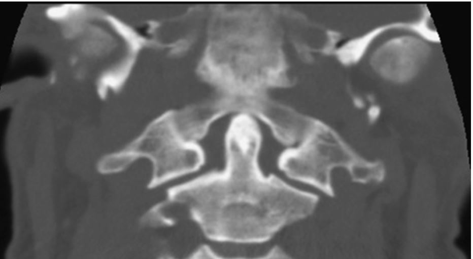
Jefferson Fracture can be tough to diagnose.

Jefferson Fracture of C1

Jefferson Fracture of C1


Note the lateral masses of C1 extend beyond the lateral borders of C2. That signifies Jefferson burst fracture. In a normal C-spine the lateral borders of C1 and C2 should line up pretty much perfectly.

Note the lateral masses of C1 extend beyond the lateral borders of C2. That signifies Jefferson burst fracture. In a normal C-spine the lateral borders of C1 and C2 should line up pretty much perfectly.


  1. Associated C2 fractures occur in approximately one half of patients.

  2. This fracture rarely leads to spinal cord injury since the fractured fragments have a tendency to fall outward away from the canal and brainstem.  (The Atlas of Emergency Radiology)


    Patients with multiple ED presentations for the same episode of back pain are at high risk for serious diagnoses.

Patients with new onset psychosis may benefit from CT imaging of the brain.

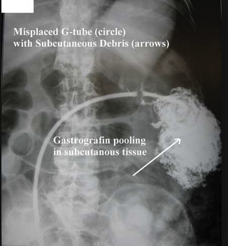
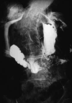
Don’t attempt to replace a g-tube if it has been placed within the last 4 weeks.  If a new g-tube (less than 4 weeks) has fallen out,  just admit the patient and consult gen surg or GI.

Sickle Cell Disease is a risk factor for rhabdomyolysis.  A marker for rhabdomyolysis in a sickle cell patient is an abnormal AST or ALT.


McKean/Kishi         Oral Boards

Case 1. 43yo male with leg pain and redness.  Patient had a fish hook injury to his leg recently in salt water.  Skin exam shows severe cellulitic changes, bullae and crepitence.

Vibrio Vulnificus infection

Vibrio Vulnificus infection

Labs c/w sepsis with elevated WBC and lactate.  X-rays show show soft tissue air.   Diagnosis was soft tissue infection from vibrio vulnificus.   Treat with 3rd Gen Cephalosporin and Doxycycline or flouroquinolone.

Dr. Lovell point: Think Vibrio vulnificus with salt water source infection and Aeromonas for fresh water source infection.  You can treat both with flouroquinolone.

Case 2. 16 yo male with syncope and facial contusion. Patient had syncope after swimming and injured his mouth. EKG showed short PR interval.  Patient had Lown-Ganong-Levine syndrome   Short PR interval with no delta wave.

The Lown–Ganong–Levine (LGL) syndrome is now believed to be due to enhanced AV nodal conduction. The proposed mechanism is an intranodal bypass tract or atrionodal tracts formerly known as James fibers. This diagnosis has been largely abandoned in the era of electrophysiology. Syndrome diagnostic criteria classically have been: a short PR interval (less than 0.12 s) and a normal QRS complex on ECG, and the clinical history of intermittent palpitations or episodes of paroxysmal SVT, atrial flutter, or atrial fibrillation.

The most important differential diagnosis is that of the WPW syndrome that has a specific treatment. The ECG finding that differs is the delta wave leading into the QRS complex seen with WPW; this is not found in LGL. (Atlas of Cardiovascular Emergencies)

LGL EKG

LGL EKG

Case 3.   31yo male presents with elbow pain.  Patient was recently scuba diving.  Vitals and physical exam of the elbow are normal.  Xrays of the elbow are normal. Labs are normal. Arthrocentesis  was done and the joint fluid analysis was normal.  Diagnosis was the decompression sickness. Treatment is hyperbaric O2.  

Dr. Lovell point: Decompression sickness and arterial gas embolism are both indications for hyperbaric oxygen.


Regan          Emergency Preparedness

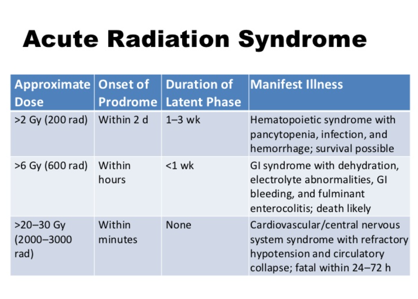
Dr. Regan discussed radiation physics.  Here is my attempt at a descriptive analogy.   Think of a Gray as 1 dollar.  Think of a rad as a penny.  There are 100 rads in a Gray. (100 pennies in a dollar).  When a rad hits the body it is then measured as a miliseivert.  Think of this as taking your dollar and bringing it to a currency exchange to purchase euros. 1 rad will buy 10miliseiverts.  300 rads or 3000 miliseiverts of radiation exposure is very dangerous. 600rads/6000 miliseiverts is almost always fatal.

Radiation Illness 2.PNG

Decontamination for radiation-exposed patients presenting to the ED is primarily removing their clothing and irrigating them with copious water.  You then test them with a Geiger counter.  They are OK to go into the hospital when you get their radiation down to 2X the ambient radiation level.  You can also Geiger count their urine and a nasal swab to check for internal radiation.  Nuclear Medicine Dept has Geiger counters. Get CBC’s on radiation-exposed patients every 2 hours. Personnel doing radiation decontamination have to be specially trained and wear a level C hazmat suit. 


Classification of Blast Injuries

Classification of Blast Injuries

High Order explosives like TNT, C4, Semtex create a well-defined pressure wave and cause primary blast injuries.  Low order explosives like pipe bombs do not create a well-developed pressure wave and don’t cause primary blast injuries.

Imaging of blast lung injury

Imaging of blast lung injury

Patients exposed to bomb blasts will develop lung finding relatively rapidly so you can order CXR or CT Chest early in ED course.

The lung is very susceptible to primary blast injury. Pulmonary barotrauma is the most common fatal primary blast injury and the most common critical injury in people close to the blast center. Pressure differentials across the alveolar–capillary interface can cause disruption, hemorrhage, pulmonary contusion, pneumothorax, hemothorax, pneumomediastinum, and subcutaneous emphysema…

In general, managing blast lung injury is similar to caring for pulmonary contusion and acute respiratory distress syndrome, except that early recognition of the syndrome may be complicated by initially benign symptoms, especially in the context of hectic mass casualty incident situations. Hypoxia is an almost universal finding.12 Monitoring of respiratory rate and room-air pulse oximetry, as well as serial chest radiographs, may be needed. Fluid administration should ensure tissue perfusion without volume overload... Keep tidal volume to 6 to 7 mL/kg ideal body weight to limit the peak inspiratory pressure and to minimize ventilator-induced lung barotrauma. Often, neuromuscular paralysis and early institution of pressure-limited ventilation (plateau pressures <30 cm H2O),13 with the lowest pressures compatible with adequate ventilation, may be the best strategy. Inverse inspiratory-to-expiratory ratio ventilation may be useful. Permissive hypercapnia is acceptable depending on cerebral perfusion pressure or increased intracranial pressure.13 Aggressive methods of oxygenation, such as extracorporeal membrane oxygenation or intravascular oxygenation, may become necessary within hours of the injury. (Tintinalli 8th edition)


Eydelman     ECMO

ECMO is basically a placenta-like form of oxygenation for critically ill patients.

ECMO gained traction in adults for management of military blast injuries and treatment of fulminant lung injury due to influenza.

Veno-venous ECMO bypasses the lungs but the heart still pumps the blood.  

Veno-Arterial ECMO bypasses the lung and the machine pumps the blood.  V-A ECMO can perfuse in cardiac arrest.

The literature is not super strong in support of ECMO but it is trending toward better outcomes. A 2016 study showed 50% survival for out of hospital V-Fib arrest who received ECMO therapy.

There are some EM centers doing ECMO for selected cardiac arrest patients.  You can also use ECMO for cardiogenic shock.  Other indications: electrical storm, heart block, ARDS, severe tox cases, hypothermia/hyperthermia, and blast lung injury.

As emergency physicians, we can advocate on behalf of our patients for doing ECMO in select situations.  This means consulting with ICU and CV surgery specialists in the appropriate clinical scenario.

Critical Care in the future may see ECMO replacing intubation and positive pressure ventilation.  That is because positive pressure ventilation is so deleterious to patients.


Rehman       5 Slide F/U   PCP Pneumonia

No patient details, just the take home lessons.


PCP is the most common opportunistic infection among AIDS patients.21 The causal agent is known as P. jirovecii (previously known as P. carinii). Approximately 70% of HIV-infected patients acquire PCP at some time during their illness, and PCP is often the initial opportunistic infection that establishes the diagnosis of AIDS. This infection is the most frequent serious complication of HIV infection in the United States and the most common identifiable cause of death in patients with AIDS. The classic presenting symptoms of PCP are fever, cough (typically nonproductive), and shortness of breath (progressing from being present only with exertion to being present at rest).

Symptoms are often insidious and accompanied by fatigue. Chest radiographs most often show diffuse interstitial infiltrates (Table 154-3), but negative radiographic findings exist in 15% to 25% of patients with PCP.22 The lactate dehydrogenase level is elevated in patients with PCP, but this test has low sensitivity and specificity, impairing utility. Arterial blood gas analysis usually demonstrates hypoxemia and an increase in the alveolar-arterial gradient. Suspect early PCP if a patient demonstrates a decrease in pulse oximetric values with exercise; even an ED "walk" can detect this exercise desaturation. Presumptive diagnosis of PCP is often made in the ED if there is hypoxemia without any other explanation. Inpatient diagnostics include bronchoscopy with specimen analyses.

Initial PCP therapy is trimethoprim-sulfamethoxazole (Table 154-2) PO or IV for 3 weeks (2 double-strength tablets three times daily). Adverse reactions, including rash, fever, and neutropenia, occur in up to 65% of AIDS patients. Pentamidine is an alternative agent (Table 154-2). Give steroids to patients with a partial pressure of arterial oxygen of <70 mm Hg or an alveolar-arterial gradient of >35 mm Hg, usually oral prednisone starting at 40 milligrams twice daily and tapering over 21 days (Table 154-2).

Seventy percent of patients have reinfection within 18 months; thus, prophylactic therapy is key. Oral trimethoprim-sulfamethoxazole, one double-strength tablet daily, is the preferred agent. Prophylaxis for all patients with CD4+ T-cell counts of <200 cells/mm3 is another common approach to mitigate PCP. Repeat PCP infections are often less responsive to therapy.  (Tintinalli 8th edition)

Dr. Lovell comment: Dr. Triteman is an ID specialist here a ACMC who has a HIV Clinic.  You can contact him to arrange follow up for newly diagnosed HIV patients who are stable for discharge.


Ahmad            Five Slide F/U  Back Pain

No patient details, just the take home lessons.

Bowel perforation from blunt abdominal trauma can present to the ED days after injury.

If a patient has bowel perforation from blunt abdominal trauma, you have to consider that the patient may have some underlying bowel pathology such as crohn’s or ulcerative colitis.








Conference Notes 4-3-2019

Airway Day

Dodd Avoiding Airway Nightmares

Oxygenation and ventilation keep the patient alive. These are different than intubation which places a tube in the patient’s trachea. Remember when you are attempting to intubate that you also need to focus on oxygenation and ventilation. Preoxygenate with high flow O2 and ventilate with spontaneous respirations if adequate or assist with BVM, BiPap, or even LMA. Don’t let the procedure of intubation distract you from maintaining adequate oxygenation and ventilation for the patient.

Intubation safety is about preparation and mindset. Have 2 back up plans. Discuss your primary approach and back up plans with your team before you start your first attempt at intubation. Make sure your team has a shared mental model of how you are going to manage the airway.

Optimize your first attempt. If the first attempt fails, the incidence of bad outcomes increases.

Specificity of the Lemon assessment is 86%. Sensitivity however is low &lt;70%. Basically if you have signs of a difficult airway it will be difficult. If you have no signs of a difficult airway the Lemon assessment may fail you and the airway may s…

Specificity of the Lemon assessment is 86%. Sensitivity however is low <70%. Basically if you have signs of a difficult airway it will be difficult. If you have no signs of a difficult airway the Lemon assessment may fail you and the airway may still be difficult.

Simplified airway algorithm. Optimal first attempt>>> if failure>>>B-V-M and second attempt>>>if failure>>>Intubating LMA>>>if failure>>>Cricothyrotomy

Pre-Oxygentate with a non-rebreather mask using “flush rate” wide-open wall oxygen. In addition to a NRB mask, also apply nasal canula O2 at 15L for passive oxygenation. Preoxygenate patients semi-upright. The semi-upright position improves functional residual capacity and decreases atelectasis.

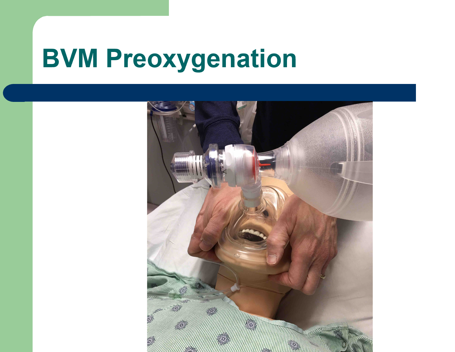
Bag Valve Mask is a critical life-saving skill. Use the Thenar Grip Method

Thenar Grip with patient semi-upright. Patient has apneic oxygenation with nasal cannula in addition to oxygen through BVM at wide open “flush rate”. A peep valve is in place on the BVM. Peep valves improve pre-oxygenation.

Thenar Grip with patient semi-upright. Patient has apneic oxygenation with nasal cannula in addition to oxygen through BVM at wide open “flush rate”. A peep valve is in place on the BVM. Peep valves improve pre-oxygenation.


Dr. Dodd recommends making your first attempt with macintosh style video laryngoscopy blade and a bougie. Then placing ET tube over the bougie into the trachea.

Here’s a summary of the BEAM Trial demonstrating that using a bougie as your first attempt improves first pass success. One caveat: you can’t use the bougie with a hyperangulated VL blade. You have to use the glidescope stylet when using the hyperan…

Here’s a summary of the BEAM Trial demonstrating that using a bougie as your first attempt improves first pass success. One caveat: you can’t use the bougie with a hyperangulated VL blade. You have to use the glidescope stylet when using the hyperangulated blade.

Lovell Difficult Airway

Unfortunately I missed this outstanding lecture.

Kennedy/Putman/Multiple Faculty Members Airway Workshop

Conference Notes 3-27-2019

Jurkovic Safety Lecture Trauma Bypass

These are the Criteria for a medical center to have a Level 1 Trauma designation.

These are the Criteria for a medical center to have a Level 1 Trauma designation.

Since 2016 CFD criteria for a patient meeting trauma criteria includes patients 55yo and older who fall from standing position and hit their head.

Bypass means that BLS and stable ALS patients get diverted unless the other hospitals are on bypass.

Patients that fall into these categories cannot be bypassed to another institution.

Patients that fall into these categories cannot be bypassed to another institution.

Bypass is a courtesy. BLS and stable ALS patients can still go to the waiting room. Cases that are exempt from bypass are Trauma, STEMI, CVA, full arrest, respiratory distress and those with unstable vital signs. These patients are still going to arrive in our ED whether or not we are on bypass.

In consultation with the House Supervisor, and the ED Lead Physician, it is the role of the ED Charge Nurse to activate bypass. All monitored beds in the hospital need to be occupied. Also any Internal Disaster would prompt a bypass decision.

On average, bypass diverts 1.7 patients/hour. The effect on ED patient volume is not that significant.

IDPH is investigating how hospitals are using bypass to see if an alternative system is possible.

Walchuk/Ebeledike Oral Boards

Case 1. 54 yo female patient presents after resuscitated cardiac arrest. Patient became nauseated at a restaurant and was found unresponsive in bathroom. Exam shows urticaria. Patient became hypotensive again in ED. Diagnosis was anaphylaxis and treatment initiated including epi drip.

Anapylaxis is IGE mediated.

If you have suspected allergic reaction and two systems are involved or the patient is hypotensive, diagnose anaphylaxis and give Epi. There is no contraindication to Epi in the setting of anaphylaxis.

If you have suspected allergic reaction and two systems are involved or the patient is hypotensive, diagnose anaphylaxis and give Epi. There is no contraindication to Epi in the setting of anaphylaxis.

Anaphylaxis treatments. First line therapy is EPI. Everything else second line to EPI. One additional pearl, if patient is on a beta blocker and has hypotension refractory to EPI, you can try glucagon 1mg IV Q 5 minutes.

Anaphylaxis treatments. First line therapy is EPI. Everything else second line to EPI. One additional pearl, if patient is on a beta blocker and has hypotension refractory to EPI, you can try glucagon 1mg IV Q 5 minutes.

Case 2. 15 yo female patient presents with knee injury while playing soccer.

Patient’s knee exam was as pictured. Xray confirmed lateral patellar dislocation.

Patient’s knee exam was as pictured. Xray confirmed lateral patellar dislocation.


Case 3. 28 yo female present with rash. Patient had EM rash.

Erythema multiforme. Common precipitating factors are infection, especially with Mycoplasma and herpes simplex virus; drugs, especially antibiotics and anticonvulsants; and malignancies. However, the cause is often unknown.3 Most likely, erythema mu…

Erythema multiforme. Common precipitating factors are infection, especially with Mycoplasma and herpes simplex virus; drugs, especially antibiotics and anticonvulsants; and malignancies. However, the cause is often unknown.3 Most likely, erythema multiforme is the result of a hypersensitivity reaction, with immunoglobulin and complement components demonstrated in the cutaneous microvasculature on immunofluorescent studies of skin biopsy specimens, circulating immune complexes found in the serum, and mononuclear cell infiltrate noted on histologic examination.3 (Tintinalli 8th edition)

Systemic steroids are commonly used for localized disease and provide symptomatic relief but are of unproven benefit in influencing the duration and outcome of erythema multiforme.7 Many authorities recommend a short, intensive steroid course of prednisone, 60 to 80 milligrams PO once a day, particularly in drug-related cases, with abrupt cessation in 3 to 5 days if no favorable response is noted. Systemic analgesic agents and antihistamines provide symptomatic relief. (Tintinall 8th edition)

Erythema multiforme (EM) is an acute inflammatory skin disease (Figure 249–1) with a broad range of severity, from a minimal, nuisance-level event to a severe multi-system illness. It is divided into two distinct sub-types, considering the extent of involvement, presence of epidermal detachment, and the development of mucous membranes lesions. Erythema multiforme minor, the less severe form of the illness, is a localized papular eruption of the skin, with an acral distribution and involving target lesions and/or raised, edematous papules. Erythema multiforme major is the more severe form of EM with multi-system involvement and widespread vesiculobullous lesions and erosions of the mucous membranes; specifically, EM major includes involvement of one or more mucous membrane areas and epidermal detachment less than 10% of total body surface area.

Some authorities include Stevens-Johnson syndrome (SJS) as a severe form of EM major while others consider it a less severe form of toxic epidermal necrolysis. Perhaps the most appropriate classification approach for the emergency physician is as follows: Stevens-Johnson syndrome noted with less than 10% of the body surface area with epidermal detachment; the “overlap” presentation of Stevens-Johnson syndrome and toxic epidermal necrolysis noted with 10% to 30% epidermal detachment; and toxic epidermal necrolysis noted with greater than 30% epidermal detachment.1 In either consideration, SJS is a serious dermatologic illness with significant, widespread skin involvement, more extensive epidermal detachment, and mucous membrane lesions. This taxonomic controversy has no meaning for the emergency physician; what is important for the emergency physician is the recognition of a significant, potentially life-threatening, multi-system dermatologic condition. (Tintinalli 8th edition)

Lambert Pelvic Ultrasound

Yolk sac is the first structure visible of an IUP at about 5-6 weeks. Fetal cardiac activity comes later at about 8.5 weeks.

Yolk sac is the first structure visible of an IUP at about 5-6 weeks. Fetal cardiac activity comes later at about 8.5 weeks.

Live IUP has to have gestational sac with fetal pole and cardiac activity in the uterus. The endometrial stripe needs to surround the gestational sac/fetal pole/heart.

IUP has a yolk sac and fetal pole without cardiac activity within the endometrial echo of the uterus.

If the pregnancy is not within the endometrial echo then the pregnancy is extrauterine (ectopic).

You should have Gyne consult formally on a patient in ED if the patient has abnormal vitals, significant pain, fluid in the pelvis or adnexal mass identified on ultrasound.

When US reveals an unequivocal IUP and no other abnormalities, ectopic pregnancy is effectively excluded unless the patient is at high risk for heterotopic pregnancy. An embryo with cardiac activity seen within the uterine cavity is referred to as a viable IUP. When an embryo without cardiac activity is visualized within the uterus, the diagnosis of fetal demise can be entertained, provided that the crown–rump length is at least 5 mm. Briefly, transvaginal scanning can usually visualize the early sonographic signs of pregnancy, the gestational sac, yolk sac, and fetal pole, at 4.5, 5.5, and 6.0 weeks, respectively. Visualization by transabdominal scanning can be done approximately 1 week later.

No further diagnostic testing is needed when sonographic findings confirm or are highly suggestive of ectopic pregnancy. An empty uterus with embryonic cardiac activity visualized outside the uterus is diagnostic of ectopic pregnancy. This is seen in <10% of ectopic pregnancies using transabdominal scanning, but in up to 25% of cases when the transvaginal approach is used. When a pelvic mass or free pelvic fluid is seen in conjunction with an empty uterus, ectopic pregnancy is considered highly likely (Figure 98-2). The combination of an echogenic adnexal mass with free fluid in the setting of an empty uterus confers a risk of ectopic pregnancy near 100%, whereas a large amount of free fluid alone has a 86% risk (Table 98-4). In addition to a living extrauterine pregnancy, an extrauterine gestational sac is highly predictive of ectopic pregnancy (Figure 98-3). Any adnexal mass (other than a simple cyst) seen with US also has high positive predictive value for the diagnosis of ectopic pregnancy.20,21 It has also been suggested that increased thickness of the endometrial stripe is predictive of ectopic pregnancy when no other diagnostic findings are noted on US. However, the wide overlap between endometrial stripe thickness in normal and ectopic pregnancy limits the usefulness of this observation.22 (Tintinalli 9th edition)

ectopic ultrasound.PNG


Lambert and Team Ultrasound Ultrasound Lab


Conference Notes 3-6-2019

Wong Pediatric Chest Pain

Chest pain in children is rarely cardiac in nature. Cardiac issues in kids rarely present with chest pain.

Asthma and reactive airway disease are common causes of chest pain.

Pediatric coronary arteries rarely get stented because the stented artery will not grow with the child. Consequently the child who has a coronary stent, as they mature and grow, will have a fixed size coronary artery due to the stent and have coronary insufficiency the rest of their life.

Anomalous coronary artery can result in coronary insufficiency. Anomalous coronaries branch off the wrong part of the aorta and branch off at an acute angle. Both result in reduced flow.


Anomalous Coronary Artery

Anomalous Coronary Artery

Kawasaki’s aneurysms can cause chest pain.

Aneurysms in an 18yo male due to Kawasaki’s disease

Aneurysms in an 18yo male due to Kawasaki’s disease

Cocaine and marijuana can cause pediatric chest pain.

Arrhythmias can be a cause of chest pain.

Pericarditis and myocarditis. Myocarditis is a high risk diagnosis that can have serious morbidity and mortality. Think about myocarditis with in patients with fever, tachycardia, and recent viral illness associated with their chest pain.

Hypertrophic cardiomyopathy can cause chest pain. The hypertrophic septum can block the LV outflow tract and decrease coronary flow. HOCM causing chest pain will have significant murmur.

Aortic stenosis or pulmonary stenosis are valvular causes of chest pain and will have a murmur.

Aortic dissection can cause chest pain. It is exceedingly rare in kids. Consider it in kids with Marfan’s, Ehlers Danlos, or family hx of Marfan’s Ehlers-Danlos, or other connective tissue disorder

The high-risk diagnoses for pediatric chest pain are anomalous coronary artery, hypertrophic cardiomyopathy and myocarditis. Anomalous coronary artery can be very difficult to diagnose. You won’t be able to definitively diagnose it in the ED if the patient does not have EKG changes. If their pain has resolved the EKG changes may have resolved as well. Consider anomalous coronary artery if the patient has exertional chest pain. Hypertrophic cardiomyopathy causes syncope or sudden death more commonly than chest pain. If it is causing exertional chest pain there will be a prominent murmur. Myocarditis may have tachycardia, EKG changes, signs of heart failure, elevated troponin, or recent history of viral illness.

The incidence of PE in pediatric patients is increasing. There is a bimodal age distribution (neonates and teenagers). Screen with a d-dimer level over 0.75.

Congenital heart disease, Long QT and Brugada do not present with chest pain.

Myocarditis is the highest risk diagnosis that if sent home is most likely to have a bad outcome. Myocarditis can be identified by abnormal EKG, tachycardia, and elevated troponin. The patient may have an elevated troponin or history of recent viral illness.

ACA Guideline for kids age 5-18: Ask about exertional chest pain. It is a Red Flag historical finding. Ask for family hx of cardiomyopathy, early CAD, and sudden death. Perform a physical exam for murmur and lung sounds. Get an EKG.

Get an echo for exertional chest pain patients, patient with high risk past medical or family history, murmur, or abnormal EKG. Echo may not need to be emergent. Asymptomatic patients in the ED can be discharged home with activity limitation and pediatric cardiology follow up.

Kane, DA. Congenit Heart Dis. 2010;5(4):366–373.

Kane, DA. Congenit Heart Dis. 2010;5(4):366–373.


Holter monitor will capture every beat for 24 hours. Event monitors are worn for 30days and capture 15 min recordings retrospectively from the time the patient voluntarily triggers the device when having some symptoms.

Kids with psychogenic chest pain referred to cardiology usually end up getting extensive workups with echo, event monitors and stress tests.

Ahmad/Johns Oral Boards

Case 1. 23 yo female presents with chest pain and shortness of breath. Dad notes a change in patient’s mental status. Patient is tachycardic and hypotensive. EKG shows electrical alternans

Electrical Alternans

Electrical Alternans

Large pericardial effusion with RV collapse

Large pericardial effusion with RV collapse

Diagnosis was pericardial tamponade due to autoimmune disease. Treatment is IV fluid resuscitation and consulting for or performing pericardiocentesis.

Case 2. 39 yo male presents with a seizure. Blood sugar is normal. Patient received IM midazolam in the prehospital phase with continued seizure. IV Ativan in ED did not terminate seizure. Patient had recent history of positive PPD for employment screening. Patient has been taking isoniazid. Patient was treated with standard medications for status epilepticus with no improvement. B6 (pyridoxine) was given next which terminated seizure. Diagnosis was seizures secondary to isoniazid.

These symptoms may progress to the three classic features of acute isoniazid overdose: seizures, metabolic acidosis, and protracted coma.46,47 Seizures typically follow acute isoniazid ingestions of greater than 20 to 30 milligrams/kg. Isoniazid-induced seizures are generalized tonic-clonic in nature and are often refractory to standard anticonvulsive therapy with benzodiazepines and barbiturates. The mechanism for isoniazid-induced seizures is a functional deficiency of pyridoxine (vitamin B6) and inhibition of the synthesis of γ-aminobutyric acid, the primary CNS inhibitory neurotransmitter. Seizures with therapeutic doses of isoniazid have been reported in patients,48 presumably due to very low vitamin B6 levels.49 Although the metabolic acidosis that accompanies isoniazid-induced seizures is likely due to motor activity, the lactic acidemia may not resolve as rapidly as with other more typical epileptic seizures.

Consider isoniazid overdose in patients with refractory seizures.44 Isoniazid-induced seizures are treated with a combination of benzodiazepines and pyridoxine. The dose of pyridoxine is a gram-for-gram equivalent to the amount of isoniazid ingested.50 For patients who ingest an unknown quantity of isoniazid, the recommended dose of pyridoxine is 5 grams IV in adults and 70 milligrams/kg (maximum 5 grams) in pediatric patients. Pyridoxine may be administered at a rate of approximately 1 gram IV every 2 to 3 minutes until the seizures stop or the maximum dose has been given. After the seizures have ceased, the remainder of the pyridoxine dose should be given over the following 4 to 6 hours to limit recurrent seizures.

Adequate single-dose therapy of pyridoxine should be effective to stop most seizures, but patients who do not receive adequate pyridoxine dosing may have repeat seizures. Pyridoxine may also assist in reversing isoniazid-induced comas. (Tintinalli 8th edition)


Case 3. 79 yo male patient presents with abdominal pain. Abdominal exam showed peritonitis.

Free air under both diaphragms

Free air under both diaphragms

Diagnosis was perforated viscous causing abdominal pain. Treatment is IV crystalloids, IV antibiotics, and surgical consultation.


Davis/Miner/Mullen/Lorenz BounceBacks

For ETOH dependent patients who are withdrawing, follow the CIWA score and if it is increasing consider higher level of care dispo such as step-down. If a patient has a history of prior ICU admit consider step-down admission.

Dr. Ryan comment: If a patient has 2 medical problems such as alcohol withdrawl and pancreatitis you may want to consider a step-down admission.

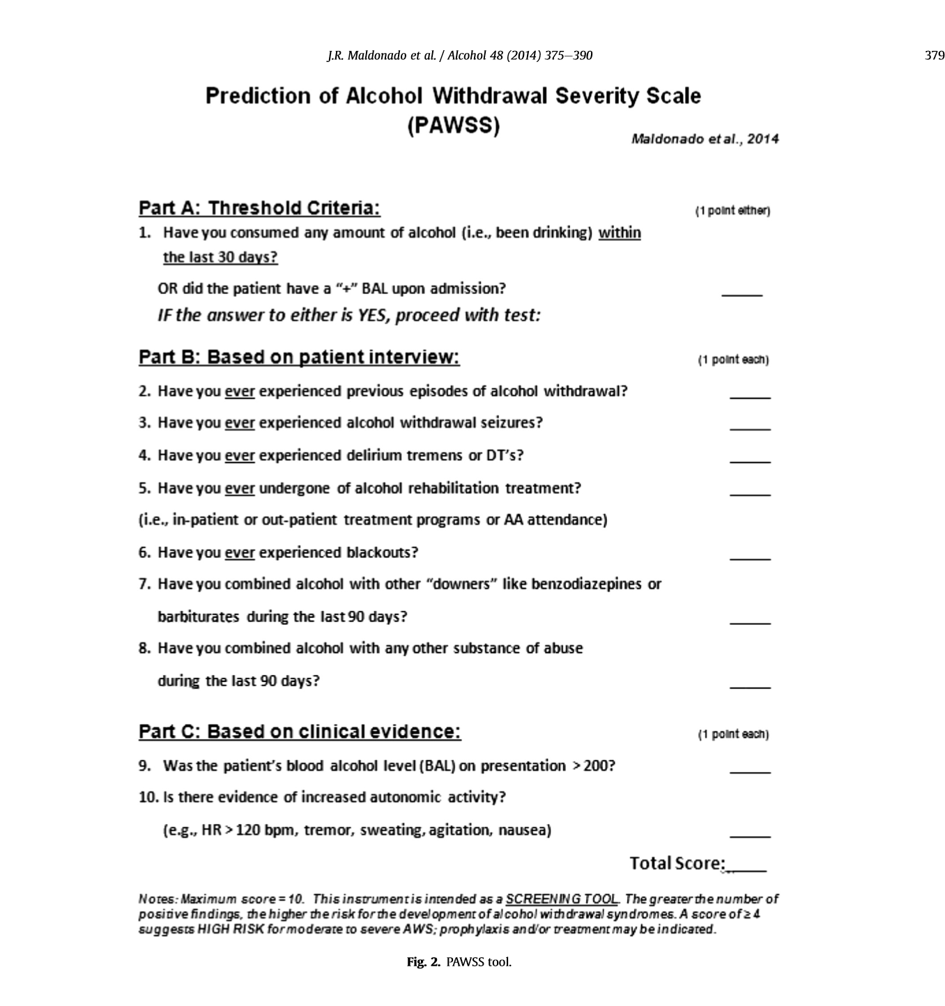
PAWS score on MD Calc can be used to risk stratify alcohol withdrawal patients. At scores &gt;/=4 consider step-down or ICU admission.

PAWS score on MD Calc can be used to risk stratify alcohol withdrawal patients. At scores >/=4 consider step-down or ICU admission.


Sending home first trimester pregnant patients with vaginal bleeding has risks. To mitigate risk, be sure to give very clear return instructions to the patient so that they come back right away for abdominal pain, vaginal bleeding, lightheadedness, or any other symptoms. Dr. Ryan comment: The patient going home has to understand that pain, bleeding, or lightheadedness is not expected for a normally progressing pregnancy and any of these symptoms should prompt return to the ED.

Patients with fever and encephalopathy need to be considered for LP. Consider meningitis if you don’t have another clear cut-source of infection and the patient’s mental function is significantly different from baseline.


Tyler Fluid Management in Septic Patients

Too much IV fluids is a risk for increased morbidity and mortality in sepsis patients.

50% of sepsis patients are fluid-responsive. The other 50% non-responders are further along on the Starling curve and additional fluids don’t help.

You can assess fluid responsiveness in the ED most simply by assessing for improvement of blood pressure/MAP with a 250ml fluid bolus or 3minutes of passive leg raise.

There aren’t many other ways to accurately assess volume status in septic patients. Dr. Tyler recommended that the best was the calculating the VTI at the LV outflow tract using bedside echo. Low VTI suggests that the patient could use more fluids.

IVC evaluation on bedside U/S is not useful generally unless the IVC is extreme on either side (totally collapsing or totally plethoric). You can use the IVC as a dynamic assessment of fluids status before and after fluid administration. If the IVC looks more full after a 250ml bolus you can take that as fluid responsiveness.


IVC showing collapse which in the right clinical context as a dynamic evaluation of fluid responsiveness may indicate need for more volume.

IVC showing collapse which in the right clinical context as a dynamic evaluation of fluid responsiveness may indicate need for more volume.

Full IVC which in the appropriate clinical context may indicate patient is no longer in need of further volume resuscitation

Full IVC which in the appropriate clinical context may indicate patient is no longer in need of further volume resuscitation


Ultrasound lung windows showing B lines are useful to assess for pulmonary edema.


B Lines are a marker of pulmonary congestion/edema

B Lines are a marker of pulmonary congestion/edema

Causes of elevated lactate: sepsis, tissue hypoxia, accelerated aerobic glycolysis (example is continuous albuterol med nebs), liver dysfunction, malignancy, medications, toxic alcohols, DKA, and thiamine deficiency.

On the other hand a normal lactate does not rule out serious problems.

Dr. Tyler is giving IV fluids to his sepsis patients who are fluid responsive in 500ml to 1 liter boluses and re-assessing prior to giving more fluids up to the 30ml/kg target.

Move to norepinephrine if you assess that patient is not improving with IV fluids.

Katiyar Billing and Coding

We are the masters of acute care and resuscitation.

Billing criteria for Critical Care: High probability of significant or life threatening deterioration. If the patient cannot be managed in an office or if you did nothing, something bad would happen, that fits the definition of critical care.

In your note, document the patient was critically ill and had risk of significant deterioration. You also need to document complex decision making and your total time involved in critical care not including procedure time. You need to document re-evaluations of the patient. It is helpful to document your interpretation of pulse ox, monitor, diagnostic testing and vital signs to support your critical care billing. It is also helpful to document that you discussed case with family and consultants.

Procedures such as intubation, arthrocentesis, lumbar puncture, and chest tubes are billed separately.


You can bill critical care for patients that you intervene on in the ED and because of your care, they no longer need ICU admission and get admitted to the floor. This would include clinical situations such as severe asthma, severe COPD exacerbation, and anaphylaxis.


Copy of Conference Notes 2-20-2019

Katiyar/Davis                               Oral Boards


Case 1. 70 yo female patient presents with altered mental status.  Patient is tachycardic and hypotensive. Patient’s temperature is 106F.  Diagnosis is heat stroke with rhabdomyolysis.

 The cardinal features of heat stroke are hyperthermia (>40°C [>104°F]) and altered mental status. Although patients presenting with classic (nonexertional) heat stroke may exhibit anhidrosis, the absence of sweat is not considered a diagnostic criterion because sweat is present in over half of patients with heat stroke.15

The CNS is particularly vulnerable in heat stroke. The cerebellum is highly sensitive to heat, and ataxia can be an early neurologic finding. Virtually any neurologic abnormality may be present in heat stroke, including irritability, confusion, bizarre behavior, combativeness, hallucinations, plantar responses, decorticate and decerebrate posturing, hemiplegia, status epilepticus, and coma. Seizures are quite common, especially during cooling. Neurologic injury is a function of the maximum temperature reached and the duration of exposure.15

The distinction between exertional and classic (nonexertional) heat stroke is not clinically important, because immediate cooling and support of organ system function is the therapeutic goal for both. A delay in cooling increases the mortality rate. (Tintinalli 8th edition)

cooling techniques.PNG


Case 2. 8 yo male patient was playing in the woodshed and came into the house screaming in pain. Patient had persistent pain despite IV morphine.  There appears to be a spider bite on exam.

Diagnosis was black widow spider bite.

The bite mark itself tends to be limited to a small puncture wound or wheal and flare reaction that often is associated with a halo (Table 115–3). However, the bite from L. mactans produces latrodectism, a constellation of signs and symptoms resulting from systemic toxicity. Some cases do not progress; others show severe neuromuscular effects within 30 to 60 minutes. The effects from the bite spread contiguously. For example, if a person is bitten on the hand, the pain progresses up the arm to the elbow, shoulder, and then toward the trunk during systemic poisoning.

The myopathic syndrome of latrodectism involves muscle cramps that usually begin 15 to 60 minutes after the bite. The muscle cramps initially occur at the site of the bite, but later involves rigidity of other skeletal muscles, particularly muscles of the chest, abdomen, and face. The pain increases over time and occurs in waves that cause the patient to writhe. Large muscle groups are affected first. Classically, severe abdominal wall spasm occurs and is confused with a surgical abdomen, especially in children who cannot relate the history to the initial bite.38 Muscle pain often subsides within a few hours but can recur for several days. Transient muscle weakness and spasms is reported to persist for weeks to months.

In the past, 10 mL 10% calcium gluconate solution was given IV to decrease cramping. However, a retrospective chart review of 163 patients envenomated by the black widow concluded that calcium gluconate was ineffective for pain relief compared with a combination of IV opioids and benzodiazepines.56,138 Another study found greater neurotransmitter release when extracellular calcium concentrations were increased, suggesting that administration of calcium is irrational in patients suffering from latrodectism.191 The mechanism of action of calcium remains unknown, and its efficacy is anecdotal; therefore, we do not recommend calcium administration for pain management.

Although often recommended, methocarbamol (a centrally acting muscle relaxant) and dantrolene also are ineffective for treatment of latrodectism.138,196 A benzodiazepine, such as diazepam, is more effective for controlling muscle spasms and achieves sedation, anxiolysis, and amnesia. Management should primarily emphasize supportive care, with opioids and benzodiazepines for controlling pain and muscle spasms, because the use of antivenom risks anaphylaxis and serum sickness.

Latrodectus antivenom (Merck) is rapidly effective and curative. In the United States the antivenom formulation is effective for all species, but is available as a crude hyperimmune horse serum that is reported to cause anaphylaxis and serum sickness. The morbidity of latrodectism is high, with pain, cramping, and autonomic disturbances, but mortality is low. (Tintinalli 8th edition)


Case 3. 67 yo male with weakness and difficulty walking. Diagnosis is Guillain-Barre syndrome

Signs and symptoms of the classic form include an ascending symmetric weakness or paralysis and areflexia or hyporeflexia. Paralysis may ascend to the diaphragm, compromising respiratory function and requiring mechanical ventilation. Autonomic dysfunction may be present as well.

Cerebrospinal fluid analysis shows high protein levels (>45 milligrams/dL) and WBC counts typically <10 cells/mm3, with predominantly mononuclear cells. When there are >100 cells/mm3, other considerations include HIV, Lyme disease, syphilis, sarcoidosis, tuberculous or bacterial meningitis, leukemic infiltration, or CNS vasculitis. Electrodiagnostic testing demonstrates demyelination. Nerve biopsy reveals a mononuclear inflammatory infiltrate. If MRI is performed to rule out alternative diagnoses, it will show enhancement of affected nerves.

The first step in management is assessment of respiratory function. Airway protection in advance of respiratory compromise decreases the incidence of aspiration and other complications. A well-established monitoring parameter is vital capacity, with normal values ranging from 60 to 70 mL/kg.

Avoid depolarizing neuromuscular blockers like succinylcholine for intubation in Guillain-Barré syndrome due to the risk of a hyperkalemic response.

Both IV immunoglobulin and plasma exchange shorten the time to recovery.1,2 Neither has been shown to be superior to the other, nor are they more efficacious when used together. There are adverse effects seen with both modalities of treatment. IV immunoglobulin has been associated with thromboembolism and aseptic meningitis; plasma exchange is associated with hemodynamic instability and a small increase in the rate of relapse, though full recovery is still more likely. In general, IV immunoglobulin is more widely available and less cumbersome to administer. Corticosteroids are of no benefit and may be harmful.3 (Tintinalli 8th edition)

Tekwani Difficult Airway Lecture

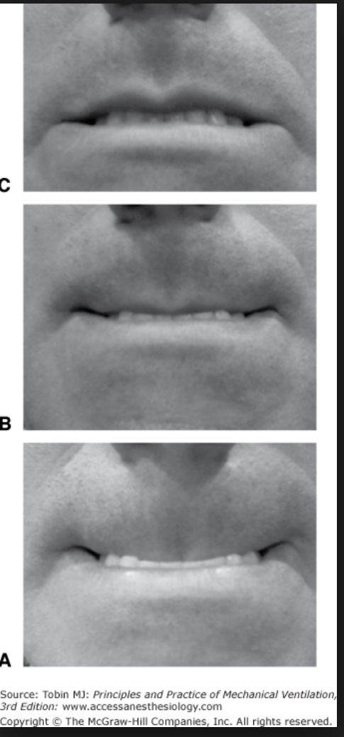
The upper lip bite test. If the patient can’t bite their upper lip even a bit (picture C), that is a sign of a difficult airway. Upper lip bite test had a sensitivity of 70% and a sensitivity of &gt;85% and an accuracy of &gt;85% for predicting a di…

The upper lip bite test. If the patient can’t bite their upper lip even a bit (picture C), that is a sign of a difficult airway. Upper lip bite test had a sensitivity of 70% and a sensitivity of >85% and an accuracy of >85% for predicting a difficult airway. 2018 Cochrane review by Roth showed that all our airway evaluation tests have relatively low sensitivity and high specificity. Not what you want in a screening test. There are no high sensitivity bedside screening tests for predicting a difficult airway. This test was the best.

With a prevalence of 10%, 10 out of 100 patients will have difficult laryngoscopy. Of these, 3 will be missed by the upper lip bite test(33% of 10). Of the 90patients without difficult laryngoscopy 7 will be unnecessarily classified as having a difficult airway.

It is the editor’s opinion that it makes sense to consider BMI, neck thickness and mobility, some form of mallampati, do a 3-3-2 and assess mandibular movement if possible with the upper lip bite test.

This review looked at many scoring scales and receding jaw and mandibular movement were consistent factors in difficult airways. One investigator used basically this combination of screening tests and had pretty spot on prediction of a difficult airway.

Inhaled nitrous oxide can be used to manage severe pulmonary hypertension.

Dr. Florek comment: Jaw thrust maneuver can be used to assist fiberoptic intubation. It opens up some additional space in the upper airway to maneuver the fiberoptic scope.

Dr. Harwood comment: In the patient with significant upper airway bleeding, use 2 yankaur suction devices to improve clearance of blood from the airway.

Girzadas Board Prep Zebras

This lecture was sent out in PDF form to all the residents.

Delbar Safety Lecture Inter Unit Transfers

“A wealth of information creates a poverty of attention” It is tough to balance enough info with too much info. Too much info in a handoff can cause the receiving physician to become distracted.

Emergency physicians and Admitting physicians have different perspectives on a specific patient. We are thinking acute care and they are thinking longer term hospital course.

We frequently don’t appreciate the stressors that admitting physicians are feeling.

Use of a standardized process, understood by both teams, that covers patient care info important to both teams, can be helpful in decreasing communication breakdowns.

Dr. Delbar reviewed the recent literature regarding inter Unit transfers.

Editor’s note: SBAR is a very widely accepted handoff tool that can be used. Giving your handoff in a standardized way covering: S situation B background A assessment and R recommendation may be helpful in improving handoff communication.

Conference Notes 2-6-2019

Patel/Burch Oral Boards

Case 1. 30 yo female patient presents with syncope. Patient was tachycardic and hypotensive. Patient had some abdominal tenderness. Further history revealed that patient had a fall about a week earlier on to her left side. On FAST, patient has free fluid in morrison’s pouch. Diagnosis was splenic rupture. Patient required IVF and IV PRBC’s to resuscitate and get to OR for surgical management.

According to the American College of Surgeons (ACS), mortality from splenic injuries accounts for approximately 10% of all trauma-related deaths [5]. In 1998, Cocoanour stated that the incidence of delayed splenic injury was approximately 1% [6]. However, in 2017, Furlan et al. reported that the incidence of delayed splenic injury has risen to 3%-15% with an inconsistent association with the severity of splenic trauma [7]. Compared to the 1% mortality associated with acute splenic injury, the mortality from delayed diagnosis of splenic injury following blunt abdominal trauma ranges from 5%-15% [3]. The trend in the increased presentation of delayed splenic injury is a risk of patient morbidity and mortality and can be attributed to the difficulty in identifying delayed splenic rupture. Difficulties with diagnosing delayed splenic injury may include atypical presentations that may mimic other pathologies, the lack of symptomatology, or equivocal imaging. Due to the variability in presentation and the difficulty with the initial assessment of delayed splenic injury, it is the opinion of the authors that delayed splenic rupture be considered in the differential diagnosis of an acute surgical abdomen regardless of the time or mechanism of the inciting event.

(Chikamuche, Cureus. 2018 Feb; 10(2): e2160.)

Delayed rupture of the spleen remains a real diagnostic entity and may occur in 1% of blunt abdominal trauma victims. The process most likely results from rupture of pre-existent subcapsular hematoma of the spleen. Such a hematoma is likely the result of parenchymal injury that may or may not be detected by CT scan. Such delayed ruptures can present atypically and after minor trauma that may be interpreted by the patient to be insignificant, and therefore, forgotten. In these instances, continued clinical vigilance and early appropriate imaging will likely decrease morbidity.

(Allen, The Journal of Emergency Medicine, Vol. 23, No. 2, pp. 165–169, 2002)

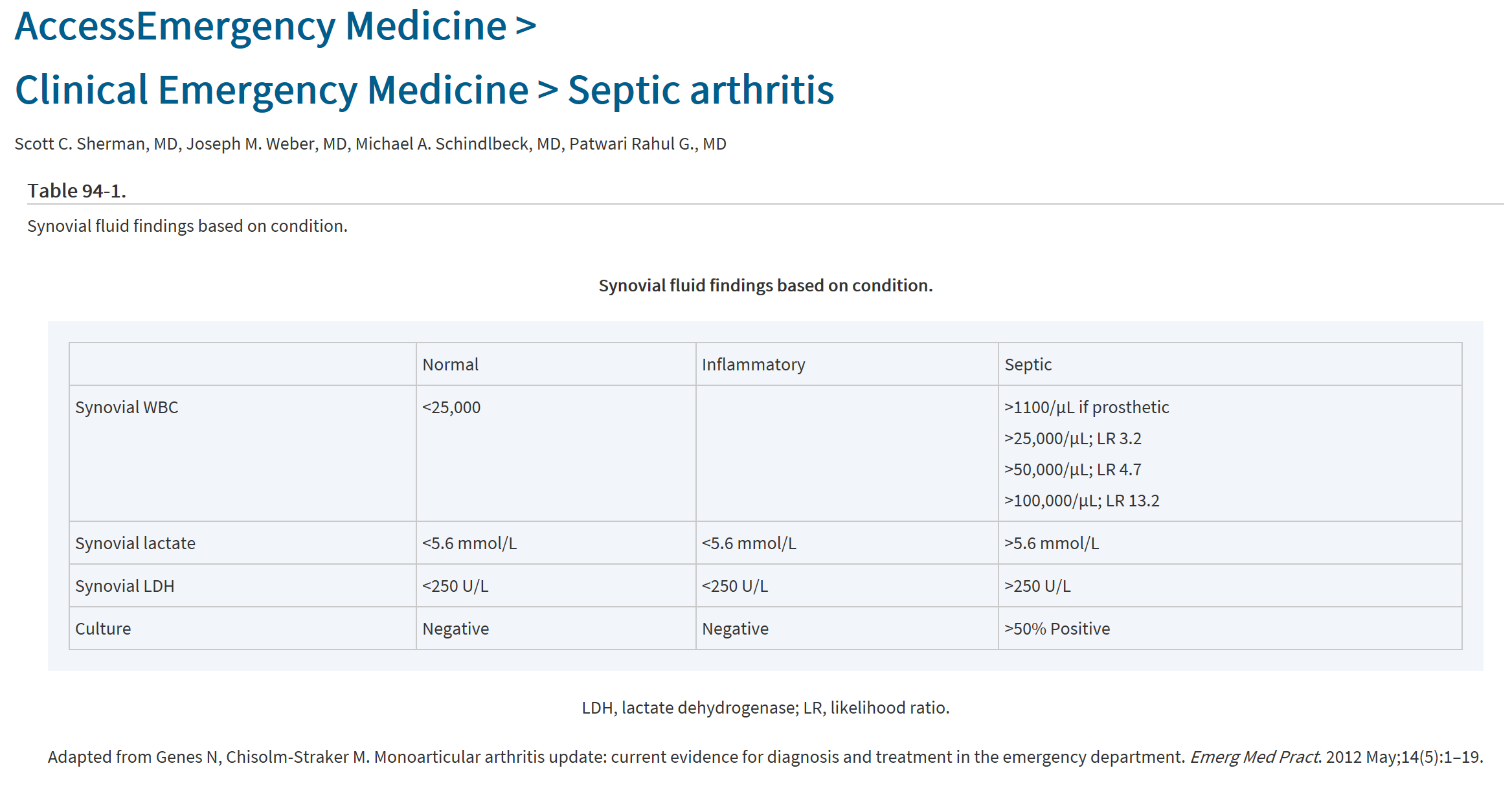
Case 2. 32 yo male patient with knee pain. Patient had concurrent paronychia on great toe of same lower extremity. Arthrocentesis of knee shows 192,000 WBC’s. Most likely patient had hematogenous spread from paronychia causing septic arthritis. Treatment requires IV antibiotics and joint irrigation.

synovial fluid septic joint.PNG

Case 3. 39yo firefighter presents with confusion with shortness of breath. Patient is tachycardic. Patient was fighting a fire when his team members pulled him from the burning building. Exam shows soot and edema in nasal passages and oropharynx. Patient required emergent intubation to protect airway. Patient’s CO level was 22%. Patient also had 2nd degree burns on 25 %BSA.

The initial diagnosis of smoke inhalation is made from a history of exposure to fire in an enclosed space and physical signs that include facial burns, singed nasal hair, soot in the mouth or nose, hoarseness, carbonaceous sputum, and expiratory wheezing. No single method for diagnosing the extent of inhalation injury exists. Measurement of arterial carboxyhemoglobin is used to document prolonged exposure to products of incomplete combustion. The chest radiograph may be normal initially. Bronchoscopy and radionuclide scanning may also be helpful in evaluating the full extent of injury.

Treat suspected inhalation injury prior to definitive diagnosis. Provide humidified oxygen (100%) by facemask. Obtain arterial blood gas concentrations, including carboxyhemoglobin levels. Control of the upper airway is achieved by prompt endotracheal intubation. Indications for intubation include (1) full-thickness burns of the face or perioral region, (2) circumferential neck burns, (3) acute respiratory distress, (4) progressive hoarseness or air hunger, (5) respiratory depression or altered mental status, and (6) supraglottic edema and inflammation on bronchoscopy. Additionally, consider the patient's anticipated clinical course. (Tintinalli 8th ed.)

Patient also required crystalloid resuscitation per the Parkland formula and transfer to a burn center. There also needed to be discussion of hyperbaric O2 therapy for CO toxicity with receiving hospital.

Regan/Cranmer Active Shooter Workshop

FBI definition: An Active shooter is an individual actively engaged in killing or attempting to kill people in a confined and populated area.

The Emergency Department is the most common site for violence/shooting in a hospital. 23% of hospital shootings occurred due to a person taking a security guard’s or police officer’s weapon.

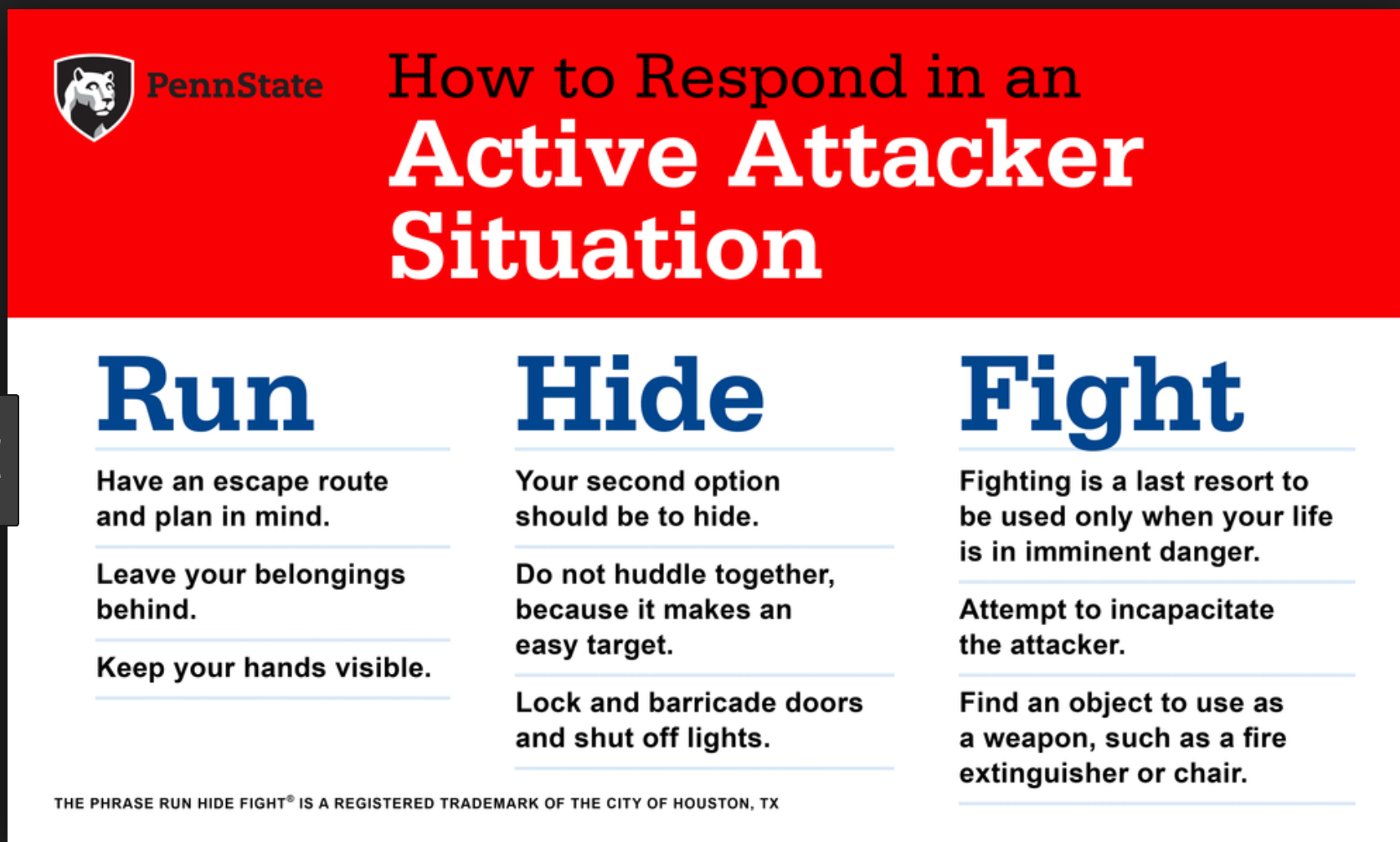
Dr. Regan says, if you hear shots and don’t see a shooter, run! Don’t wait for others or stop to get your stuff. Dr. Cranmer says, it is common to downplay or ignore popping sounds. Don’t do this, be situationally aware and pay attention to those no…

Dr. Regan says, if you hear shots and don’t see a shooter, run! Don’t wait for others or stop to get your stuff. Dr. Cranmer says, it is common to downplay or ignore popping sounds. Don’t do this, be situationally aware and pay attention to those noises. If you hear gunshots or popping noises, discontinue patient care. Don’t stop for victims or try to protect patients. If you die, you can’t help the patients later. Get out of there! Your safety takes priority over co-worker or patient safety. If you are running down a long straight hallway zig-zag down the hall to make it harder for the shooter to hit you. Do not run along the wall of a hallway. Bullets tend to ricochet and fly along the wall.

When you leave the building, keep your hands up and visible. Don’t have your cell phone in your hand. Don’t make any sudden movements. Don’t approach the police. Police are there to stop the shooter. You don’t want them to mistake you for the shooter. The meeting point outside our hospital for persons fleeing from an active shooter is the Ronald McDonald House.

If you are hiding and have a cell phone, text to a person outside the hospital to notify the police. Use any object in a room to barricade the door. Don’t open the door to let others in the room. If someone knocks on the door and says they are the police, ask them to slide their badge under the door or provide some other form of verification that they are the police before opening the door.

To report a shooter, call 41-1057 (Public Safety) and call 911.

If you have to fight the active shooter, work as a team to attack the shooter. Multiple people coming at the attacker at once is harder for them to fight back. Fight dirty and fight hard. Your life is on the line.

The inventory room is the safest room in the ED to hide/barricade in. It has heavy metal doors that can be locked. In the PED, the Inventory and storage rooms are the safer rooms with metal doors.

The notification for an active shooter overhead in the hospital will be: Security Alert-Active Shooter-Location. There also may be Everbridge text notifications.

Dr. Regan and Cranmer then covered specific scenarios and walked us through the ER to demonstrate escape routes and safe areas.

This workshop was a real eye-opener for ED staff. There was much discussion about the ethical choices of protecting your personal safety vs staying with patients. Dr. Regan and Cranmer both emphasized the importance of protecting your personal safety so you stay alive to care for patients.


Mullen Intracranial Hemorrhage

Epidural Hematoma.Blunt trauma to the temporal or temporoparietal area with an associated skull fracture and middle meningeal arterial disruption is the primary mechanism of injury. Occasionally, trauma to the parieto-occipital region or the posteri…

Epidural Hematoma.

Blunt trauma to the temporal or temporoparietal area with an associated skull fracture and middle meningeal arterial disruption is the primary mechanism of injury. Occasionally, trauma to the parieto-occipital region or the posterior fossa causes tears of the venous sinuses with epidural hematomas.

The classic history of an epidural hematoma involves a significant blunt head trauma with loss of consciousness or altered sensorium, followed by a lucid period and subsequent rapid neurologic demise. This clinical presentation occurs in a minority of cases. Traumatic blows to the thin temporal bone over the lateral aspect of the head carry the highest risk (e.g., baseball or pool stick injury). The diagnosis of an epidural hematoma is based on CT scan and physical examination findings. The CT appearance of an epidural hematoma is a biconvex (football-shaped) mass, typically found in the temporal region.

The high-pressure arterial bleeding of an epidural hematoma can lead to herniation within hours after an injury. Early recognition and evacuation reduces morbidity and mortality. Underlying injury of the brain parenchyma is often absent; full recovery may be expected if the hematoma is evacuated prior to herniation or the development of neurologic deficits. (Tintinalli 8th ed)

Subdural hematoma is caused by sudden acceleration-deceleration of brain parenchyma with subsequent tearing of the bridging dural veins. This results in hematoma formation between the dura mater and the arachnoid (Figures 257-8 and 257-9). Subdural …

Subdural hematoma is caused by sudden acceleration-deceleration of brain parenchyma with subsequent tearing of the bridging dural veins. This results in hematoma formation between the dura mater and the arachnoid (Figures 257-8 and 257-9). Subdural hematoma tends to collect more slowly than epidural hematoma because of its venous origin. However, subdural hematoma is often associated with concurrent brain injury and underlying parenchymal damage. Brains with extensive atrophy, such as in the elderly or in chronic alcoholics, are more susceptible to the development of acute subdural hematoma. Even seemingly benign falls from standing position can result in subdural bleeding in the elderly. Children <2 years old are also at increased risk of subdural hematoma.

Acute cases usually present immediately after severe trauma, and often the patient is unconscious. In the elderly or in alcoholics, chronic subdural hematomas may result in vague complaints or mental status changes. Often, there is no recall of injury. On CT scan, acute subdural hematomas are hyperdense (white), crescent-shaped lesions that cross suture lines. Subacute subdural hematomas are isodense and are more difficult to identify. CT scanning with IV contrast or MRI can assist in identifying a subacute subdural hematoma. A chronic subdural hematoma appears hypodense (dark) because the iron in the blood has been metabolized.

The definitive treatment depends on the type, size, effect on underlying brain parenchyma, and the associated brain injury. Mortality and the need for surgical repair are greater for acute and subacute subdural hematomas. Chronic subdural hematomas can sometimes be managed without surgery depending on the severity of the symptoms. (Tintinalli 8th ed.)

Subarachnoid hemorrhage. Only 1% of patients presenting to the ED with headache have subarachnoid hemorrhage. However, 10% to 14% of those complaining of the "worst headache of their life" have subarachnoid hemorrhage.7,8 Acute onset of a severe hea…

Subarachnoid hemorrhage. Only 1% of patients presenting to the ED with headache have subarachnoid hemorrhage. However, 10% to 14% of those complaining of the "worst headache of their life" have subarachnoid hemorrhage.7,8 Acute onset of a severe headache is subarachnoid hemorrhage until proven otherwise.10,11 With third-generation CT equipment, CT scan done within 6 hours of headache onset is reported to have a sensitivity approaching over 99% and specificity over 99%, with a negative predictive value of 99.4% and positive predictive value of 100%.46,48 If head CT is negative for blood but suspicion for subarachnoid hemorrhage is strong, or if the patient presents beyond 6 hours of headache onset, the next step is LP to detect blood or xanthochromia in the cerebrospinal fluid. (Tintinalli 8th ed.)


Allam Chest Radiology

Detection of free intraperitoneal air (pneumoperitoneum) is one of the principal uses of radiography in patients with abdominal pain. Pneumoperitoneum is nearly always due to perforation of the gastrointestinal tract, and virtually all patients requ…

Detection of free intraperitoneal air (pneumoperitoneum) is one of the principal uses of radiography in patients with abdominal pain. Pneumoperitoneum is nearly always due to perforation of the gastrointestinal tract, and virtually all patients require surgery. In 80–90% of cases, free intraperitoneal air is due to a perforated peptic ulcer. An upright chest radiograph is the preferred imaging test because it readily detects free air under the diaphragm. In most cases, the clinical and radiographic findings are obvious, and radiography serves to confirm the diagnosis. Diagnostic difficulty arises when the clinical presentation is muted, particularly in elderly or debilitated patients, or when the radiographic findings are subtle (Cina et al. 1994). Although radiography can detect small amounts of free air, not all cases of peptic ulcer perforation show free air—sensitivity may be as low as 60% (Emergency Radiology)

5 T’s that give you a wide mediastinum on CXR

Thyroid enlargement

Thymoma

Teratoma

Traumatic Aortic Injury

Terrible Lymphoma


Two things will “white out” a lung, pleural effusion and atelectasis. Pleural effusion shifts mediastinum away from the affected lung. Atelectasis pulls mediastinum toward the affected lung.

Two things will “white out” a lung, pleural effusion and atelectasis. Pleural effusion shifts mediastinum away from the affected lung. Atelectasis pulls mediastinum toward the affected lung.

Dr. Allam made the point that when checking NG tube placement, the NG tube should follow the spine until the NG gets below the diaphragm. If the NG tube is angling out laterally away from the spine in the chest you have to consider that it is in the…

Dr. Allam made the point that when checking NG tube placement, the NG tube should follow the spine until the NG gets below the diaphragm. If the NG tube is angling out laterally away from the spine in the chest you have to consider that it is in the lung. This CXR shows the NG tube in the left lung.

When checking CXRs with a pacemaker, follow the pacer wires from beginning to end to identify lead fracture. You have to look at the leads like you check ribs for fractures. You have to run your eyes over the entire wire.


Conference Notes 1-16-2019

Katiyar Billing and Coding Lecture

Unfortunately I missed this outstanding lecture.

Katiyar Study Guide Orthopedics

Remember that infection and cancer/leukemia are possible in all these age groups.

Remember that infection and cancer/leukemia are possible in all these age groups.

Pediatric Hip pain differential

Any visible posterior fat pad sign on the lateral elbow film is abnormal. A large anterior fat pad sign is also abnormal. If you see a posterior fat pad sign in a child think supracondylar fracture. A visible posterior fat pad sign in an adult is as…

Any visible posterior fat pad sign on the lateral elbow film is abnormal. A large anterior fat pad sign is also abnormal. If you see a posterior fat pad sign in a child think supracondylar fracture. A visible posterior fat pad sign in an adult is associated with radial head fracture.

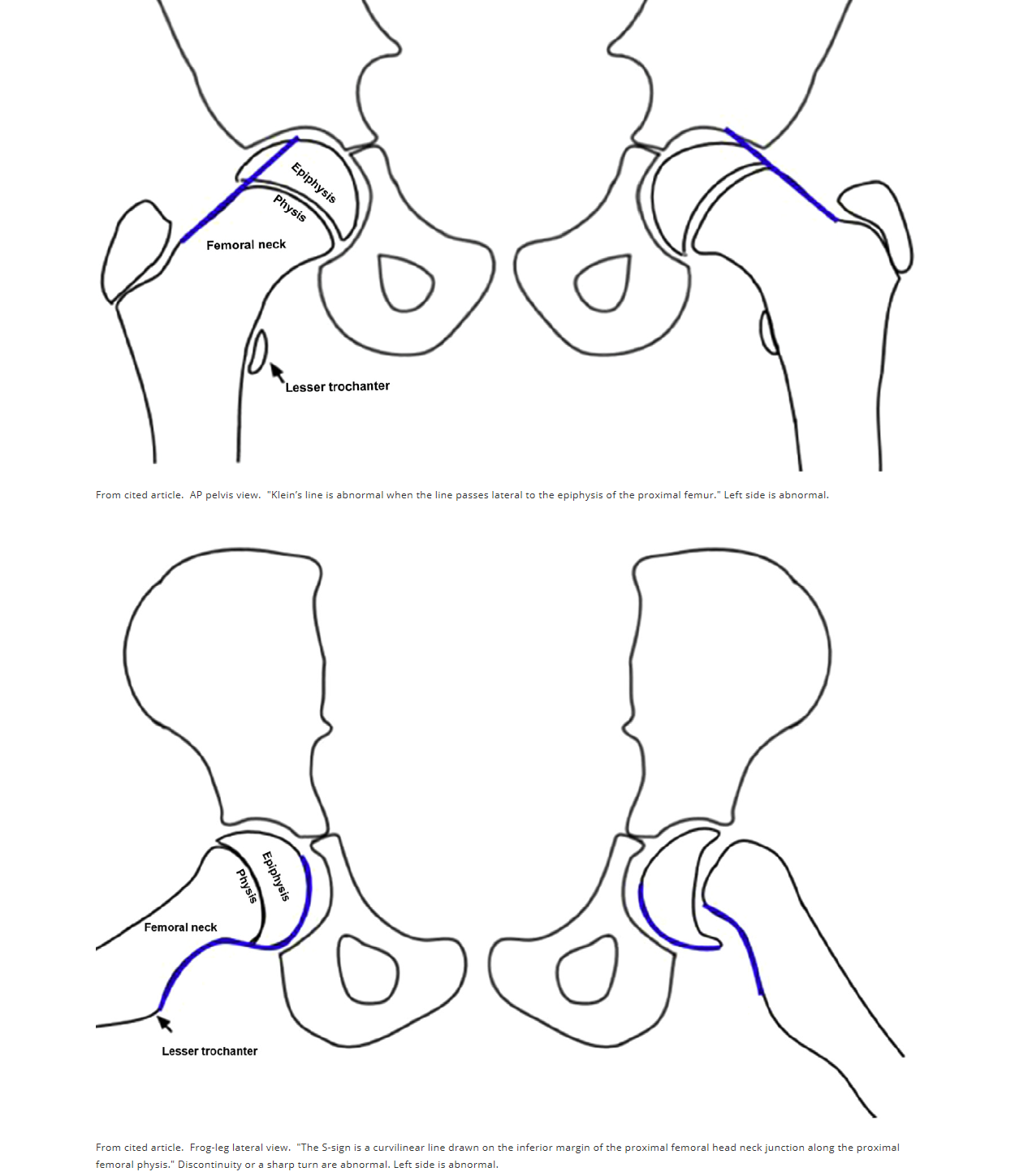
The S sign if normal is smooth with no step-off or sharp turns. An abnormal S sign has either a step off or an abrupt turn or buckle.Klein's line and S-sign A heterogeneous group of 20 orthopedic surgeons, radiologists, and pediatricians viewed 35 r…

The S sign if normal is smooth with no step-off or sharp turns. An abnormal S sign has either a step off or an abrupt turn or buckle.

Klein's line and S-sign
A heterogeneous group of 20 orthopedic surgeons, radiologists, and pediatricians viewed 35 radiographs of SCFE using Klein's line on the AP view and the S-sign on frog-leg lateral view to make the diagnosis. They found the overall diagnostic accuracy was better with the S-sign than Klein's line, 92% vs 79%. Sensitivity of the S-sign was 89%, specificity 95%. Sensitivity of Klein's line was 68%, specificity 89%. Combined S-sign + Klein's line sensitivity was 96%, specificity 85%. Take a look and review Klein's line for the AP view and the new S-sign for the frog-leg lateral.


Kennedy/Jurkovic Oral Boards

Case 1. 47 yo male presents with abdominal pain and hematemesis. Patient has history of ETOHism. On exam, patient was hypotensive and vomiting large volumes of blood. Diagnosis was life-threatening variceal bleeding. Management included crystalloid and blood product resuscitation. Intubation. octreotide, PPI, reverse coagulopathy, antibiotics, and blakemore tube. GI consult and emergent endoscopy were all also indicated. Consider vasopressin. If endoscopy is not successful, consider IR for TIPS procedure.

A restrictive transfusion threshold using hemoglobin concentrations of &lt;7 grams/dL in most patients and &lt;9 grams/dL in older patients with comorbidities who are not tolerating the acute anemia is recommended.5,31Patients with cirrhosis have an…

A restrictive transfusion threshold using hemoglobin concentrations of <7 grams/dL in most patients and <9 grams/dL in older patients with comorbidities who are not tolerating the acute anemia is recommended.5,31

Patients with cirrhosis have an impaired immune system and have an increased risk of gut bacterial translocation during an acute bleeding episode. Prophylactic antibiotics (e.g., ciprofloxacin 400 milligrams IV or ceftriaxone 1 gram IV) reduce infectious complications, rebleeding, days of hospitalization, mortality from bacterial infections, and all-cause mortality,42 and should be started as soon as possible. (Tintinalli 8th ed)

Case 2. 62yo male patient with altered mental status. BP=85/62. Heart rate 46. Patient had history of thyroidectomy and clinical picture of hypothyroidism. Diagnosis was myxedema coma with hypoglycemia, hypothermia, and bradycardia. . Patient was intubated and paced. Patient was given IV dextrose, IV Synthroid, and IV hydrocortisone. Beware of massive tongue in patients with myxedema coma. It may make intubation more difficult.

myxedema tx.PNG

Case 3. 27 yo female involved in a motorcycle crash. BP=165/105 P=105. Patient injured her wrist. X-ray shows perilunate dislocation. You can attempt reduction in the ED using finger traps. Most of these patients need surgery.

Perilunate dislocation. Note overlapping bones on AP view. On lateral view the capitate is posterior to the lunate and the lunate is in line with the radius.

Perilunate dislocation. Note overlapping bones on AP view. On lateral view the capitate is posterior to the lunate and the lunate is in line with the radius.

Perilunate dislocation highlighted on lateral view of wrist.. Note the capitate is posterior to the lunate and the lunate is still in line with the distal radius.Perilunate or lunate dislocations require emergency orthopedic/hand consultation.22 Tre…

Perilunate dislocation highlighted on lateral view of wrist.. Note the capitate is posterior to the lunate and the lunate is still in line with the distal radius.

Perilunate or lunate dislocations require emergency orthopedic/hand consultation.22 Treatment is determined by the extent of the injury. Closed reduction and long arm splint immobilization is appropriate for reducible dislocations.23 Open, unstable, and irreducible dislocations require open reduction and internal fixation, with repair of the ligaments and fractures. Some orthopedists operate on all perilunate and lunate dislocations.15 The complications include development of carpal instability patterns that lead to early degenerative arthritis, delayed union, malunion, nonunion, avascular necrosis, and, occasionally, median nerve compression from the volar dislocation of the lunate into the carpal tunnel.21

Goodmanson The Legend of the Ventilator

 Thanks to Dr. Lovell for writing the notes for this lecture!

Initial Settings: 

Lung Injury:

Mode:  AC

TV:  6-8 cc/kg IBW (protective)

Rate:  16  higher side....mid/high teens

FiO2:  start high, titrate down ASAP!

PEEP:  5 to start, may go up


-versus-

Obstructive Disease:

Mode:  VC

TV:  8 cc/kg IBW

Rate: 8-10 (lower, think permissive hypercapnea)

FiO2: start high, titrate down ASAP!

PEEP:  5 to start, may go down

Goals:  Higher flow rate, higher I:E ratio which is most impacted with slow RR


Increase oxygenation:  increase FiO2 or increase PEEP

Increase ventilation:  increase RR or TV, but really it’s all about the RR-shouldn’t exceed 8 cc/kg IBW


Airway Pressures:

Peak Pressure = Vt + Insp flow rate + airway resistance + lung compliance (focus on resistance and compliance)

Plateau Pressure = Vt + lung compliance (focus on lung compliance)


Hi peak + nml plateau = Hi resistance

Hi peak + Hi plateau = decreased compliance


Peak pressure continuously reported, but Plateau pressure obtained through inspiratory hold


The Crashing Ventilated Patient:  DOPES mnemonic


Think:

Dislodgement

Obstruction (of tube or of patient)

Pneumothorax or Patient (eg big PE)

Equipment failure

Stacked breaths (auto PEEP)


Do:

Disconnect from the ventilator (P, E, S)

Begin BVM ventilation

Feel, look, listen (D, O, P, S)

Pass a suction catheter or bougie through the ETT (O)

Consider needle/finger thoracostomy (P)

Consider manual expiration maneuver (S)


Auto-PEEP (aka breath stacking):

Look at Flow display on ventilator for incomplete emptying-expiratory flow is still occurring at the beginning of the next breath

Prevent by decreasing respiratory rate

Correct by disconnecting ventilator, manual expiration maneuver (lean on chest)

Remember, tension pneumothorax and breath stacking represent same end-pathophysiology (obstructive).  Use US if you have time, otherwise needle the chest bilaterally if patient coding.

�RzX��

Conference Notes 1-9-2019

Lorenz/Shroff STEMI Conference

There are numerous causes of ST elevation other than STEMI.

There are numerous causes of ST elevation other than STEMI.

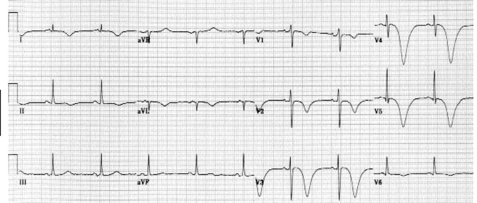
Pictured are Cerebral T waves which can be seen in subarachnoid hemorrhage. Subarachnoid hemorrhage can cause changes on the EKG such as deep t wave inversions or ST elevation.ECG Findings of Cerebral T WavesInverted, wide T waves are most notable i…

Pictured are Cerebral T waves which can be seen in subarachnoid hemorrhage. Subarachnoid hemorrhage can cause changes on the EKG such as deep t wave inversions or ST elevation.

ECG Findings of Cerebral T Waves

Inverted, wide T waves are most notable in precordial leads (can be seen in any lead).

QT interval prolongation.

Pearls

These are associated with acute cerebral disease, most notably an ischemic cerebrovascular event or subarachnoid hemorrhage.

They may be accompanied by ST segment changes, U waves, and/or any rhythm abnormality.

Differential diagnosis includes extensive myocardial ischemia.

Strongly suspect an intracranial etiology in a patient with altered mental status and these electrocardiographic findings. (Atlas of EM)


Catecholamine surge seems to be the pathophysiologic pathway of Takotsubo Cardiomyopathy. The LV apex is most sensitive to catecholamines. Takotsubo’s causes apical ballooning in response to a catecholamine surge.Features of takotsubo cardiomyopathy…

Catecholamine surge seems to be the pathophysiologic pathway of Takotsubo Cardiomyopathy. The LV apex is most sensitive to catecholamines. Takotsubo’s causes apical ballooning in response to a catecholamine surge.

Features of takotsubo cardiomyopathy.

Chest pain and shortness of breath after severe stress (emotional or physical)

Electrocardiogram abnormalities that mimic those of a heart attack

No evidence of coronary artery obstruction

Movement abnormalities in the left ventricle

Ballooning of the left ventricle

Recovery within a month

What is it?

Takotsubo cardiomyopathy is a weakening of the left ventricle, the heart's main pumping chamber, usually as the result of severe emotional or physical stress, such as a sudden illness, the loss of a loved one, a serious accident, or a natural disaster such as an earthquake. (For additional examples, see "Stressors associated with takotsubo cardiomyopathy.") That's why the condition is also called stress-induced cardiomyopathy, or broken-heart syndrome. The main symptoms are chest pain and shortness of breath. (Online Harvard Website)

Dr. Lovell comment: Pay attention to the details of managing the post-resuscitated cardiac arrest patient. Protect the airway. Avoid hyper/hypo-oxia and hyper/hypo-ventilation. Provide Targeted Temperature Management. Correct hypotension. Provide ASA and Heparin. Identify STEMI and discuss with cardiologist consideration of Cath lab.

ACC Risk Stratification Algorithm for Comatose Post-Cardiac Arrest Patients. Patients with multiple unfavorable characteristics are less likely to benefit from emergent cardiac cath.

ACC Risk Stratification Algorithm for Comatose Post-Cardiac Arrest Patients. Patients with multiple unfavorable characteristics are less likely to benefit from emergent cardiac cath.

Beware the Hyperacute T wave in the setting of chest pain. Hyperacute T waves may not always have ST elevation. The real key is the T wave’s size in relation to the QRS complex. This EKG was from a patient who went on to develop ST elevation and V-f…

Beware the Hyperacute T wave in the setting of chest pain. Hyperacute T waves may not always have ST elevation. The real key is the T wave’s size in relation to the QRS complex. This EKG was from a patient who went on to develop ST elevation and V-fib arrest. If you are suspicious of hyperacute T waves, get frequent repeat EKG’s to identify ST elevation or other evolution.

Dodd Research Lecture

Reasons to do research: Curiosity, Change the World, Change clinical care, Develop a device or drug, and Career advancement.

Dr. Dodd reviewed all the research opportunities available in our department.


Burns RUSH Exam

Mnemonic to remember all the aspects of the RUSH exam. HI-MAP ED: Heart, IVC, Morrisons, Aorta, Pneumothorax, Ectopic, DVT (common femoral veins)

You will used the phased array probe (cardiac probe) and the linear probe to get your views.

This is the order of images in the RUSH exam following the HI MAP mnemonic. You can add in suprapubic views looking for signs of ectopic pregnancy(pelvic fluid) and images of bilat femoral veins to screen for DVT.

This is the order of images in the RUSH exam following the HI MAP mnemonic. You can add in suprapubic views looking for signs of ectopic pregnancy(pelvic fluid) and images of bilat femoral veins to screen for DVT.

Lambert Transesophageal Echo in Cardiac Resuscitation

TEE has been shown to alter the resuscitation management of the cardiac arrest patient. TEE can improve the performance of CPR by identifying where chest compressions are impacting the heart. TEE will frequently give the resuscitationist information to improve the location of chest compressions on the chest. Without TEE info CPR may be only compressing the SVC or RV and not the LV.

TEE can also identify pericardial tamponade, valvular dysfunction, V-fib, and cardiac rupture.


Team Ultrasound Ultrasound Lab

blob

Write here…


blob

Write here…


blob


Conference Notes 12-19-2018

Girzadas Difficult Airway: Intubating the Morbidly Obese Patient

3 Major Concerns:

  1. Rapid Oxygen Desaturation. This is due to patients with high BMI having smaller lung volumes and lower functional residual capacity. Patients with high BMI also have a higher metabolic rate and are using oxygen more rapidly. Because of these factors they desaturate much more quickly. Your safe apnea time is about 75% less than a patient with normal BMI. You need to counter this problem by obsessive pre-oxygenation using 15L Nasal cannula and Flush Rate O2 via a NRB mask or BVM or Bipap. Have the patient’s head up as much as possible during pre-oxygenation.

One option to pre-oxygenate. Other options are NRB with wall O2 open to Flush Rate. Bipap is another excellent option if you have time and the patient can tolerate.

One option to pre-oxygenate. Other options are NRB with wall O2 open to Flush Rate. Bipap is another excellent option if you have time and the patient can tolerate.


2. More difficult airway management. This is due to larger tongue, more redundant tissue in the airway, thicker/less mobile neck. Patients have a smaller airway making visualization and intubation more difficult. Counter this problem with careful positioning. Elevate the head of the bed 25 degrees and put the patient in the sniffing position by elevating the head so the external ear canal is anterior to the sternum.

A is not good positioning. B and C both have the ear canal anterior to the sternum which will optimize your glotic view.

A is not good positioning. B and C both have the ear canal anterior to the sternum which will optimize your glotic view.

3. More difficult rescue of the failed airway. It is more difficult to BVM patients with high BMI. LMA is the preferred rescue device but airway pressures may cause the seal to fail. Finally cricothyrotomy is more difficult. You can counter this problem by planning your rescue prior to sedating the patient. Have an LMA at the bedside. Have a BVM with a Peep valve ready to go. Have an oral airway to facilitate bagging. Consider using ultrasound to identify where the cricothyroid membrane is and mark on the neck it’s location so if you have to perform a cric, you know exactly where to go.

Faculty members present felt that RSI after careful pre-oxygenation and positioning was still the best way to optimize your first pass success in the morbidly obese patient. Remember though that if you have significant concerns about how difficult the airway may be, you can always opt for topical anesthetic of the airway and ketamine sedation to avoid RSI.

Ahmad Study Guide Trauma

The safest initial therapy for symptomatic sucking chest wounds is the careful application of a petrolatum gauze–based dressing taped on three sides (Figure 53-4). Apply three or four layers of petrolatum gauze over the wound. The dressing should ex…

The safest initial therapy for symptomatic sucking chest wounds is the careful application of a petrolatum gauze–based dressing taped on three sides (Figure 53-4). Apply three or four layers of petrolatum gauze over the wound. The dressing should extend 6 to 8 cm beyond the margins of the wound so that it will not be sucked into the pleural cavity in the spontaneously breathing patient. Cover the petrolatum gauze with dry 4 × 4 gauze squares. Apply tincture of benzoin around three sides of the dressing. Apply tape to secure the three sides of the dressing to the chest wall. (Reichman’s EM Procedures 3rd Ed.) Editor’s note: Similar to the above picture, I would think that one of those pink defib conduction pads that you use for the hand-held defibrillator taped on three sides would work as well.

Small (<20%) pneumothoraces don’t necessarily need a chest tube. You can put the patient on supplemental oxygen and check another CXR in 6 hours. If it has improved, the patient can be discharged home.

Fracture of the sternum has been historically considered a marker of serious life-threatening injury, particularly cardiovascular injury. However, in clinical practice, it is the type of associated injury that determines morbidity and mortality. Patients with isolated sternal fractures and otherwise negative workup (including chest CT, echocardiogram, cardiac US, and cardiac enzymes at the time of presentation and 6 hours afterward) can safely be discharged home.42 Current experience with sternal fractures as a result of motor vehicle crashes notes a 1.5% incidence of cardiac dysrhythmias requiring treatment and a mortality rate of <1%.43 Such data suggest that sternal fractures are not an indicator of significant blunt myocardial injury. Patients with sternal fractures presenting with normal vital signs and an initial normal ECG should have a repeat ECG in 6 hours and, if unchanged, require no further workup for cardiac injury.44,4 (Tintinalli 8th edition)

Whether a normal chest radiograph excludes an aortic injury is a matter of controversy. One careful review of radiographs in patients with aortic injury did not find any normal radiographs. However, this study excluded technically suboptimal radiographs, was conducted by expert trauma radiologists, and the radiographic findings were subtle (Figures 14 and 15) (White et al. 1994). In two large clinical series, 5–7% of aortic injury cases had radiographs that were interpreted as negative for aortic injury (Fabian et al. 1997, Hunt et. al. 1996). However, the quality of the radiographic technique in these cases was not mentioned, and the radiographic criteria used to assess aortic injury were not stated. Several other case series also report normal mediastinal appearance in patients with aortic injury (Exadaktylos et al. 2001, Woodring 1990). Only when a chest radiograph is of sufficient quality to clearly determine that the mediastinal contours are normal could it be considered adequate to exclude a mediastinal hematoma (Mirvis 2006). However, in one study, 10% of aortic injury cases do not have a mediastinal hematoma on CT and so even a normal, technically optimal chest radiograph should not be used alone to exclude aortic injury (Cleverley et al. 2002).

Although of limited value in excluding aortic injury, the chest radiographic signs of mediastinal blood should be recognized and, when present, should prompt a rapid definitive investigation for aortic injury.

Definite signs of hemomediastinum in a patient with aortic injury.The mediastinum is widened and the aortic knob is distorted by surrounding blood (arrow).Mediastinal blood causes widening of the right paratracheal stripe (white arrowheads) and disp…

Definite signs of hemomediastinum in a patient with aortic injury.

The mediastinum is widened and the aortic knob is distorted by surrounding blood (arrow).

Mediastinal blood causes widening of the right paratracheal stripe (white arrowheads) and displacement of the left paraspinal line (black arrowheads), which extends up to the aortic knob.

The trachea is displaced to the right (white asterisks) and the left mainstem bronchus is displaced inferiorly (blackasterisks). This is due to blood surrounding the aorta. The faint shadow of the SVC is visible to the right of the paratracheal stripe. (The aortogram of this patient is shown in Figure 5.)

[From: Schwartz DT, Reisdorff EJ: Emergency Radiology. McGraw-Hill, 2000.]

CaptureAortic injury findings.PNG


Allam Chest Radiography

CaptureRight lung fissures.PNG


Calcified lung nodules are almost always benign.

Cavitary lesions can be lung cancer, metastasis, abscess, TB, Fungal or Wegener’s.

Opacities are bright areas on CXR. Lucencies are dark areas on CXR. Infiltrates is a term that is not well-defined and Dr. Allam advised us to avoid that term.

The causes of opacities on CXR include pus (pneumonia), blood, or water.

CaptureLung anatomy.PNG
Lingular pneumonia. If the left heart border is obscured, it is due to lingular pneumonia. If the left diaphragm is obscured it is due to left lower lobe pneumonia or atelectasis.

Lingular pneumonia. If the left heart border is obscured, it is due to lingular pneumonia. If the left diaphragm is obscured it is due to left lower lobe pneumonia or atelectasis.

Honeycombing pattern c/w Pulmonary Fibrosis. Interstitial pulmonary fibrosis (IPF), also called Usual Interstitial Pneumonitis, results in a restrictive lung disease that diminishes lung compliance and restricts diffusion of oxygen across the tissue…

Honeycombing pattern c/w Pulmonary Fibrosis. Interstitial pulmonary fibrosis (IPF), also called Usual Interstitial Pneumonitis, results in a restrictive lung disease that diminishes lung compliance and restricts diffusion of oxygen across the tissue to the blood. Over time, the disease leads to impaired exercise tolerance, chronic hypoxia, and cough. Treatment is aimed at immunomodulation through steroids and cytotoxic agents to halt disease. While late IPF is easily identified on chest radiographs, early disease is difficult to distinguish from other interstitial processes, such as CHF.

Pearls

Pulmonary fibrosis has a classic appearance of interstitial markings more prominent at the bases.

If pulmonary fibrosis is suspected, further evaluation should be done via high-resolution protocol chest CT to further determine the etiology. (Atlas of Emergency Radiology)

Deep sulcus sign

Deep sulcus sign indicative of pneumothorax in supine patients. The air accumulates inferiorly rather than superiorly as it would in an upright patient.

Deep sulcus sign indicative of pneumothorax in supine patients. The air accumulates inferiorly rather than superiorly as it would in an upright patient.

Humphrey/Delbar/Kentor Chest Trauma

East Guidelines for Blunt Cardiac Injury

Level 1

  1. An admission electrocardiogram (ECG) should be performed on all patients in whom BCI is suspected (no change).

Level 2

  1. If the admission ECG reveals a new abnormality (arrhythmia, ST changes, ischemia, heart block, and unexplained ST changes), the patient should be admitted for continuous ECG monitoring. For patients with preexisting abnormalities, comparison should be made to a previous ECG to determine need for monitoring (updated).

  2. In patients with a normal ECG result and normal troponin I level, BCI is ruled out. The optimal timing of these measurements, however, has yet to be determined. Conversely, patients with normal ECG results but elevated troponin I level should be admitted to a monitored setting (new).

  3. For patients with hemodynamic instability or persistent new arrhythmia, an echocardiogram should be obtained. If an optimal transthoracic echocardiogram cannot be performed, the patient should have a transesophageal echocardiogram (updated).

  4. The presence of a sternal fracture alone does not predict the presence of BCI and thus should not prompt monitoring in the setting of normal ECG result and troponin I level (moved from Level 3).

  5. Creatinine phosphokinase with isoenzyme analysis should not be performed because it is not useful in predicting which patients have or will have complications related to BCI (modified and moved from Level 3).

  6. Nuclear medicine studies add little when compared with echocardiography and should not be routinely performed (no change).

    Trauma attending comment: Not recommended to get a troponin unless there is a new EKG abnormality, arrhythmia, or hemodynamic changes. Editors comment: This is a controversial topic. I think many clinicians would consider getting a troponin in a patient with concern for possible blunt cardiac injury.

CaptureNexus chest rule.PNG

Trauma Attending comment: Treat hemothorax with a 32-36F chest tube. Indications for going to the OR is 1500ml of blood out immediately or 250ml/hr for four hours.

High flow nasal cannula O2 may benefit patients with flail chest by providing some level of Peep. Bipap can also be used to support alert, cooperative flail chest patients.

There are a few well-supported contraindications to performing an ED thoracotomy. A thoracotomy should not be performed in trauma patients who have no vital signs in the field.15,39,40 In the absence of field vitals, the survival rates are extremely low and the few who survive have severe neurologic impairment. Outside of patients who collapse in the ED, victims of blunt trauma with or without field vitals should not routinely undergo an ED thoracotomy.40,41 It is also contraindicated when prehospital CPR exceeds 10 minutes without a return of spontaneous circulation after blunt trauma, when prehospital CPR exceeds 15 minutes without a return of spontaneous circulation after penetrating trauma, and when the patient presents to the ED in asystole without a pericardial tamponade.39-43 An ED thoracotomy should not be performed regardless of the indications if a Trauma Surgeon or other qualified Surgeon is not available to take the patient to the Operating Room for definitive management. Do not perform an ED thoracotomy with the anticipation of transferring the patient to another facility if they can be resuscitated. In the near future, resuscitative endovascular balloon occlusion of the aorta (REBOA) may make some indications for a thoracotomy into contraindications (Chapter 74). (Reichman’s EM Procedures 3rd ED.)

Trauma Attending comment: It should be very rare for an emergency physician to perform an emergency thoracotomy. There are many risks to the patient and the physician when performing this procedure.

Harwood comment: Never do an emergency thoracotomy for blunt trauma or GSW to the chest, or non-chest trauma. But if there is an isolated stab wound to the region of the heart and patient has signs of life that may be the one time to go for it.

Trauma Attending comment: If you do open the chest and find a pericardial tamponade. Be very careful opening the pericardium to avoid cutting the phrenic nerve. Repair the ventricular injury with stables rather than sutures or foley balloon tamponade. Sutures are difficult to place in the heart and a foley balloon tends to tear through the hole making it larger.

Patients with aortic injuries that survive to the hospital get delayed repair. The Trauma surgeons fix all the other injuries first then a day or two later do an endovascular repair.

Conference Notes 12-12-2018

Regan/Twanow Oral Boards

Case 1. 65 yo male presents with difficulty breathing. The patient is tachycardic and tachypneic. Patient has history of COPD. Patient has had worsening cough over last few days. Patient also had left sided chest pain. Patient has valid DNR/DNI order. CXR shows a left sided pneumothorax. Patient was treated with a chest tube and standard therapy for COPD.

CapturePneumo.PNG

COPD is the most common risk factor for pneumothorax. Usually pneumothoraces in COPDr’s are due to apical blebs that rupture. Dr. Lovell comment: Be cautious not to diagnose an intact bleb as a pneumothorax. You could inadvertently place a chest tube into an intact bleb and cause a pneumothorax. You may need a CT to differentiate pneumothorax from a bleb.

Case 2. 32 yo female with hip and pelvic pain due to an MVC. Patient is tachycardic and hypotensive.

Captureopenbook pelvic fracture.PNG

Patient had an open book pelvic fracture. Pelvic binder applied. Crystalloid infused and Massive transfusion protocol activated. Transfuse in a 1:1:1 ratio of prbcs, platelets, and plasma. Pelvic ring fractures have the highest risk of hemorrhage. After intra-abdominal hemorrhage has been ruled out, IR should be utilized to stabilize patients with hemorrhage from pelvic fracture. IR stops arterial hemorrhage related to pelvic fracture but not venous hemorrhage.

Case 3. 43yo male presents with bleeding gums and oral pain. Patient is febrile.

Vincent angina is a polymicrobial infection, typically limited to the gingiva, and characterized by foul breath, cervical lymphadenopathy, and fever. In immunocompromised individuals, it may extend to include a necrotic gray pseudomembrane on the ph…

Vincent angina is a polymicrobial infection, typically limited to the gingiva, and characterized by foul breath, cervical lymphadenopathy, and fever. In immunocompromised individuals, it may extend to include a necrotic gray pseudomembrane on the pharynx. (Current Diagnosis and Treatment Emergency Medicine)

Treat with Augmentin,clindamycin or doxycycline and chlorhexidine rinses.

Chastain Study Guide Endocrine Emergencies

Hypokalemia is the most dangerous electrolyte abnormality when treating DKA. Start repleting K with a K < 5.3. If K<3.5, don’t give insulin until you have started KCL infusion.

Anti-seizure medications, steroids, neuroleptics, ASA, betablockers, and calcium channel blockers can all affect glycemic control.

When treating DKA in kids, if they become altered, think cerebral edema. If iatrogenic hypoglycemia has been ruled out, start mannitol even prior to CT.

Patients with adrenal crisis will have hypotension, hypoglycemia, hyponatremia/hyperkalemia, weakness and diarrhea.

Patients with adrenal crisis will have hypotension, hypoglycemia, hyponatremia/hyperkalemia, weakness and diarrhea.

When choosing a steroid to administer for adrenal insufficiency, hydrocortisone has equal glucocorticoid and mineralocorticoid activity. Hydrocortisone is the steroid drug of choice for cases of adrenal crisis or insufficiency because it provides both glucocorticoid and mineralocorticoid effects. IV hydrocortisone (100-milligram minimum bolus) can be administered. (Titinalli 9th ed.)

Dexamethasone is the only steroid that does not interfere with the cortisol assay or cosyntropin test.

Thyrotoxicosis is a hypercoaguable state. So have your guard up for PE in patients with thyrotoxicosis and hypoxia.

Overview of therapy for thyroid storm. (Tintinalli 9th ed.)

Overview of therapy for thyroid storm. (Tintinalli 9th ed.)

Jones First Trimester Vaginal Bleeding

To optimize patient safety, consider every female ED patient pregnant until proven otherwise.

Next approach every pregnant ED patient as if they have an ectopic pregnancy until proven otherwise.

The most common site of ectopic pregnancy is the ampulla. Ectopic pregnancy is the leading cuase of first trimester maternal death.

Capturerisk for ectopic.PNG

Write here…

Emergency physicians should be extremely cautious about ordering methotrexate to treat ectopic pregnancy. You need to have very careful consultation with OB-Gyne consultant prior to administration. It may be preferable to defer entirely to OB-Gyne s…

Emergency physicians should be extremely cautious about ordering methotrexate to treat ectopic pregnancy. You need to have very careful consultation with OB-Gyne consultant prior to administration. It may be preferable to defer entirely to OB-Gyne specialist. Harwood comment: The failure rate of methotrexate is 14% with rupture. In the setting of a pelvic ultrasound showing no IUP, the new ACOG Guideline recommends not diagnosing ectopic pregnancy until the level of quantitative beta-hcg is above 3000.

Definitions

Spontaneous abortion: Estimated 20% of pregnancies terminate in abortion. One-half occur before 8 weeks’ gestation and one-fourth before 16 weeks’ gestation. Many go unnoticed and unrecognized. This is a common cause for visit to the emergency department.

Complete abortion: Fetal demise and all products of conception are spontaneously expulsed.

Missed abortion: Fetal demise and failed expulsion of the products of conception from the uterus, with a closed cervix. If the condition lasts longer than 4-6 weeks, the patient is increased risk for infection and DIC.

Incomplete abortion: Incomplete expulsion of the products of conception. There is retained products of conception. The cervix is open.

Threatened abortion: Gestation has not reached the stage of viability (< 20 weeks). Patient may have pelvic pain and some vaginal bleeding, or any of the above symptoms. US may show a gestational sac and evidence of fetal cardiac activity. (Current Diagnosis and Treatment, Pediatric EM)

Harwood comment: There is no real role for 50microgram dosing of RH negative pregnant patients in the ED with vaginal bleeding or trauma. Give all RH negative patients with an indication for rhogam 300 micrograms.

Kentor Anaphylaxis in the ED

Unfortunately I missed a large portion of this outstanding lecture.

CaptureAnaphylaxis.PNG

Write here…

Captureana;hylaxistx.PNG


Is there such a thing as biphasic anaphylaxis?
This was a retrospective review of 83 patients at a single center with severe anaphylaxis, requiring ICU admission. Of these, 99% received antihistamines, 90% steroids, and just 80% epinephrine. One patient out of 83 (1.2%) had probable biphasic allergic reaction, and this was merely skin changes, not life-threatening and not anaphylactic, according to the study definition. Of the 3 possible and 1 probable biphasic allergic reactions, they occurred at an average of 14 hours from the initial reaction. This study had some issues. The most important is that of patients with severe anaphylaxis, 20% didn’t receive the most important therapy - epinephrine. So, even with suboptimal treatment, the incidence of biphasic allergic reaction within 72 hours was very low and the single probable reaction was mild. Another issue is the retrospective nature was limited in determining what was and was not an allergic reaction, such as rash or hypotension for other reason, such as sepsis. My take home point is that true biphasic anaphylactic reaction is rare or non-existent. Most patients simply need to be observed a few hours to ensure they don’t have persistent anaphylaxis, especially those with ingestion of a food as the cause of the reaction.

Another Spoonful
The Skeptics Guide to EM did an in-depth, outstanding summary of biphasic anaphylaxis. Their bottom line: “Prolonged observation is likely unnecessary,” once symptoms resolve in the ED. Don’t miss this post.

Source
Low Incidence of Biphasic Allergic Reactions in Patients Admitted to Intensive Care after Anaphylaxis. Anesthesiology. 2018 Nov 5. doi:

(Journal Feed 12-6-2018)

Lorenz/Shroff Christmas Cases

Indications for emergent esophageal FB removal are complete obstruction, battery in the esophagus, and sharp object in esophagus.

The initial fluid rate used for burn resuscitation has been updated by the American Burn Association to reflect concerns about over-resuscitation when using the traditional Parkland formula. The current consensus guidelines state that fluid resuscitation should begin at 2 ml of lactated Ringer’s x patient’s body weight in kg x % TBSA for second- and third-degree burns. The calculated fluid volume is initiated in the following manner: one-half of the total fluid is provided in the first 8 hours after the burn injury (for example, a 100-kg man with 80% TBSA burns requires 2 × 80 × 100 = 16,000 mL in 24 hours). One-half of that volume (8,000 mL) should be provided in the first 8 hours, so the patient should be started at a rate of 1000 mL/hr. The remaining one-half of the total fluid is administered during the subsequent 16 hours. (ATLS 10th edition)

ATLS manual 10th edition

ATLS manual 10th edition

Calculating burn area based on rule of 9’s. ATLS manual 10th edition

Calculating burn area based on rule of 9’s. ATLS manual 10th edition

Putman ENT Emergencies

Mumps

Capturemumps.PNG

VIRAL PAROTITIS (MUMPS)

Viral parotitis is an acute infection of the parotid glands, characterized by unilateral or bilateral parotid swelling. It is most often caused by the paramyxovirus and may be caused less commonly by influenza, parainfluenza, coxsackie viruses, echoviruses, lymphocytic choriomeningitis virus, and even human immunodeficiency virus.25 It is most common in children under the age of 15 years old, but since November 2014, clusters of mumps have been reported in adult members of professional hockey teams. The virus is spread by airborne droplets, incubates in the upper respiratory tract for 2 to 3 weeks, and then spreads systemically. Vaccine protection is not 100%, and outbreaks occur in settings of close contact, such as schools, colleges, sports teams, and camps.26

After a period of incubation, one third of patients experience a prodrome of fever, malaise, headache, myalgias, arthralgias, and anorexia during a 3- to 5-day period of viremia.25 The classic salivary gland swelling then follows. Unilateral swelling is typically followed by bilateral parotid involvement. The gland is tense and painful, but erythema and warmth are notably absent. Stensen's duct may be inflamed, but no pus can be expressed.25

Diagnosis is clinical and treatment is supportive. Salivary gland swelling typically lasts from 1 to 5 days. The patient is contagious for 9 days after the onset of parotid swelling, and children with mumps should be excluded from school or day care for this interval.

Mumps is usually benign in children but can be severe in adults. Unilateral orchitis affects 20% to 30% of males (with a predisposition of ≥8 years of age), whereas oophoritis affects only 5% of females. Other complications of the mumps virus include mastitis, pancreatitis, aseptic meningitis, sensorineural hearing loss, myocarditis, polyarthritis, hemolytic anemia, and thrombocytopenia.25 Immunocompetent patients with isolated viral parotitis or orchitis can be managed as outpatients. Admit patients with systemic complications.(Tintinalli 8th ed)

Peritonsillar abscess drainage

Some tips: the carotid is posterior-lateral to the tonsil, so keep your needle directed posteriorly and medial to the molars.

The cavitary U/S probe can be very helpful to localize the abscess.

Using an 18 gauge 3 cm spinal needle is useful because compared to a standard 18g needle, the longer needle keeps the syringe at or distal to the patient’s teeth. This improves visibility of the peritonsillar area, gives an increased range of movement, and has more utility in the setting of trismus. You have to use an 18g needle to be able to aspirate pus. Smaller needles may not be able to aspirate pus.

You can get good exposure by having the patient use a laryngoscope or lighted disposable vaginal speculum to hold down their own tongue.

Trim the needle cap and place it over the needle to act as a depth gauge (Figure 175-5A). The needle should project only 1 cm from the distal end of the needle cap. Alternatively, apply a piece of tape onto the needle to mark a point 1 cm from the t…

Trim the needle cap and place it over the needle to act as a depth gauge (Figure 175-5A). The needle should project only 1 cm from the distal end of the needle cap. Alternatively, apply a piece of tape onto the needle to mark a point 1 cm from the tip of the needle (Figure 175-5B). The guard (cap or tape) serves as a marker for the maximum allowable depth to insert the needle during the procedure. Limiting of the depth of insertion of the needle will prevent injury to the carotid artery that is located approximately 1.5 to 2 cm posterior and lateral to the tonsil.

Conference Notes 12-5-2018

Barounis/Dodd/Ketterhagen Central Line Simulation Lab

Our awesome EM Intensivists taught us how to optimally place a central line. I can’t do justice to all the teaching they did but here are just a few fine points I picked up.

When putting your sterile gown on by yourself, Velcro the neck first before you put your arms through the sleeves.

The plastic adhesive strips in the sterile probe cover package can be used to tape the probe cord to the drape so the probe doesn’t fall off the patient.

Line up bevel of needle with the numbers on the barrel of the syringe so you know where the bevel is.

When you are locating the IJ in the neck, keeping the probe pointed at the floor instead of aiming it toward the neck will help keep the orientation of the IJ and IC more true.

Once you have punctured the skin, move the ultrasound probe with your needle so that you always have the tip in view and can visualize exactly where you enter the vessel.

When making the cut with a scapel put the blunt side of the scapel towards the wire.


Hawkins Case Presentation and Ortho Xrays

45 yo male presents with altered mental status. Patient was reported to be intoxicated and vomiting. In ED patient was found to have head laceration and rash. He was hypotensive and tachycardic.

Patient was in detox recently and was on multiple medications including antabuse.

DDX=disulfiram reaction, ETOH intoxication, serotonin syndrome, ICH, toxic ingestion, dehydration, sepsis, anaphylaxis.

Patient had improving blood pressure and rash with IV fluids. Mental status worsened with a GCS of 7-8. Patient is next intubated.

Disulfiram reactions can result in tachycardia, hypotension and cardiovascular collapse.

Other tha ETOH, disulfiram reactions can also be caused by chlorpropamide, flagyl and griseofulvin.

The duration of the disulfiram-alcohol reaction varies from 30 to 60 minutes in mild cases to several hours and is largely dependent on the amount of alcohol that needs to be metabolized. Due to vomiting and volume depletion, serum glucose, electrolytes, and kidney function should be evaluated. Since only small amounts of ethanol can precipitate a disulfiram–ethanol reaction, it may be useful to confirm the presence of ethanol with a blood concentration. Patients with cardiovascular instability should have an ECG. Symptomatic and supportive care is the mainstay of treatment. Most patients with hypotension respond to intravenous 0.9% sodium chloride. Refractory hypotension is rare, but if necessary, a vasopressor can be administered. A direct-acting adrenergic agonist such as norepinephrine should be used since disulfiram inhibits dopamine β-hydroxylase (DBH), an enzyme necessary for norepinephrine synthesis. As such, indirect vasopressors, such as dopamine, that require functioning norepinephrine synthesis may be less effective.

For further symptomatic care, antiemetics can be administered, and for cutaneous flushing, a histamine (H1) receptor antagonist, such as diphenhydramine, can be given.101 Most patients with a disulfiram–ethanol reaction have mild symptoms, are hemodynamically stable, and can be safely discharged following resolution of symptoms.

Fomepizole may halt the accumulation of acetaldehyde and thus cease severe disulfiram–ethanol reactions. Fomepizole, an inhibitor of alcohol dehydrogenase, may terminate the progression of the disulfiram reaction by blocking ethanol metabolism to acetaldehyde (Antidotes in Depth: A30). In a study of alcoholics, fomepizole decreased acetaldehyde concentrations and improved clinical symptoms in those expe­ri­enc­ing a disulfiram–ethanol reaction.59 A recent case series reported two patients who developed severe disulfiram–ethanol reactions with hypotension and tachycardia unresponsive to fluids who were treated successfully with a single dose of fomepizole.97 One patient improved clinically 90 minutes after administration of fomepizole and the other within 30 minutes. (Goldfrank’s Toxicologic Emergencies)


This is an avulsion of the lateral capsule and is called a Segond fracture. It has a very high (&gt;90%) association with a tear of the anterior cruciate ligament. (Atlas of Emergency Radiology)

This is an avulsion of the lateral capsule and is called a Segond fracture. It has a very high (>90%) association with a tear of the anterior cruciate ligament. (Atlas of Emergency Radiology)

Segond Fracture is associated with an ACL tear


Montaggia vs Galeazzi

Both Monteggia and Galeazzi fracture-dislocations require emergent orthopedic consultation and are treated with immobilization in a long-arm splint (with elbow flexed at 90 degrees). The forearm is placed in a neutral position for a Monteggia fractu…

Both Monteggia and Galeazzi fracture-dislocations require emergent orthopedic consultation and are treated with immobilization in a long-arm splint (with elbow flexed at 90 degrees). The forearm is placed in a neutral position for a Monteggia fracture and supinated for a Galeazzi fracture. Treatment is usually surgical for both injuries, although children may be treated by reduction and casting. (The Atlas of Emergency Medicine)


Katiyar Toxicologic Emergencies Hallucinogens

Hallucinogenic compounds all have similar chemical structure.

Hallucinogenic compounds all have similar chemical structure.


Hallucinogenic botanicals include morning glory and Hawaiian baby woodrose.

Magic Mushrooms are a source of psilocybin.

Ayahuasca and the Yakee plant are plants in the amazon that contain the compound N,NDMT a potent short-acting hallucinogen.

Toad licking can cause hallucinogenic effect. (Colorado toad). Toad licking also can cause arrythmias.

in one species of toad, Bufo alvarius (Sonoran Desert toad or Colorado River toad).77 Although bufotenine has been classified as a Schedule I substance by the DEA for many years, 5-MeO-DMT was not scheduled until 2009.91 Like DMT, 5-MeO-DMT is rapidly metabolized by intestinal monoamine oxidase enzymes; oral ingestion of toad venom or skins would thus have limited potential as a route of recreational use.21 Methods for extracting and drying B. alvarius secretions for smoking and insufflation are available on the Internet. Death has resulted from wrongful use of Bufo secretions for purposes of aphrodisia.30,55 The toad venom glands also produce cardioactive steroids, called bufadienolides, which cause digoxinlike cardiac toxicity, and in some species, can secrete tetrodotoxin.87,139 (Goldfrank’s Toxicologic Emergencies)


Peyote and mescaline are other hallucinogens. Some cacti available on line contain peyote or mescaline.

Salvia is from the mint family and can be purchased on the internet or at a garden store. The leaves contain hallucinogens.

Nutmeg contains a hallucinogen.

Most hallucinogens affect the serotonin receptors. There may also be sympathomimetic effects.

Hallucinogens are not routinely identified on urine toxicology screens.

Treat hallucinogenic overdoses with benzodiazepines, cooling, and quiet environment.


Katiyar Toxicologic Emergencies Hypoglycemic Agents

Ackee fruit from Jamaica can cause hypoglycemia.

Sulfonylurea medications increase insulin secretion from pancreatic beta cells.

Metformin inhibits gluconeogenesis, enhances glucose transport into muscles. Decreases glucose being released from liver.


Mild changes in renal function or a new drug interaction can be causes of new onset hypoglycemia.

A single large injection of insulin is more dangerous than multiple smaller doses because a large dose creates a “depot effect”. The “depot effect” will result in prolonged release of insulin.


Insulinoma and sulfonylureas will result in elevated c-peptide levels. Synthetic insulin does not have c-peptide.


Treat sulfonylurea toxicity with glucose and food. Also start octreotide. Check the blood sugar Q1 hour. Keep for OBS or admit


Estoos/Kishi/Miner Trauma Lecture: Neck and Above


In the hypotensive trauma patient, consider hemorrhage as #1 cause. After hemorrhage, consider obstructive causes of shock (tamponade or tension pneumothorax).


When exposing the patient make sure you examine the whole body back and front looking for subtle injuries. At the same time consider environmental exposures such as acids, bioterrorism agents, or radioactive contamination. After fully exposing the patient, keep patient warm with blankets and heat to avoid hypothermia.

The GCS is a critical means of communicating the neurologic status of the patient between caregivers as well as a way to monitor the patient’ neurologic status.

The GCS is an objective measurement of clinical status, correlates with outcome, is a reliable tool for interobserver measurements, and is effective for measuring patient recovery or response to treatment over time. However, the scale has several li…

The GCS is an objective measurement of clinical status, correlates with outcome, is a reliable tool for interobserver measurements, and is effective for measuring patient recovery or response to treatment over time. However, the scale has several limitations. It measures behavioral responses, not the underlying pathophysiology. Patients with similar GCS scores may have dramatically different underlying structural injuries and require different clinical interventions (Figure 257-2). It is not as useful as a single acute measure of severity as it is as a tool to measure disease progression over time. The GCS may additionally be affected by drugs, alcohol, medications, paralytics, or ocular injuries. Finally, the scale lacks the granularity necessary to assess mTBI. (Tintinalli 8th edition)


Key ED goal for head trauma management is to avoid hypotension and hypoxia. Elevate the head of the bed. Mannitol or hypertonic saline may be used to lower ICP. Avoid mannitol in patients with renal failure. Avoid hypertonic saline in patients with CHF.

Basilar skull fracture=temporal bone fracture.

Subdural hematomas have significant underlying brain injury. The blood collection can cross suture lines. It is venous bleeding from bridging veins.

Subdural hematomas have significant underlying brain injury. The blood collection can cross suture lines. It is venous bleeding from bridging veins.


Traumatic subarachnoid hemorrhage

Traumatic subarachnoid hemorrhage

Traumatic subarachnoid hemorrhages as opposed to aneurysmal subarachnoid hemorrhage are more peripherally located.


Epidural hematoma

Epidural hemaoma

Epidural hemaoma

Traumatic intraparenchymal hemorrhages are often in frontal or temporal lobes.

DAI has a benign appearing CT head/MRI with a severely affected GCS/mental status exam. Some DAI patients will improve significantly within a year or so. Prognosis is difficult to predict.

Patients with head injury have 50% increased mortality with single episode of hypotension.

Do not use therapeutic hypothermia in traumatic head injury. Recent RCT showed no benefit and trauma surgeons emphasized that hypothermia increases traumatic bleeding.

When intubating patients with significant head injury and you don’t have to do a crash or life-saving emergent intubation, be very careful to avoid any desaturation or hypotension during the intubation.

Unfortunately I missed a significant portion of this outstanding lecture.


Conference Notes 11-28-2018

Lorenz/Shroff STEMI Conference

cardshok.PNG

Be very cautious giving IV beta blockers to patients with decreased cardiac function. Patients with diminished cardiac function can deteriorate with IV beta blockers. Determining a patient’s cardiac function can be challenging in the acute setting. Consider checking a bedside echo prior to giving a rate control agent. Start with a low dose and re-evaluate closely. During the discussion of this clinical situation, the Cardiologist at the meeting voiced a contrarian view and felt this was an overly cautious approach and that most patients can tolerate reasonable doses of rate control agents.

Tekwani comment: Get an ABG or VBG early on to see the lactate. It may clue you in to a hypoperfused state or cardiogenic shock. Harwood comment: A VBG also gets you a rapid potassium level.


scad.PNG

Write here…

scad 2.PNG


Cardiology comments: We try to avoid cath in patients with coronary artery dissection because cath can propagate the dissection. Most patients are treated with anticoagulation. I f a patient has a new right bundle branch block and left anterior fasicular block, be concerned for a proximal LAD occlusion and need for a pacemaker. Post-partum patients who develop coronary artery dissection should be counselled to not have further pregnancies. Cardiac surgery can be helpful at times to manage SCAD. There was consensus among the cardiologists that managing coronary artery dissections is very treacherous and cardiac cath is not a slam dunk. If you have a post-partum patient presenting to the ED with a STEMI you should activate the cath lab but discuss with the cardiologist the possibility of SCAD.

If you have concerns for ACS get repeat EKG’s every 10 minutes or so to try to pick up a STEMI. Inferior STEMI’s can initially have subtle inferior ST elevation, so be alert to ST depression in leads 1 and AVL which can be more eye catching. If Lead 3 is elevated more than Lead 2 or AVF it suggests right coronary involvement. Avoid Nitro in a patient with a normal BP and concern for right coronary infarct.

rv inf.PNG


Felder/Tran Oral Boards

Case 1. 19yo female with chest pain for 2 days. Patient is tachycardic. Patient had been forcefully vomiting in the days prior to presentation.

bh.PNG

Diagnosis was Boerhaave’s syndrome. Patient required IV antibiotics and Thoracic surgery consultation.

Case 2. 11 yo male with behavior changes. Vitals are normal. Patient had recent sore throat with incomplete antibiotic therapy. Patient recently moved from Saudi Arabia. Patient had choreiform movements and emotional lability. Diagnosis was Sydenham’s Chorea and Acute Rheumatic Fever. Treatment is 500mg of PCN BID chronically.

Capture.PNG


Sydenham chorea, which is due to an autoimmune insult to the basal ganglia,11 occurs in 10% to 15% of patients and may be the only manifestation of ARF; involuntary choreiform movements and facial grimacing are exacerbated by stress and disappear wi…

Sydenham chorea, which is due to an autoimmune insult to the basal ganglia,11 occurs in 10% to 15% of patients and may be the only manifestation of ARF; involuntary choreiform movements and facial grimacing are exacerbated by stress and disappear with sleep. Mild cases may present with restlessness and clumsiness. The motor movements may be unilateral. Symptoms are often preceded by behavioral disturbances including emotional lability, personality changes, anxiety, and poor school performance. Some patients may have “Sydenham speech” that is characterized by bursts of dysarthric speech. The time to development of chorea (1–6 months) is longer than for arthritis or carditis: streptococcal antibodies may be decreasing or undetectable at presentation. Physical findings of chorea include irregular contractions of the hands when squeezing the examiner’s finger (milkmaid’s grip), spooning and pronation of the hands when the arms are extended, and wormian movements of the tongue upon protrusion. Alterations in handwriting may be noted. The duration of chorea varies, but it is a self-limited process. Recurrence has been reported in 20% to 60% of patients and usually occurs within 2 years of initial presentation.11 Sixty-three to 94% of patients with Sydenham chorea will also have cardiac involvement.11 (Tintinalli 8th edition)


Case 3. 64 yo female with facial weakness. Patient has DM and HTN. Exam also identified vesicles on the external ear and ear canal.

Rhs.PNG


RAMSEY HUNT SYNDROME (HERPES ZOSTER OTICUS)

Ramsey Hunt syndrome is a herpes zoster infection of the geniculate ganglion. Signs and symptoms include unilateral facial nerve palsy, severe pain, and a vesicular eruption on the face. Ramsey Hunt syndrome may be indistinguishable from Bell's palsy if paralysis precedes the vesicular eruption. Cranial nerve VIII may also be involved with associated vertigo, nausea, and hearing loss. As opposed to classic Bell's palsy, when active herpes zoster is suspected, treatment is with both steroids (prednisone 1 milligram/kg per day PO for 7 days) and antivirals (famciclovir 500 mg PO three times a day for 7 days or valacyclovir 1 gram PO three times a day for 7 days).5 (Tintinalli 8th edition)


Tekwani Difficult Airway Conference

3 indications to Intubate: Protect airway, Can’t oxygenate or ventilate, Anticipated clinical course.

The LEMON mnemonic is a well accepted algorithm for the initial evaluation of the airway and predicting a difficult airway.  It is not 100% sensitive for identifying a difficult airway.

The LEMON mnemonic is a well accepted algorithm for the initial evaluation of the airway and predicting a difficult airway. It is not 100% sensitive for identifying a difficult airway.

Write here…

Thenar Grip Technique for bag-valve-mask

Thenar Grip Technique for bag-valve-mask


If you foresee a difficult airway, call for help. Ask Anesthesia or the MICU Intensivist to come down to back you up.
If you have a failed airway where basically you can’t intubate, place a LMA and proceed to cricothyrotomy.

Pre-oxygenate with NRB mask with O2 at “flush rate” for at least 3 minutes or use BIPAP. Preoxygenate the patient sitting up if possible to optimize pulmonary function. Also use apneic oxygenation at 15L NC in addition to your NRB or BIPAP.

Dr. Patel comment: You can use HI FLOW nasal cannula O2 for apneic oxygenation.

When intubating, start bagging the patient as soon as their O2 sat drops to the 95% range.

Case 1. If an asthmatic has a PCO2 of 42 or higher you need to be thinking they are fatiguing. You need to escalate care and be prepared for intubation.

After intubating an asthmatic patient set your vent to a Tidal volume of 6 ml/kg. Set ventilation rate at 12. Set O2 sat at 100% initially then titrate down to 40% quickly if possible. Set Peep at 0-5. I:E 1:5. High inspiratory flow rate 80-100 liter per minute. You will have to tolerate an elevated PCO2 so you don’t run into breath stacking, elevated peak and plateau pressures, or barotrauma.

Case 2.

ACE-I are the #1 cause of angioedema. Calcium channel blockers are the #2 cause of angioedema.

When giving ketamine for induction of a patient with a presumed difficult airway give 2mg/kg but give it slowly over a few minutes. If you give this dose rapidly, you can cause apnea.


Carlson Toxicology Case Conference: Antidotes

Antidote=Anything that improves survival from a toxin.

When calculating the Osmoles for a boards question you can simplify the equation by rounding 18 to 20, 2.8 to 3 and 4.6 to 5. You will get close enough to the right calculation.

When calculating the Osmoles for a boards question you can simplify the equation by rounding 18 to 20, 2.8 to 3 and 4.6 to 5. You will get close enough to the right calculation.


Ethylene glycol and methanol will increase the anion gap. The antidote for ethylene glycol poisoning is fomepizole.

The antidotes for severe lead poisoning are BAL and EDTA. Oral succimer can also be used for mild cases or if you can’t use BAL due to peanut allergy. Editor note: mnemonic for BAL-EDTA is LED. BAL ends with L and EDTA starts with ED.

The antidote for Gyromitra mushroom poisoning is B6 (pyridoxine). Patients can seize from Gyromitra poisoning. B6 is also used for seizures due to INH poisoning.

The antidote for TCA cardiac toxicity is NA-Bicarb.

If you see this EKG in the setting of an overdose, think TCA and give a trial of NABicarb. If it works, it will narrow the QRS complex and narrow the terminal R wave of AVR. A Wide R wave in AVR and wide QRS complexes generally are clues on the EKG …

If you see this EKG in the setting of an overdose, think TCA and give a trial of NABicarb. If it works, it will narrow the QRS complex and narrow the terminal R wave of AVR. A Wide R wave in AVR and wide QRS complexes generally are clues on the EKG to a TCA overdose.


The antidote for calcium channel blocker overdose is Insulin at 0.5 U/kg bolus followed by 0.5U/kg/hr drip. You can also give calcium gluconate. Calcium gluconate is preferred over Calcium chloride due to less risk of soft tissue damage if the calcium extravasates from the vein.

Jimsonweed has anticholinergic effects. The treatment is benzodiazepines. A direct antidote is physostigmine if needed.

Antidotes for sulfonylurea overdose are glucose and octreotide. Glucagon may help if there is some glycogen stores remaining.

Oleander, foxglove, and lily of the valley are all botanical glycosides and the antidote for them is digibind (FAB fragments). You can also try atropine.

The antidote for hydrofluoric acid burn is calcium gluconate. The fluoride burns through tissue and binds calcium. You can give calcium gluconate by making a gel with surgilube, or give subQ injections with a TB syringe or for severe burns give intra-arterial calcium gluconate.

Methylene blue is the antidote for methemoglobinemia. Dapsone can cause methemoglobinemia.

The antidote for organophosphate poisoning is atropine and 2-PAM. The atropine reverses the muscarinic symptoms, specifically it dries secretions. 2PAM re-activates the acetylcholinesterase to stop the nicotinic effects.

The antidote for iron toxicity is deferoxamine. Side note by Dr. Carlson: The antibiotic omnicef is chemically similar to deferoxamine and can give small kids “brick red” poop. This can be concerning for parents.

The antidote for carbon monoxide is hyperbaric oxygen.

co.PNG

Conference 11-21-2018

Katiyar Billing and Coding

Unfortunately I missed this outstanding lecture.

Williamson/Florek Oral Boards

Case 1. 45 yo male presents with fever and shortness of breath. Patient is tachycardic. Patient has history of type 2 diabetes. Exam demonstrates scrotal swelling, erythema, and crepitence. The patient required IV antibiotics (Vanco/Zosyn/Flagyl) and emergent surgical debridement.

Dr. Williamson made the point that diabetics with fournier’s gangrene may have sensory neuropathy that diminishes pain perception and may not be aware of an early perineal infection.

FOURNIER'S GANGRENE

Fournier's gangrene is a polymicrobial, synergistic, infective necrotizing fasciitis of the perineal, genital, or perianal anatomy. This process typically begins as a benign infection or simple abscess that quickly becomes virulent, especially in an immunocompromised host, and results in microthrombosis of the small subcutaneous vessels, leading to the development of gangrene of the overlying skin.

Patients with diabetes and alcohol abuse are disproportionately affected with Fournier's gangrene.6 Mortality rates have varied from 3% to 67%,7 but contemporary estimates range from 20% to 40%.8,9,10,11,12,13 Age over 60 and complications during treatment are the most important predictors of death.12,13

In advanced Fournier's gangrene, the local signs and symptoms are usually dramatic, with marked pain and swelling. Crepitus and ecchymosis of the inflamed tissues are common features. Prompt recognition of Fournier's gangrene in its early stages may prevent extensive tissue loss that accompanies delayed diagnosis or treatment. Treat with aggressive fluid resuscitation, gram-positive, gram-negative, and anaerobic antibiotic coverage (see also chapter 151, "Sepsis"). Recommended agents include piperacillin-tazobactam, 3.375 to 4.5 grams IV every 6 hours, or imipenem, 1 gram IV every 24 hours, or meropenem, 500 milligrams to 1 gram IV every 8 hours, plus vancomycin.7,8,9 Urgent urologic consultation is required for wide surgical debridement.7 The addition of clindamycin, 600 to 900 milligrams IV every 8 hours, or metronidazole, 1 gram IV, then 500 milligrams IV every 8 hours, to the antimicrobial regimen may be of benefit.7 Hyperbaric oxygen therapy in the pre- and postoperative setting is a treatment option but does not improve mortality.14 Admission to the intensive care unit postoperatively is typically required.7 (Tintinalli 8th edition)

Case 2. 2 yo female patient presents with decreased level of consciousness. Patient is hypotensive and tachycardic. The patient took some of mom’s medications. X-rays show radio-opaque pills.

Radio-opaque iron tablets in the stomach.The preferred method of GI decontamination is whole bowel irrigation (WBI).6Dr. Wilimson made the point to not do whole bowel irrigation in patients who have GI bleeding or signs of shock.Activated charcoal d…

Radio-opaque iron tablets in the stomach.

The preferred method of GI decontamination is whole bowel irrigation (WBI).6

Dr. Wilimson made the point to not do whole bowel irrigation in patients who have GI bleeding or signs of shock.

Activated charcoal does not adsorb iron. WBI with polyethylene glycol electrolyte lavage solution (PEG-ELS) should be initiated if pills are seen on abdominal radiographs. PEG-ELS is given by nasogastric tube at a rate of 25 mL/kg/h in small children and 1 to 2 L/h in adolescents and adults. The end point of therapy is a clear rectal effluent. It is also useful to obtain an abdominal radiograph after completion of WBI to confirm the absence of pills. Active GI bleeding, ileus, and bowel obstruction are contraindications to WBI.6 (Pediatric EM 4th Edition)

Either a 50mg/kg ingestion of iron or a serum iron level of 500 are indicators of severe toxicity.

Even moderately poisoned children require meticulous supportive care to ensure a positive outcome. For patients in shock, large volumes of intravenous fluids and sodium bicarbonate are required to maintain fluid, electrolyte, and acid–base status.

Editor’s note: Dr. Carlson disagreed with the use of sodium bicarbonate. She felt sodium bicarb was not indicated for standard management of iron toxicity.

Chelation with intravenous deferoxamine is used for significant iron ingestions. Indications are the presence of significant symptoms or signs of iron poisoning, a serum iron concentration greater than 500 μg/dL, or metabolic acidosis.

Deferoxamine should be administered at a rate of 15 mg/kg/h. Administration of intravenous deferoxamine to patients with intravascular volume deficits risks nephrotoxicity. It is important to provide a bolus of crystalloid before initiating the deferoxamine infusion. The duration of chelation therapy is variable; there are no reliable end points.7 Serum iron determinations during the course of iron poisoning do not reflect clinical toxicity, and they are often unreliable during deferoxamine therapy.

Using a return of urine color to normal is not recommended as an end point for chelation therapy. It has never been validated, and pigmentation of urine (vin rose urine) is concentration and pH dependent. The most useful criterion for continued chelation is the presence of a metabolic acidosis despite satisfactory perfusion. This indicates the presence of non–transferrin-bound iron in the plasma. Deferoxamine is rarely required beyond the initial 24 hours after iron ingestion.

Hypotension is a potential side effect of intravenous deferoxamine therapy if it is given too rapidly. In a dog model, hypotension has been observed at infusion rates of 100 mg/kg/h. It is not reported at the usually recommended rate in humans, 15 mg/kg/h. Delayed pulmonary toxicity with symptoms resembling those of acute respiratory distress syndrome has been reported in patients who received prolonged chelation (>24 hours).8

Renal failure can be seen in ill hypovolemic patients. For patients undergoing chronic therapy, visual and hearing deficits, and Yersinia infections have been reported. (Pediatric EM 4th Edition)

Dr. Carlson comment: the TIBC level does not have a role in determining iron toxicity.

Case 3. 38 yo male with right arm pain. Patient fell from ladder and injured right arm. Patient has a laceration in area of injury. Xrays showed the injury below.

Galeazzi Fracture, fracture of the distal radius with radial-ulnar dislocation. This injury requires surgical management.

Galeazzi Fracture, fracture of the distal radius with radial-ulnar dislocation. This injury requires surgical management.

Menon Global Health in New Zealand

It’s a big decision to go to New Zealand to do locums. You have to weigh the upsides and downsides for you and your family.

Downsides to going: You will make less money (@$100,000) and the cost of living is higher. Taxes are 30% in New Zealand and you also get taxed again in the US for whatever you make over $100,000. You obviously need to move far away. You will be placed in a rural environment because that is where they need the docs. You have to commit for at least a year.

Reasons to do this: You get to do something really cool. You get significant time off (6 weeks paid, 10 days paid holiday). You get to practice your craft in a different environment. You get to experience a different healthcare system and a different way of life.

The work: EM is a relatively young specialty in NZ. Because of that, consultants like anesthetists and pediatricians frequently get involved in your cases. NZ has a national formulary so medications are affordible for patients. Malpractice risk is quite low. No night shifts for attendings! There are no respiratory therapists. Hemodialysis is rare. The pain control culture is very different. Patients want very little pain medication. There is much less imaging than in the US. Getting a CT is kind of a big deal.

Hawkins/Pastore Global Health in Dominican Republic

Top 3 causes of death in Dominican Republic: Ischemic heart disease, stroke, road injury.

Our residents and faculty took part in this global Health experience through a faith-based clinic and surgery center (Institute for Latin American Concern) affiliated with Creighton Medical School and Advocate.

Visiting health professionals taking part in this global health experience live with local families near the clinic.

Drs. Hawkins and Pastore took back the awesome experience of living and practicing medicine in another country and a gratitude for what we have here in the US.

Ahmad Determinining Capacity and Leaving AMA

AMA myths debunked: Insurance does cover AMA dispo’s. You can give prescriptions to patients signing out AMA.

You as the physician can explore with the patient what factors are pushing them toward leaving AMA. Sometimes you may be able to address their concerns so they can stay.

AMA discussions do not need to be confrontational. You will be better served if you can be supportive and collaborative with the patient. Maybe think about it as managing/optimizing the AMA process instead of having a conflict with the patient.

Patients who have decisional capacity can not be kept against their will unless they are suicidal, homicidal, or psychotic.

Snip20181121_6.png


To proceed with an against-medical-advice discharge, assess the patient's capacity, with special attention to barriers limiting capacity. Alcohol use and psychiatric diagnoses are not absolute barriers to discharge against medical advice, with the exception of suicidal and homicidal patients. Document the patient's behavior that clearly demonstrates there was no impairment of capacity by intoxication or mental illness. Educate the patient about the risks associated with refusing to complete evaluation and/or treatment. Discuss the patient's reasons for leaving, because these often present opportunities for negotiation and convincing the patient to continue care.9 Use plain language and avoid medical terms. Given the medical-legal and patient risks of against-medical-advice discharge, make a substantial effort to convince the patient to remain but do not resort to threats. Incorrect statements such as "insurance will not pay for this visit if you leave against medical advice" may further damage the patient–provider relationship and discourage the patient from returning.26,27 Model documentation of an against-medical-advice discharge should contain the following elements21,28:

  • Documentation of capacity (ideally with examples and examination clearly noted)

  • Discussion of the risks reviewed with the patient including what diagnoses were being considered

  • Explicit documentation in the chart that the patient was leaving against medical advice and what treatments, procedures, and courses of actions were refused by the patient

  • Offers made of alternative treatments or courses of action

  • Efforts to involve family, friends, or clergy in the decision

  • Explanation of any potentially problematic entries in the chart such as nursing notes or abnormal laboratory values—for example, if the patient has an elevated serum alcohol level, document that the patient is clinically sober and has capacity, if true

  • Patient's signature on the against-medical-advice form, and if patient refuses to sign, document that fact

  • Documentation of treatment and follow-up provided

  • Documentation that the patient was told he or she is welcome to return at any time

While the most important part of documenting an against-medical-advice discharge is the discussion with the patient addressing the items above, having the patient sign an actual against-medical-advice form may help provide further liability protection in three ways: "1) it may terminate the providers legal duty to treat a patient; 2) creation of the affirmative defense of 'assumption of risk'; and 3) the creation of a record of evidence of the patient's refusal of care."29

When a patient leaves against medical advice, reasonable treatment should be provided as appropriate for the patient's medical condition and concordant with the patient's wishes. For example, provide antibiotics for infection, aspirin for chest pain, or stabilization for fractures. Tell the patient to return at any time. Provide a listing of resources for close follow-up and instruct the patient on signs and symptoms to prompt a return visit to the ED should the patient change his or her mind.9,21 (Tintinalli 8th edition)

Example of documentation describing patient’s decision making to sign out AMA: “It is my medical opinion that the patient appears to currently have capacity to refuse care. He is alert, able to reason through the information I am providing him and seems to understand the serious risks of refusing care up to and including death. He is able to communicate his refusal to me and does not appear to be actively suicidal or have worsening depression influencing his decision making capacity.”

Delbar Pancreatitis and Biliary Tract Disease

The level of lipase elevation does not correlate with severity of pancreatitis. CT imaging is not required for most cases of pancreatitis.
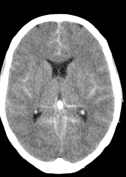
Treat pancreatitis with aggressive LR administration. Give 1-2 liters as a bolus then continue with a rate of 150-200ml/hour.

Patients generally need a total of 2.5 to 4 L of fluid over the first 12 to 24 hours.19,22 The specific rate of fluid delivery depends on the patient’s clinical status. In the situation of renal or heart failure, deliver fluid more slowly to prevent complications such as volume overload, pulmonary edema, and abdominal compartment syndrome. Crystalloids are the resuscitation fluids of choice. Normal saline in large volumes may cause a nongap hyperchloremic acidosis and can worsen pancreatitis, possibly by activating trypsinogen and making acinar cells more susceptible to injury.19,39 A single randomized study showed a decreased incidence of systemic inflammatory response syndrome in patients who received lactated Ringer’s instead of 0.9% normal saline.39 Regardless of which fluid is selected, monitor vital signs and urine output for response to hydration. (Tintinalli 8th edition)
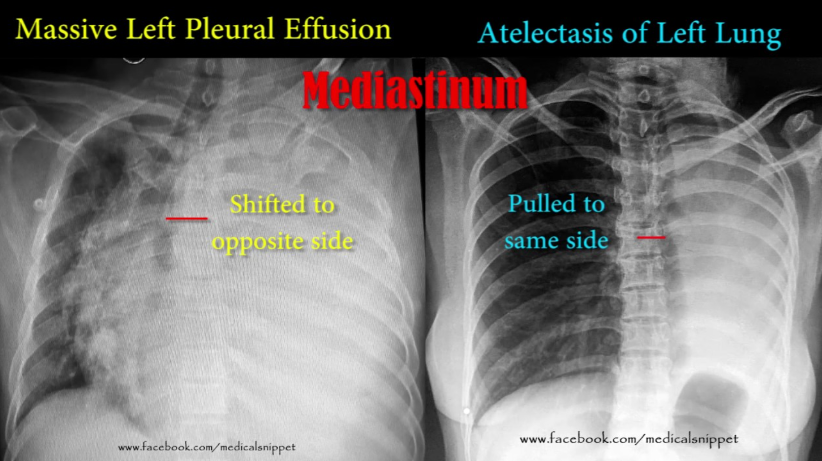
Mild pancreatitis does not have organ dysfunction. If a patient has SIRS or organ dysfunction they have moderate or severe pancreatitis. The three most common organ dysfunctions associated with pancreatitis are renal, cardiovascular, and pulmonary.
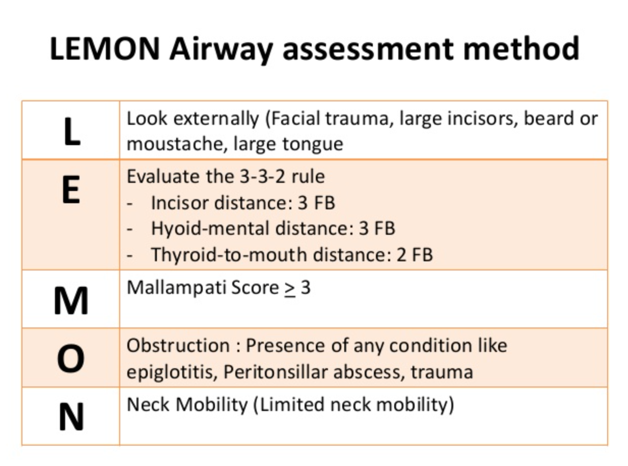
Sonographic Murphy’s sign is 97% specific. So if the patient has tenderness with the probe over a gallbladder with stones, they have cholecystitis.

Choledocolithiasis and Cholangitis both need GI and Gen Surg on consult. Patients with cholangitis also need IR to get source control by placing a percutaneous drain.

Abughnaim Healthcare Disparities

Health Disparities= Higher burden of illness for a specific group

Healthcare Disparity= Difference in access to care and quality of healthcare between groups.

Social determinants of health include many things outside of the healthcare system such as housing, income, racism, pollution, social and family support, the legal system, etc.

Consider the social differential diagnosis for different complaints. Lack of access, need to provide childcare, poverty, side effects of medications, can’t miss work, need to serve as a care giver to another person, prison, and homelessness are just some examples.

Some social determinants of health

Some social determinants of health

ED Care Managers can work through the social differential and figure out how to support a patient to improve their overall health and be able to be compliant with the treatment plan. Care Managers can arrange disposition from the ED to a SNF. They can arrange financial assistance. They can set up follow up appointments.


Conference Notes 11-14-2018

Logan M&M

No case details, just the take home points.

If you have to intubate a patient who has pulmonary hypertension or RV failure consider using push dose pressors to optimize BP prior to intubation and do an awake intubation with ketamine instead of RSI.

If a trauma patient says, “ Don’t let me die!” that usually portends badness. Be aggressive in managing these patients.

Be disciplined when doing your secondary survey. Even examine the patient’s mouth. You have to be very careful not to miss other injuries.

Always have a back-up plan when intubating a patient. Have all the tools you need to perform both your primary plan and your back up plan ready to go at the bedside.

Remember that narcan can cause your patient to vomit.

NALOXONE (NARCAN)

Naloxone is a pure opioid antagonist that works by competitively inhibiting narcotics at the opioid receptor. Intravenous administration reverses the respiratory depressive effects of opioids within 1 to 2 minutes. Its clinical duration of effect is approximately 20 to 30 minutes. Long-acting narcotics may cause resedation. The opioids and their metabolites are active longer than the reversal agent. Carefully monitor a patient receiving naloxone for resedation and respiratory depression. The drug can be delivered via multiple routes (i.e., endotracheally, intramuscularly, intravenously, subcutaneously, and sublingually). The administration of 1 to 2 mg intravenously in adults and 0.1 mg/kg in children will reverse most respiratory arrest situations. Administer additional doses every 2 to 3 minutes to a total of 10 mg. Actively seek another etiology of the sedation and respiratory depression, other than narcotics, if the respiratory depression is not reversed after 10 mg of naloxone. Use caution as naloxone can result in opioid withdrawal in those with physical dependence or intoxicated with narcotics.

Small aliquots of 40 µg titrated to effect may be delivered in a situation where the patient is slightly oversedated and rapid full reversal of the narcotic is not desired. Mix 0.4 mg of naloxone with 9 mL of normal saline to produce a concentration of 40 µg/mL. Administer 1 to 2 mL aliquots every 1 to 3 minutes to alleviate respiratory depression yet maintain the narcotic analgesic effect. (Reichman’s Emergency Medicine Procedures)

Don’t be afraid to call for help or back up. Being over-confident and getting in over your head can be problematic.

Be sure to talk with EMS when they drop off the patient in the ED. They can have critical info to give you that will make a difference in your managment.

For pediatric lacerations: LET applied with tegaderm works better than LET applied with gauze. Intra-nasal versed works well for sedation. Child life specialists can be very helpful to distract pediatric patients during laceration repair.

Lovell Procedural Sedation

For procedural sedation, we are usually shooting for moderate sedation. Sometimes we need to go to deep sedation for more painful procedures. Ketamine is in a different category altogether which is dissociative sedation. The patient retains blood pr…

For procedural sedation, we are usually shooting for moderate sedation. Sometimes we need to go to deep sedation for more painful procedures. Ketamine is in a different category altogether which is dissociative sedation. The patient retains blood pressure and airway reflexes with ketamine.

Moderate sedation is characterized by a depressed level of consciousness and a slower but purposeful motor response to simple verbal or tactile stimuli. Moderate sedation most closely matches the formerly used term "conscious sedation." Patients at this level generally have their eyes closed and respond slowly to verbal commands. Moderate sedation can be used for procedures in which detailed patient cooperation is not necessary, and muscular relaxation with diminished pain reaction is desired. During moderate sedation, the patient is usually able to maintain a patent airway with adequate respirations.9 Depending on the agent, the incidence of hypoxia and/or hypoventilation during moderate sedation is 10% to 30%.10,11,12 Procedures performed using moderate sedation include reduction of dislocated joints, thoracostomy tube insertion, and synchronized cardioversion. Agents used for moderate sedation in adults include propofol, etomidate, ketamine, methohexital, and the combination of fentanyl and midazolam.

Dissociative sedation is a type of moderate sedation. Dissociation is a state in which the cortical centers are prevented from receiving sensory stimuli, but cardiopulmonary activity and responses are preserved. Ketamine is the agent most commonly used for dissociative sedation.13

Deep sedation is characterized by a profoundly depressed level of consciousness, with a purposeful motor response elicited only after repeated or painful stimuli. Deep sedation may be required with procedures that are painful and require muscular relaxation with minimal patient reaction. The risk of losing airway patency or developing hypoxia or hypoventilation is greater with deep sedation than with moderate or minimal sedation.10,14,15 Examples of ED procedures sometimes requiring deep sedation are reducing fracture dislocations, open fracture reductions, and burn wound care. Deep sedation generally is achieved in the ED with the same agents as moderate sedation, but with larger or more frequent doses. (Tintinalli 8th edition)

Remember that propofol and etomidate do not provide analgesia. Propofol tends to have associated hypotension. Etomidate can stimulate vomiting. Etomidate can also cause myoclonus. Etomidate is associated with adrenal suppression but this is not a significant concern for brief procedural sedation.

Procedural sedation has low risk of aspiration. ACEP guidelines say NPO status has no relevance to procedural sedation.

FASTING STATE

There is no primary evidence that the risk of aspiration during procedural sedation is increased with recent oral intake.24,25,26 Current guidelines regarding the safe fasting period prior to procedural sedation were developed by expert consensus,27 and the American Society of Anesthesiologists guidelines for fasting prior to general anesthesia are of limited relevance to the risk of aspiration with ED procedural sedation.25 Thus recent food intake is not a contraindication.27 If the risk of aspiration is concerning, waiting 3 hours after the last oral intake before performing procedural sedation is associated with a low risk of aspiration, regardless of the level of sedation.27 (Tintinalli 8th edition)

Critical Questions and Recommendations

Question 1: In patients undergoing procedural sedation and analgesia in the ED, does preprocedural fasting demonstrate a reduction in the risk of emesis or aspiration?

Level B recommendations: Do not delay procedural sedation in adults or pediatrics in the ED based on fasting time. Preprocedural fasting for any duration has not demonstrated a reduced risk of emesis or aspiration when administering procedural sedation and analgesia. (ACEP Guideline)

Complications are primarily determined by the interaction of the depth  of sedation and the patient's current medical condition. A common tool  for assessing the patient's underlying medical condition is the American  Society of Anesthesiologists' p…

Complications are primarily determined by the interaction of the depth of sedation and the patient's current medical condition. A common tool for assessing the patient's underlying medical condition is the American Society of Anesthesiologists' physical status classification system.16 The risk of a significant complication from ED procedural sedation and analgesia in American Society of Anesthesiologists class I (healthy normal patient) and II (patient with mild systemic disease) is low, usually less than 5%.1,5,6,7,8 The risk of an adverse procedural sedation and analgesia event is correspondingly higher in patients with an American Society of Anesthesiologists class of III (patient with severe systemic disease) or IV (severe systemic disease that is a constant threat to life).17,18 (Tintinalli 8th edition)

If you get over these doses, toxicity will manifest as either CNS symptoms/signs or Cardiovascular toxicity. Treat toxicity with benzos for seizures, ACLS medications for shock, and intralipid.

If you get over these doses, toxicity will manifest as either CNS symptoms/signs or Cardiovascular toxicity. Treat toxicity with benzos for seizures, ACLS medications for shock, and intralipid.

EM Faculty Sedation Workshop

Putman/DenOuden Sedation Debrief

Do a formal Time-Out with every sedation. Use the time-out to go over your mental checklist assuring you have everything you need (suction, ambu bag, capnometry, medications, reversal agents, rescue devices, etc).

Whatever sedation agent you choose, be prepared for the possible side effects and complications known for that medication.

After sedation is complete and the patient is awake, inform the patient of how the sedation went and whether there were any difficulties such as apnea or need for assisted ventilations or hypotension.

Patient can go home following procedural sedation if they score a 9. Basically, normal mental status, normal vitals, can walk, talk, and cough.

Patient can go home following procedural sedation if they score a 9. Basically, normal mental status, normal vitals, can walk, talk, and cough.


Conference Notes 11-7-2018

Wing M&M

No case details, just a few take home points.

If a patient has signs of cardiac strain from PE or has other high risk factors (PESI score), consider admitting patient to the ICU or Step-down rather than the floor.

Snip20181107_1.png
The reported sensitivity and specificity of CT in demonstrating right heart dysfunction are around 81% and 47% respectively 5.Described features include:abnormal position of the interventricular septum 1 flattening of the interventricular septum par…

The reported sensitivity and specificity of CT in demonstrating right heart dysfunction are around 81% and 47% respectively 5.

Described features include:

abnormal position of the interventricular septum 1 flattening of the interventricular septum paradoxical interventricular septal bowing, i.e. towards the left ventricle

right ventricular enlargement (right ventricle bigger than the left ventricle)

pulmonary trunk enlargement (bigger than the aorta)

features of right heart failure: inferior vena caval contrast reflux, 1 dilated azygous venous system, dilated hepatic veins +/- with contrast reflux

D sign of LV

D sign of LV

McConnell’s sign

McConnell’s sign

Snip20181107_7.png

All the above indicators should prompt consideration of admission to a higher level of care (Step down or ICU)

Putman HEENT Study Guide

Don’t try to close lacerations near the medial canthus on either the upper or lower eyelids. There can be an injury to the tear duct. Consult Ophthalmology.

Don’t try to close lacerations near the medial canthus on either the upper or lower eyelids. There can be an injury to the tear duct. Consult Ophthalmology.

TRAUMATIC IRITISPatients with posttraumatic iritis usually present 1 to 2 days after blunt trauma to the eye, complaining of photophobia, pain, and tearing. They often have marked blepharospasm and perilimbal injection (ciliary flush). Test for pain…

TRAUMATIC IRITIS

Patients with posttraumatic iritis usually present 1 to 2 days after blunt trauma to the eye, complaining of photophobia, pain, and tearing. They often have marked blepharospasm and perilimbal injection (ciliary flush). Test for pain on accommodation by having the patient first look across the room at a distant object and then quickly focus on the examiner’s finger held several inches away. If near gaze causes pain, there is a high probability of iritis. The pupil may be large or small. Posttraumatic miosis develops secondary to spasm of the pupillary sphincter muscle, whereas posttraumatic mydriasis results when sphincter fibers are ruptured. Slit lamp examination will usually reveal cells in the anterior chamber, the hallmark of iritis.
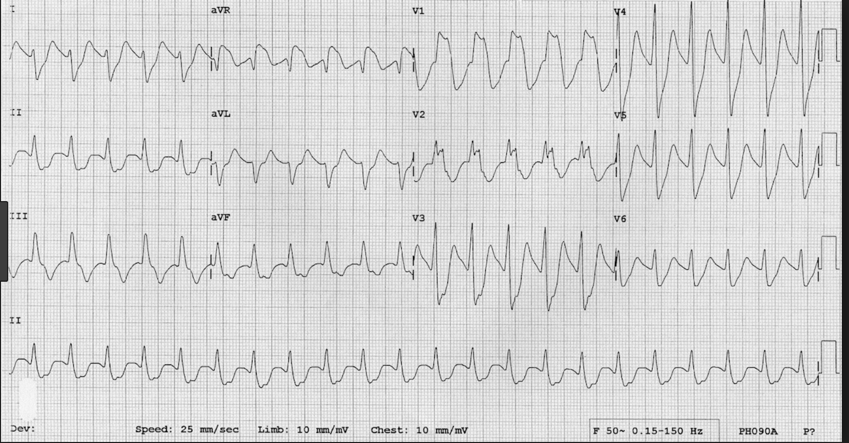
Treat with a long-acting topical cycloplegic, such as 5% homatropine, four times a day for 1 week, oral anti-inflammatory medication, and dark sunglasses to decrease pain. Symptoms may persist for up to 1 week. Although ocular steroids decrease inflammation, prescribe them only after consultation with the ophthalmologist who will see the patient in follow-up.

We commonly associate HSV keratitis with a dendritic pattern on the cornea

HSV keratitis on the surface of the cornea

HSV keratitis on the surface of the cornea

If HSV involves the deeper layers of the cornea, you can also see a disciform HSV keratitis.

Disciform HSV keratitis

Disciform HSV keratitis

Malignant otitis externa can be seen in diabetic patients. The most common complication is paralysis of the 7th cranial nerve.

Malignant otitis externa can be seen in diabetic patients. The most common complication is paralysis of the 7th cranial nerve.

Walchuk/Robinson Oral Boards

Case 1. 61yo female brought in by EMS with “stroke” symptoms. Dexi=155. Patient is altered and has slurred speech. Last time patient was normal is unclear. On further history, patient states she has been dizzy for a week. Patient is on phenytoin for seizures. Her phenytoin level is markedly elevated to 63. Treatment of phenytoin toxicity is supportive. Very severe toxicity may benefit from dialysis.

Phenytoin has a long and erratic absorption phase after oral overdose, so the decision to discharge or medically clear a patient for psychiatric evaluation cannot be based on one serum level. After acute ingestions, serum level should be measured every few hours. Patients with serious complications after an oral ingestion (seizures, coma, altered mental status, or significant ataxia) should be admitted for further evaluation and treatment. Those with mild symptoms should be observed in the ED and discharged once their levels of phenytoin are declining and they are clinically well. Mental health or psychiatric evaluation should be obtained, as indicated, in cases of intentional overdose. (Tintinalli 8th edition)

Snip20181107_3.png
Snip20181107_8.png

Case 2. 61 yo male brought in by EMS after a motorcycle crash. It was low speed accident and patient struck his head on the other vehicle. Patient has bilateral hand weakness. CT head and CT cervical spine show no acute abnormalities. MRI of the cervical spine shows:

Central Cord Syndrome is the most common spinal cord syndrome. It is seen most commonly in elderly patients with hyper-extension cervical spine injuries.

Central Cord Syndrome is the most common spinal cord syndrome. It is seen most commonly in elderly patients with hyper-extension cervical spine injuries.

Signs of Central Cord Syndrome

Signs of Central Cord Syndrome

The patient required C-spine stabilization and Neurosurgery consultation. Steroids are no longer recommended for central cord syndrome.

Case 3. 18yo male with right wrist injury from playing football.

Distal Radial-Ulnar Joint (DRUJ) dislocation should be suspected with any widening of the space between the distal radius and ulna. The lateral view shows to distal ulna displaced posterior to the radius.

Distal Radial-Ulnar Joint (DRUJ) dislocation should be suspected with any widening of the space between the distal radius and ulna. The lateral view shows to distal ulna displaced posterior to the radius.

Treatment is closed reduction, splinting the wrist, and orthopedic follow up. In some cases surgery is required.

Davis/Shroff/Friend ED BounceBacks

No case details, just a few take home points.

3 cases were presented. Each patient returned to the ED with a change in clinical picture. If the patient has an unclear diagnosis (belly pain, vague neurologic symptoms, back pain) and you are discharging them home, be sure to give clear discharge instructions describing signs/symptoms that should prompt return to the ED.

Dr. Williamson comment: These cases demonstrate the importance of communication with the patient. You want the patient to feel totally comfortable about returning to the ED for further evaluation.

Dr. Ryan comment: I tell the patient, “if your pain is getting worse, or you get a fever, or some new problem develops, that should not be happening. If it does happen, that is a sign that something is wrong and you need to come back to the ER.” Also give patients a time frame on when they should be feeling better. If they are not feeling better by then, they need to return for further evaluation.

Ebeledike/Johns Safety Lecture Choosing In-Patient Level of Care

Choosing the appropriate level of inpatient care for a specific patient (Floor, Telemetry, Step Down, or ICU) can be challenging.

Examples of diagnoses suitable for telemetry: Stable NSTEMI, syncope presumed to be cardiac, arrhythmia/heart blocks, pacemaker or ICD problem.

Examples of Patients suitable for Step-Down: Patient on a single pressor, chronically ventilated patients, patients requiring a high level of nursing care, and patients with significant risk of deterioration.

In general, if you are concerned that a patient may deteriorate or decompensate, strongly consider placing them in Step-Down rather than the floor.

If the patient is critically ill put them in the ICU.

Dennis Ryan comment: Consult with the ICU attending to collaborate on whether a patient belongs in Step-Down or the ICU to better determine level of care.

Editor’s comment: Suggested simplified algorithm. Stable patients go to the floor/med-tele/telemetry. If you are worried the patient may deteriorate consider Step-down. Critically ill patients go to ICU.

Lorenz/Shroff Visual Diagnosis

The Chiefs presented multiple clinical pictures for pattern recognition. This outstanding presentation moved too fast for me to capture.

Conference Notes 10-31-2018

Twanow M&M

No case details just take home points.

Top 10 Lessons in residency:

10. If you want to be fast in the ED, focus on dispo’s. Make your dispo’s prior to seeing new patients.

9. Be kind to yourself. We have a hard job. Beating yourself up after a less than optimal case is not productive and can be self-destructive.

8. If you feel like something is wrong with a patient, listen to your gut instincts and work them up or re-evaluate them.

7. If your patient is not responding to therapy, you may be missing something. There may be another diagnosis that has not been identified.

6. Stay late for the right things (education, critical patient care) but know when to call it a day. It’s good to put in the effort to learn and care for patients. On the other hand you have to recognize when you are fatigued and not able to be the best for yourself and your patients.

5. Don’t send unstable patients to the CT scanner.

4. You will be in uncomfortable clinical situations at times but know who your backup is.

3. Some patients can be challenging to get along with, but beware, these challenging patients can still be sick with serious disease.

2. Our patients are our responsibility. This includes new patients, signed out patients, difficult patients, all of them.

  1. Be sure to take care of your colleagues. We all need each other to do this job.

Carlson Salicylate Toxicology

Many OTC products contain salicylate. Oil of wintergreen (methylsalicylate) has very high levels of salicylate.

Get serial serum levels of salicylate when managing salicylate overdose patients.

Salicyate causes neurostimulation resulting in tinnitus and increased respiratory rate and vomiting. It increases capillary permeability and can cause pulmonary edema. It uncouples oxidative phosphorylation and will result in lactic acidosis.

Snip20181031_3.png

Andrea made the point that if a salicylate toxic patient becomes lethargic or somnolent you’ve got big problems. Lethargy is a sign of brain dsyfunction, which is the main cause of deaths from salicylate.

Chronic salicylate toxicity is more lethal than acute poisoning. Chronic poisoning has high brain tissue levels despite modest blood levels.

Beware of brain symptoms such as lethargy and somnolence. Andrea made the point that salicylate-toxic patients most often die from a CNS death.

Beware of brain symptoms such as lethargy and somnolence. Andrea made the point that salicylate-toxic patients most often die from a CNS death.

Guidelines for hemodialysis in salicylate overdose. (EXTRIP Guidelines)

Guidelines for hemodialysis in salicylate overdose. (EXTRIP Guidelines)

Activated charcoal if given very early, before symptoms develop, binds salicylate very well. If a patient is symptomatic it is likely too late to benefit from activated charcoal.

Treat non-cardiogenic pulmonary edema with peep. You can try BiPap. You can intubate but it is dangerous because of the acidosis. It is difficult for a ventilator to keep up with the patient’s minute ventilation. If you have to intubate, use larger tidal volumes (around 8ml/kg) and high respiratory rate (40). If you can avoid using a neuromuscular blocker that would be optimal. Give 2 amps of bicarb prior to intubation to help manage the acidosis and possibly avoid a peri-intubation arrest.

Urinary Alkalinization: Put 3 amps of bicarb in a 1L bag of D5W and run at 250ml/hr. Add 20-40 meq of potassium to each liter. Shoot for a urine ph >7.5.

Carlson/Pastore Oral Boards

Case 1. 46yo female presents with suicide attempt. Patient is unresponsive. Pupils are pinpoint. Patient responded to narcan. Further history identified that patient ingested Zohydro (extended release hydrocodone). As ED course progressed, patient became re-sedated requiring re-dosing of narcan and starting a narcan drip.

Methadone, fentanyl, tramadol and buprenorphine will not show up positive on drug screen.

Synthetic opioids, such as dextromethorphan, fentanyl, meperidine, methadone, propoxyphene, and tramadol, show little or no cross-reactivity in opiate immunoassays. Urine immunoassays specific for meperidine, methadone, and propoxyphene are available. Given the increasing importance of buprenorphine as maintenance therapy for opioid dependency, it is worth noting that the combination of high potency and low cross-reactivity means that buprenorphine will generally not be detected by opiate immunoassays. Immunoassays for specific detection of buprenorphine have therefore been developed. (Goldfrank’s Toxicology)

Case 2. 19yo female presents with nausea/vomiting and abdominal pain. Patient is pregnant. U/S of pelvis shown below.

Patients with molar pregnancy may have larger uterus and may have very high beta-hcg.

Patients with molar pregnancy may have larger uterus and may have very high beta-hcg.


Patients with molar pregnancy are at risk for trophblastic malignancy and need follow up. There is also risk of ovarian torsion.

Symptoms include vaginal bleeding in the first or second trimester (75% to 95% of cases) and hyperemesis (26%). Gestational trophoblastic disease, or molar pregnancies that persist into the second trimester, are associated with pre-eclampsia. When pregnancy-induced hypertension is seen before 24 weeks of gestation, consider the possibility of a molar pregnancy. The uterus is excessive in size for gestational age and shows a placenta with many lucent areas interspersed with brighter areas on US study. Because not all molar pregnancies are found on US, all tissue extracted from the uterus on suction curettage or during pelvic examination should be sent for histologic examination. If trophoblastic disease is suspected because of abnormally high β-hCG levels, a uterine size either larger or smaller than expected, and US findings suggestive of the diagnosis, obtain obstetric consultation. Treatment is by suction curettage in the hospital setting because of risk of hemorrhage. β-hCG levels that fail to decrease after evaluation are evidence of persistent or invasive disease necessitating chemotherapy. Metastasis to lung, liver, and brain may occur, but the prognosis for most patients is very good. (Tintinalli 8th ed.)

A molar pregnancy is caused by an abnormally fertilized egg. Human cells normally contain 23 pairs of chromosomes. One chromosome in each pair comes from the father, the other from the mother.

In a complete molar pregnancy, an empty egg is fertilized by one or two sperm, and all of the genetic material is from the father. In this situation, the chromosomes from the mother's egg are lost or inactivated and the father's chromosomes are duplicated.

In a partial or incomplete molar pregnancy, the mother's chromosomes remain but the father provides two sets of chromosomes. As a result, the embryo has 69 chromosomes instead of 46. This most often occurs when two sperm fertilize an egg, resulting in an extra copy of the father's genetic material. (Mayo Clinic online)

Case 3. 25 yo male presents with “allergic reaction” for 2 weeks. Vitals normal. Patient has pruritic rash.

Rash c/w scabies

Rash c/w scabies

Typical locations of scabies rash

Typical locations of scabies rash

A single application of 5% permethrin cream is curative for children older than 2 months. The cream may be applied to the face and scalp and needs to be left for 8 hours. Permethrin has been found to have a 97.8% cure rate with one application.10 Permethrin may cause burning and stinging as well as exacerbation of itching, although it is generally very well tolerated and has low potential for toxicity.11 The long incubation period makes treating the entire family advisable. Bedding and clothing should be laundered in hot water and dried using the hot cycle. Clothing and other materials that cannot be laundered should be removed and stored for several days to a week to avoid reinfestation.13 (Pediatric Emergency Medicine)

From Pediatric Emergency Medicine 5th Ed

From Pediatric Emergency Medicine 5th Ed

Lovell Recruiting Season Update

Berklehammer Inflammatory Bowel Disease

Snip20181031_7.png
Toxic megacolon can be seen with either ulcerative colitis or crohn’s disease. Bowel is &gt;6cm in diameter. There is loss of haustra.

Toxic megacolon can be seen with either ulcerative colitis or crohn’s disease. Bowel is >6cm in diameter. There is loss of haustra.

Crohn’s disease patients can get a stricture at the terminal ileum resulting in bowel obstruction.

Crohn’s disease patients can get a stricture at the terminal ileum resulting in bowel obstruction.

Pyoderma gangrenosum (PG) is an inflammatory condition of all ages but is most common among 20- to 50-year-old females. Lesions can be located anywhere (most commonly on the lower extremities) and begin as a papulopustule surrounded by erythema. Thi…

Pyoderma gangrenosum (PG) is an inflammatory condition of all ages but is most common among 20- to 50-year-old females. Lesions can be located anywhere (most commonly on the lower extremities) and begin as a papulopustule surrounded by erythema. This pustule erodes to form a necrotic ulcer. Similar satellite pustules and ulcers form around the original lesion and eventually coalesce into a large ulcer. The surrounding border is “rolled,” due to the convex elevation, and has a violaceous hue. The ulcers are exquisitely tender to movement and palpation. On the extremities, the ulcers can rapidly involve muscles and tendons. Ostomy sites are a common location and make care very difficult.

Half of cases are idiopathic; the other half are associated with inflammatory bowel disease, hematologic diseases (leukemia, myelodysplasia, monoclonal gammopathy), and the arthritides. Since diagnosis is based on examination, dermatopathology, and exclusion of other causes, it is difficult to confirm. (Atlas of Emergency Medicine)

In patients who have crohn’s disease, be very cautious of complaints of back pain or buttock pain. Patient’s with crohn’s can get deep tissue abscesses from fistulas that directly spread to back or buttock musculature.


Conference Notes 10-10-2018

Paquette/Friend Oral Boards

Case 1. 45 yo female with left knee injury while doing yoga.

Lateral tibial plateau fracture

Lateral tibial plateau fracture

Tibial plateau fractures can be difficult to diagnose. Soft tissue injuries associated with tibial plateau fractures may influence outcomes. Anterior cruciate ligament and medial collateral ligament injuries are associated with lateral plateau fractures, whereas posterior cruciate and lateral collateral ligament injuries occur with medial plateau fractures. A Segond's fracture (see below) is pathognomonic for an anterior cruciate ligament injury, and it is important recognize and treat the ligament injury, rather than just the plateau fracture.12 Potential complications of tibial plateau fractures include popliteal artery injury with high-energy displaced fractures, the development of deep venous thrombosis, and osteoarthritis. (Tintinalli 8th ed)

Segond Fracture

Segond Fracture


Case 2. Pregnant female patient presents with abdominal pain and syncope.

Fast exam shows free fluid in Morrison’s pouch.  In the setting of early pregnancy with abdominal pain this finding is highly suggestive of ruptured ectopic pregnancy.

Fast exam shows free fluid in Morrison’s pouch. In the setting of early pregnancy with abdominal pain this finding is highly suggestive of ruptured ectopic pregnancy.

Patient was diagnosed with ruptured ectopic pregnancy and was taken to the OR.

Case 3. 40yo male presents with altered mental status and hypotension. EKG is shown below.

3rd degree heart block

3rd degree heart block

Patient ingested a toxic dose of digoxin.

Digoxin poisoning can induce nearly every form of dysrhythmia or conduction disturbance. Classic ECG findings include supraventricular tachydysrhythmias (atrial flutter or fibrillation) combined with variable AV nodal blockade resulting in slow ventricular rates (Figure 59-1). Bidirectional ventricular tachycardia is nearly pathognomonic for serious digoxin toxicity. Additional ECG findings include sinus bradycardia, ventricular bigeminy, and ventricular fibrillation. (Tintinalli 8th ed)

Bidirectional ventricular tachycardia which is highly specific for digoxin toxicity

Bidirectional ventricular tachycardia which is highly specific for digoxin toxicity

Chinwala M&M

To protect the anonymity of the case, I will only give some take home points.

Be sure to re-eval sign-out patients who are altered or intoxicated.

If you initiate a treatment, be sure you re-evaluate the patient in a timely fashion to assess how the treatment is working and how the patient is doing.

Lorenz/Shroff Toxicology Escape Room

Anion Gap calculation= “ABC” NA-(Bicarb +Chloride)

Osmolar Gap calculation streamlined= 2NA + BUN/3 +Glucose/20 + ETOH/5 (ETOH is actually 4.6 and the way to remember this is 4 6packs in a case of beer.)

ASA, Lithium, Toxic Alcohols (ALT)= things you can dialyze.

TCA Overdose EKG. Wide terminal R wave in AVR and wide QRS complex in all leads. Treat with IV bicarb.

TCA Overdose EKG. Wide terminal R wave in AVR and wide QRS complex in all leads. Treat with IV bicarb.

Ricin is a protein that inhibits ribosomes. It is used as a bioterrorism agent. It is dervied from the castor plant.

Ricin is a protein that inhibits ribosomes. It is used as a bioterrorism agent. It is dervied from the castor plant.

Foxglove is a botanical cardiac glycoside. Basically it can cause digoxin toxicity. Treat with FAB fragments.

Foxglove is a botanical cardiac glycoside. Basically it can cause digoxin toxicity. Treat with FAB fragments.

Amanita phylloides is the most toxic mushroom. Dr. Carlson noted the “death cup” at the base of the mushroom.

Amanita phylloides is the most toxic mushroom. Dr. Carlson noted the “death cup” at the base of the mushroom.

Dr. Carlson recommended using IV NAC for all patients with acetaminophen toxicity for practical reasons. Pregnant patients need IV NAC to get better fetal treatment.

Dr. Lovell comment: Critical Factoid for figuring out how many grams are in a given volume of solution. %=grams/deciliter. So 0.9% NS has 0.9 grams of saline in a deciliter and by extrapolation, 9 grams of saline in a liter of fluid.

Conference Notes 10-3-2018

Girzadas/Chinwala Oral Boards

Case 1. 55yo male presents bradycardic and hypotensive after beta blocker overdose.

Tintinalli algorithm for different modalities to treat beta blocker overdose.

Tintinalli algorithm for different modalities to treat beta blocker overdose.


Case2. 7 yo male with GSW to the right thigh. Patient is hypotensive and tachycardic. Patient was resuscitated with IV crystalloid and IV PRBC transfusions. Patient had abnormal ABI and soft signs of vascular injury. Patient had CTA showing vascular injury an patient was taken to the OR.

Snip20181003_5.png
Tintinalli algorithm for management of penetrating vascular trauma.

Tintinalli algorithm for management of penetrating vascular trauma.


Case 3. 4yo male who had non-fatal drowning.


If normal O2 sat, normal mental status, normal lung exam after 4-6 hours patient can go home.

If normal O2 sat, normal mental status, normal lung exam after 4-6 hours patient can go home.

Pecha Kucha

Robinson Foley Catheters

The most frequent complication of urethral catheterization is infection. Foleys are the #1 cause of nosocomial infections so use them judiciously.

Patients are colonized within about a week of an indwelling foley catheter. So diagnose UTI only in symptomatic patients and/or with positive culture results.

Dr. Lovell comment: If the nurse can’t place the foley, the emergency physician needs to attempt placement of foley prior to consulting GU.

Florek Tracheostomy Problems in the ED

Risk of tracheo-innominate fistula is highest at 7-14 days after surgery.

Lubricate the Shiley prior to placing it in the tracheostomy site.

You can attempt to oxygenate patients with a mask over the patient’s mouth or over the trach site.

Some patients are neck breathers and can’t exchange air through the mouth (laryngectomy, laryngeal mass)

Most common cause of tracheostomy bleeding is mucosal irritation. Worst cause is tracheoinnominate fistula.

To control TI fistula first overinflate the shiley balloon to tamponade the bleeding. If that is ineffective you can insert your finger in the tracheostomy site and compress against the sternum

To control TI fistula first overinflate the shiley balloon to tamponade the bleeding. If that is ineffective you can insert your finger in the tracheostomy site and compress against the sternum

Tracheoinnominate fistulas are quite rare, occurring in less than 2% of cases, but they carry a mortality rate of 25% to 50%.9 They may present as the classic “exsanguinating bleed” but often present with a less impressive sentinel bleed. Any bleeding of more than a few milliliters of blood should raise concern for a possible fistula of the innominate artery. Prompt critical resuscitation measures and emergent consultation with a Vascular Surgeon and Otolaryngologic Surgeon is required. Definitive management is surgical. Techniques for temporarily controlling bleeding from the innominate artery include local digital pressure, hyperinflation of the tracheostomy tube cuff, and traction on the tracheostomy tube. An alternative method is to deflate the tracheostomy tube cuff, reposition the cuff at the bleeding site, and then reinflate or hyperinflate the cuff. When bleeding occurs, the tracheostomy tube should not be removed until the airway is secured by another means from above (orally or nasally). (EM Procedures Reference)


Erbach Dialysis Access Complications
50% of dialysis catheters develop an infection within 6 months.

AV grafts have 10% rate of infection, and AV fistulas have a 5% rate of infection

Bleeding from dialysis graft, treat with direct pressure or quickclot gauze. Consult with Vascular Surgery. Below are some other suggestions from Tintinalli.

Snip20181005_1.png


Friend G-tubes

If G-tube is clogged, first try with flushing with warm water. If that won’t work, your can try cola.

If a g-tube site is less than 4 weeks old don’t replace the tube and just consult GI. In the first 4 weeks, the site is not fully mature and replacing the tube could end up in the wrong place. After 4 weeks we need to get the tube replaced as soon as possible to avoid closure of g-tube site.

Don’t replace J-tubes.

Gastrograffen in SubQ

Gastrograffen in SubQ

Intraperitoneal gastrograffen

Intraperitoneal gastrograffen

Lorenz Insulin Pumps

Insulin pumps give a basal rate of insulin and bolus dosing when a patient eats. Patients can also have long term glucose monitors for multiple days in the skin that communicate directly with the insulin pump or an apple watch.

Patients who have pumps who are hypoglycemic, treat first with glucose then second disconnect the pump.

You can see a patient’s bolus history by reviewing their pump data.

Chinwala VP Shunts

Obstruction is more common in the first year after placement. Proximal obstruction is due to choroid plexus and distal obstruction is due to thrombosis.

Staley The Febrile Neonate

Temp of 38C at home or in the ED is considered positive for fever in the infant.

In neonates with fever, the risk for serious bacterial infection is about 13%.

The clinical appearance of a neonate does not predict serious bacterial illness. You will need to rely on tests to identify serious bacterial illness in this age group.

For patients 0-28 days of age with a fever of 38C or higher, do full septic workup. Get a CXR only for patients with respiratory symptoms such as cough or increased work of breathing. Get a NP swab to screen for RSV. Give ABX within 1 hour for these very young children. (Amp and Cefotaxime) add Vanco if the infant is critically ill or mom was treated for Group B strep.

If LFT’s are elevated, get HSV serology and start IV Acyclovir. Elevated LFT’s are a sign of HSV infection.

For patients aged 29-60 days with fever, get a CBC, blood culture, UA and urine culture. Urine studies are the highest yield tests. Again, limit CXR’s to patients with respiratory symptoms. Get a Procal, CRP and viral testing. If biomarkers are negative don’t do LP in this age group. If you don’t do LP, discharge without antibiotics. If biomarkers (WBC, Procal, CRP) are elevated, do an LP and start antibiotics. If UA is positive get a blood culture (bacteremia 10%) and consider LP (meningitis 1/200). If you do an LP and decide to treat with antibiotics, give ceftriaxone. Any discharged patient needs arranged close follow up.

Infants less than 60 days who are fussy at home and have temps close to febrile (close to 38C) should be observed in ED for a couple of hours and have temp rechecked. Consider getting a CBC, blood culture, UA, and urine culture in these borderline kids.

In infants under 60 days, a temp >/=40C indicates a 40% risk of serious bacterial illness.

Tips for doing LP’s in infants: put the CPR compression board under the patient to keep the patient from sinking into the bed. After you insert the LP needle through the skin, you can remove the stylet and advance the needle without the stylet so that you can identify CSF as soon as you enter the CSF space.

Insert the LP needle between the L4 and L5 spinous processes in the intervertebral space in the midline of the back, and direct the needle toward the umbilicus. This interspace is easily located because it lies in line between the iliac crests. Introduce the needle with the bevel of the needle up. Insert the needle until the characteristic "pop" identifies introduction into the subarachnoid space. An alternative method is to remove the stylet from the needle49 after the needle pierces the skin. Advance the needle, without the stylet, incrementally until CSF flows. Occasionally rotating the lumbar needle clockwise or counterclockwise up to 360 degrees may help improve flow if the bevel of the needle is sideways. When removing the lumbar needle, replace the stylet. (Tintinalli 8th ed)

Barounis PE and Pulmonary Embolism Response Team

From Dr. Barounis: Here are some takeaways from today's lecture, please share with other residents. I included a lot of the literature that I had obtained for the lecture so feel free to review yourself and see if you come up with different takeaways.

Imaging:

CT scans, not V/Q's for sick people please!

Massive PE:

1. Massive PE is life-threatening and immediate thrombolysis is recommended by all major societies. At ACMC surgical embolectomy can be considered if immediately available as an alternative in candidates for surgery, or when thrombolysis has been attempted and unsuccessful.

DOSE 10mg bolus, followed by 90mg over 2 hours. Consider MOPETT dosing in high bleeding risk patients (see below).

2. Hold heparin when starting tPA as there appears to be no benefit with likely a higher likelihood of bleeding.

3. Obtain a fibrinogen level prior to beginning thrombolysis (same time you are getting PT/INR/PTT). If fibrinogen is dropping consider stopping tPA, hold heparin.

4. Try high flow nasal cannula, and avoid intubation when possible prior to thrombolysis given high propensity for cardiac arrest during intubation. If diagnosis is uncertain, will require clinician judgement. Most patients die from shock, RV failure >>> hypoxemia

5. CALL 40-0702 for help This will activate the PE response team

6. Start pressors early, probably can avoid volume loading altogether. (RV perfused by MAP, not DBP)


Submassive PE:

1. Thrombolysis is indicated to reduce risk of Hemodynamic decompensation and development of respiratory failure, in the select cohort of patients at high risk of deterioration (prognostic features below)

2. The data on ultrasound-assisted thrombolysis has not been consistently shown to improve long-term outcomes, see the ULTIMA trial/ SEATTLE II trial (included below) based on RV/LV ratio @ 3 and 6 months. It did show reduce RV/LV ratio at 24 hours. ULTIMA is the only RCT comparing EKOS to heparin. The PEITHO trial also showed no reduction in PH, or death at 24 month f/u in patients who received tenecteplase vs. placebo. IT APPEARS THERE IS SHORT TERM BENEFIT, but LONG TERM patients own fibrinolytic system appears to do the job.

3. Ultrasound assisted thrombolysis has not shown improvement in clot resolution over plain catheter directed thrombolysis in the only RCT comparing the two (see below, engelberger). Each ekos catheter is around $1,100.

4. Lower dose tPA appears safe and equally effective, and is appealing in patients with submassive PE to avoid hemorrhagic complications. Currently this is done using CDT with ekos at ACMC, and future studies may prove that lower dose controlled thrombolysis will be equally efficacious?? MOPETT dosing tPA for submassive PE with high risk features, or even stable massive PE

5. Poor prognostic features: tachycardia, tachypnea, hypoxemia, lactic acidosis (>2), PESI SCORE, BOVA SCORE, poor cardiopulmonary reserve.

6. Patients with concomitant DVT and PE have higher inpatient mortality than PE alone.


PERT TEAM:

Call 40-0702; please obtain the labs provided in order set (top righ thand side of PERT DOCUMENT included below).

Best,

Dave

Proposed Management Algorithm for Massive and Sub-massive PE

Proposed Management Algorithm for Massive and Sub-massive PE


Conference Notes 9-19-2018

Hart/Nakitende U/S Monopoly

Snip20180919_1.png
You can measure the width of the pericardial fluid to grade the severity of a pericardial effusion. More than a centimeter (10mm) is significant.

You can measure the width of the pericardial fluid to grade the severity of a pericardial effusion. More than a centimeter (10mm) is significant.

An echo finding suggestive of tamponade is incomplete filling of RV in diastole. The RV wall will be scalloped.

Large pericardial effusion with incomplete filling/scalloping of RV suggestive of tamponade

Large pericardial effusion with incomplete filling/scalloping of RV suggestive of tamponade

When using echo to differentiate acute PE vs chronic pulmonary hypertension, the RV wall in acute PE will be thin while the RV wall in chronic pulmonary hypertension will be hypertrophied.

D-sign showing PE. Elevated pressure in the RV flattens the LV septal wall making the LV look like the letter D on a parasternal short view.

D-sign showing PE. Elevated pressure in the RV flattens the LV septal wall making the LV look like the letter D on a parasternal short view.


McConnell sign. The apex of the RV contracts OK despite overall RV hypokinesis. This is a specific sign of PE.

McConnell sign. The apex of the RV contracts OK despite overall RV hypokinesis. This is a specific sign of PE.

Snip20180919_7.png

Bartgen/Erbach Oral Boards

Case 1. 75yo female with fever, cough, and wheezing. O2 sat 92%. CXR shows pneumonia. Patient has SIRS. IV fluids and IV antibiotics started. Patient deteriorated late in her ED course with worsening weakness. Son noted that patient has had weakness at the end of each day for weeks now. Patient was diagnosed with Myasthenic crisis precipitated by pneumonia/sepsis. Treatment with plasmaphoresis was arranged. IVIG and steroids are also indicated. Airway needed to be managed due to expected course of illness with worsening weakness.

Dr. Bartgen made the following points about nueromuscular blocking agents in the setting of myasthenic crisis. I used a Tintinalli reference to encapsulate his comments:

The most significant ED complication of myasthenia gravis is respiratory failure, which is usually precipitated by infection, surgery, or the rapid tapering of immunosuppressive drugs. Although intubation should be considered in patients with a low forced vital capacity or in the presence of abnormal blood gas analysis, this decision is made primarily on clinical grounds. Patients may have increased sensitivity to nondepolarizing agents based on their concurrent use of acetylcholinesterase inhibitors. Additionally, they can have either resistance or prolonged duration from depolarizing agents. Because of the increased sensitivity of myasthenia gravis patients to neuromuscular junction inhibitors and an unpredictable reaction to succinylcholine in particular, avoid the administration of depolarizing or nondepolarizing paralytic agents in preparation for intubation.27 Patients with myasthenia are extremely sensitive to these agents, and the paralytic effects can be expected to persist at least two to three times longer than in normal patients. Consider using short-acting agents such as fentanyl or propofol in smaller doses, as it is important to avoid further respiratory depression. Sugammadex may be used to reverse rocuronium if necessary.28 If paralytic agents are absolutely necessary, consider using one with a shorter half-life, such as etomidate, at one-half the dose of these agents, although this recommendation is anecdotal. (Tintinalli 8th ed.)

Case 2. 70yo female with cough, shortness of breath and O2 sat of 85%. Patient was cleaning bathroom with a mix of clorox bleach and lime-away. The combo of these cleaning supplies caused strong fumes that overcame the patient. Mixing bleach with either an acid or ammonia can cause the release of chlorine or chloramine gas. The patient was treated with intubation. Steroids should be given to intubated patients with lung injury due to chlorine gas.

Case 3. 90yo female presents with scalp rash for about 6 weeks. The rash had a boggy consistency. Diagnosis was a kerion which is more common in pediatric patients and the elderly.

Kerion, treat with griseofulvin or fluconazole. Kerion causes hair loss which can help differentiate it from other scalp lesions.

Kerion, treat with griseofulvin or fluconazole. Kerion causes hair loss which can help differentiate it from other scalp lesions.

Tinea capitis (scalp) presents as a pruritic, erythematous, scaly plaque. This may develop into a delayed-type hypersensitivity reaction, where the initial erythematous, scaly plaque becomes boggy with inflamed, purulent nodules and plaques (kerion). The hair follicle is frequently destroyed by the inflammatory process in a kerion, leading to a scarring alopecia. Systemic antifungals are required to treat tinea capitis infections. Due to the long-term treatment requirement and associated side effects, referral to a dermatologist is recommended. (Tintinalli 8th ed.)

Dr. Napier comment: Consider checking LFT’s as a baseline prior to starting griseofulvin.

Ginsburg Endovascular Treatment of PE

RV/LV ratio 0.9 or greater is a sign of RV strain on CTPE study.

Massive PE patients are candidates for systemic TPA. Sub-massive PE patients are candidates for catheter directed thrombolysis. Patients with a low risk of bleeding who have RV dysfunction and elevated troponin are probably the most likely to benefi…

Massive PE patients are candidates for systemic TPA. Sub-massive PE patients are candidates for catheter directed thrombolysis. Patients with a low risk of bleeding who have RV dysfunction and elevated troponin are probably the most likely to benefit from catheter direct thrombolysis in the submassive group.

High risk PESI scores warrant consideration of ICU admit.

High risk PESI scores warrant consideration of ICU admit.

Risk of ICH with systemic TPA is 3%. Risk of ICH on heparin is 0.3%

50% of patients with massive PE have a contraindication to TPA.

This slide just gives an idea of the total incidence of PE and PE deaths in the US.  As a comparison  MVC’s account for 30-40,000 deaths/year and drug overdoses account for approximately 80,000 deaths/year.

This slide just gives an idea of the total incidence of PE and PE deaths in the US. As a comparison MVC’s account for 30-40,000 deaths/year and drug overdoses account for approximately 80,000 deaths/year.

Schroeder Management of DKA

Snip20180919_12.png

Type 1 DM has a genetic component but it is not all genetic. Only 40% of identical twins will have Type 1 DM.

Snip20180919_14.png

DKA patients are more dehydrated than they look because they are losing intracellular water with glucose in the urine rather than water and sodium in the stool.

Expect DKA patients to be potassium depleted. Begin replacing potassium very early in the management of DKA.

Younger kids, those with limited access to care, and low BMI kids are more likely to present in DKA and are more likely to have a delay in diagnosis.

If a patient has an insulin pump and presents in DKA, turn off the pump and treat with IV insulin. It is better to have complete control of the insulin the patient is receiving.

There is no indication to give an initial bolus of insulin. Just start a continuous drip drip after first priming the line with insulin. The IV tubing adheres the initial insulin and if you don’t first prime the line with insulin, it can take hours before the patient starts receiving insulin because the infusion is getting absorbed by the IV tubing.

10ml/kg bolus is probably fine and safe as your initial fluid administration. Use LR to avoid hyperchloremic acidosis associated with NS.

PECARN IV Fluid in DKA Study (NEJM 2018)

Critical Results:

  • 1,389 distinct episodes of DKA were evaluated in 1,255 patients, but only 1361 episodes of DKA were included in the primary analysis. 132 children had a second episode of DKA and underwent randomization as well.

  • There were a total of 48 episodes (3.5%) resulting in a GCS <14 with 22 episodes (1.6%) requiring hyperosmolar therapy for cerebral edema, and 12 episodes (0.9%) of clinically apparent brain injury.

  • There were no statistically significant differences in the percentage of episodes among the 4 groups where the GSC decreased to less than 14, the magnitude of decline or duration GCS remained less than 14

  • Incidence of a GCS decline <14 & clinically apparent brain injury was actually lower in fast rehydration groups (21 vs 27 & 4 vs 8 episodes respectively), but both were not statistically significant

  • Memory assessed by forward and backward digit-span scoring did not significantly differ between the 4 groups

  • Hyperchloremic acidosis was more common among patient receiving 0.9% NaCl vs 0.45% NaCl and more common in patients receiving fluid at a rapid rate vs slow rate

DKA Protocol for ACMC/Hope Children’s Hospital

DKA Protocol for ACMC/Hope Children’s Hospital

Tekwani Medical Student Rotation Review


Conference Notes 9-5-2018

Lorenz       Important Recent EM Papers

HEART Score was validated on 2440 patients in the Netherlands.

Snip20180905_1.png

Procamio Study  June 2016

Procainamide works better than Amiodarone for stable v-tach with less adverse events.

Revert Study August 2015

Initially attempt to treat SVT with postural modification to valsalva maneuver. Put head of bed at 40 degrees and have patient blow in syringe for 15 seconds. Then lay patient flat and raise legs.  Then sit them back up.

Try this modified valsalva maneuver to terminate SVT. It works 25% of the time.

Try this modified valsalva maneuver to terminate SVT. It works 25% of the time.

Dr. Lovell comment: If patient has alot of anxiety about adenosine, consider propofol to sedate patient.

Aromatherapy vs Zofran Study      August 2018

Olfactory distraction works for nausea. For patients who don't have an IV and have nausea, Inhaled isopropyl alcohol from an alcohol wipe works as well or better than zofran.   Consider this for a patient with nausea. Patients get relief for 30-60minutes.

Haldol for Gastroparesis    Acad Emerg Med 2017

5mg IM was very effective for gastroparesis symptoms.

Dr. Lovell comment: Haldol treats both pain and nausea.  Watch for QT prolongation

Treating Hypokalemia with Low-dose insulin

In patients with CKD/ESRD,  5u of insulin works just as well as 10u of insulin with much less hypoglycemia.

Flomax for Kidney Stones 2018

For stones in the distal ureter, 4-7mm, flomax worked better, faster, with less painful passage of stone. Number needed to treat is high though.  Avoid flomax in patients with risk factors for CAD.

BICAR-ICU Study

Bicarb for anion gap acidosis in the ICU did not lessen mortality or organ failure but it did decreased the need for dialysis.

Dr. Lovell: This study is a game changer for acidotic patients.  Most of these patients with anion gap acidosis had sepsis.

PEITHO   Systemic TPA for PE

Normotensive PE patients with RV strain on echo who got TPA and heparin did not have improved mortality compared to heparin alone. TPA patients did have greater adverse bleeding outcomes.

Procedure Lab